. 2012;7(7):e41771.
doi: 10.1371/journal.pone.0041771. Epub 2012 Jul 24.

Growth of mouse oocytes to maturity from premeiotic germ cells in vitro

Affiliations

Growth of mouse oocytes to maturity from premeiotic germ cells in vitro

Zhi-Peng Zhang et al. PLoS One. 2012.

Abstract

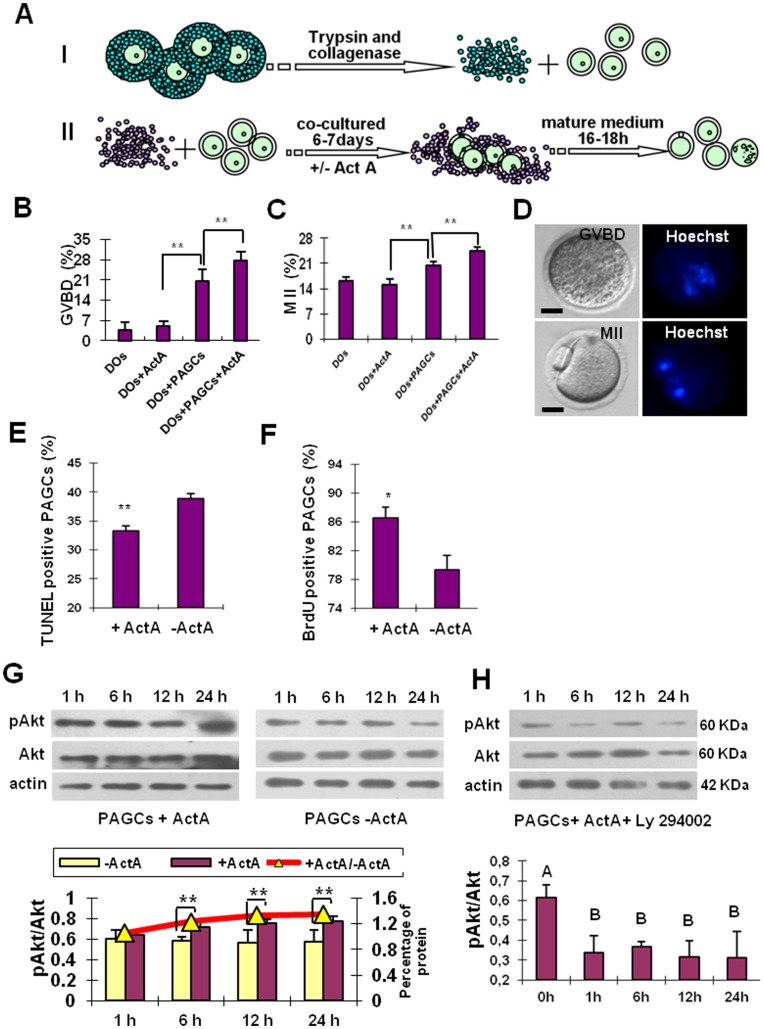
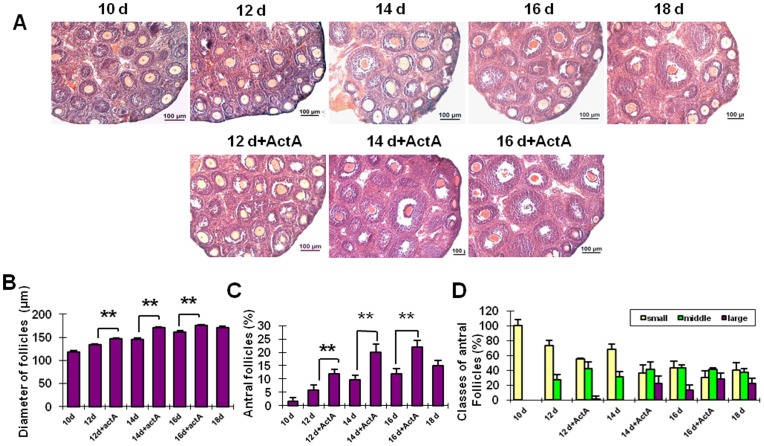
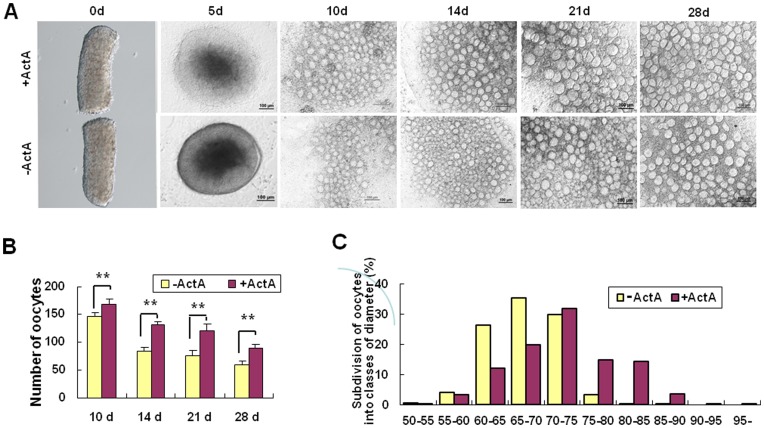
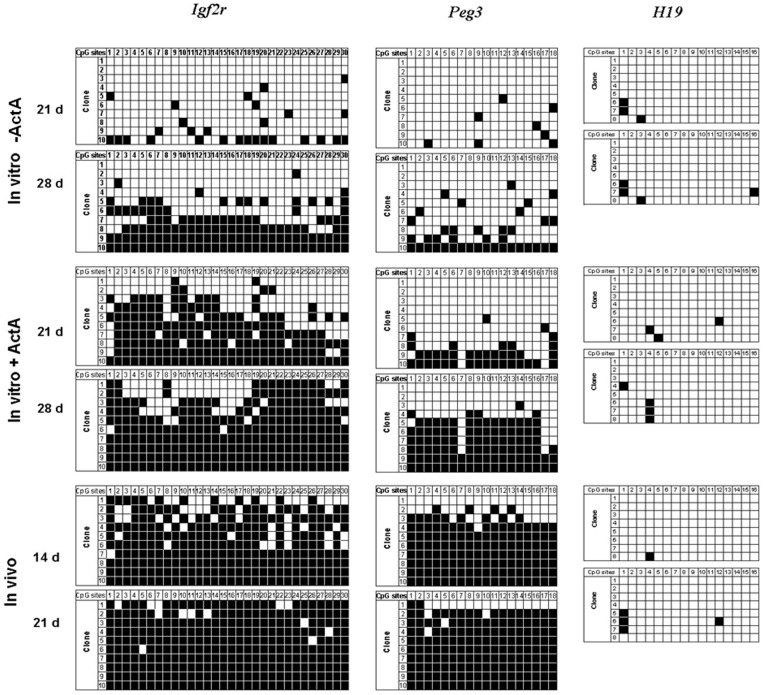
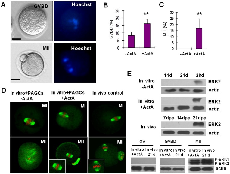
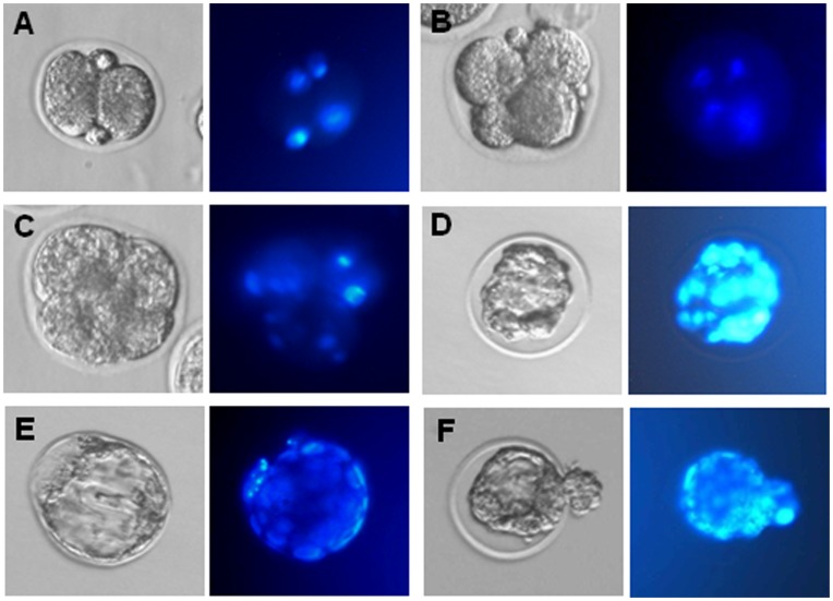
In the present study, we established an in vitro culture system suitable for generating fertilizable oocytes from premeiotic mouse female germ cells. These results were achieved after first establishing an in vitro culture system allowing immature oocytes from 12-14 day-old mice to reach meiotic maturation through culture onto preantral granulosa cell (PAGC) monolayers in the presence of Activin A (ActA). To generate mature oocytes from premeiotic germ cells, pieces of ovaries from 12.5 days post coitum (dpc) embryos were cultured in medium supplemented with ActA for 28 days and the oocytes formed within the explants were isolated and cocultured onto PAGC monolayers in the presence of ActA for 6-7 days. The oocytes were then subjected to a final meiotic maturation assay to evaluate their capability to undergo germinal vesicle break down (GVBD) and reach the metaphase II (MII) stage. We found that during the first 28 days of culture, a significant number of oocytes within the ovarian explants reached nearly full growth and formed preantral follicle-like structures with the surrounding somatic cells. GSH level and Cx37 expression in the oocytes within the explants were indicative of proper developmental conditions. Moreover, the imprinting of Igf2r and Peg3 genes in these oocytes was correctly established. Further culture onto PAGCs in the presence of ActA allowed about 16% of the oocytes to undergo GVBD, among which 17% reached the MII stage during the final 16-18 hr maturation culture. These MII oocytes showed normal spindle and chromosome assembly and a correct ERK1/2 activity. About 35% of the in vitro matured oocytes were fertilized and 53.44% of them were able to reach the 2-cell stage. Finally, around 7% of the 2-cell embryos developed to the morula/blastocyst stage.


Conflict of interest statement

Competing Interests: The authors have declared that no competing interests exist.

Figures

Figure 1
Figure 1. In vitro culture system of immature oocytes from preantral follicles.
(A) Schematic drawing of the main isolation and culture procedures. Oocytes (diameter 55–65 µm) were collected from 12–14-day-old EGFP transgenic mouse and preantral granulosa cells (PAGCs) were collected from 12–14-day-old mouse. PAGCs were cultured as described in M&M, denuded oocytes were divided into four groups: oocytes cocultured with PAGCs in the presence (DOs+PAGCs+ActA) or absence of 100 ng/ml ActA (DOs+PAGCs) for 6–7 days, and oocytes cultured alone in the presence (DOs+ActA) or absence of ActA (DOs) for the same period. Finally, GV oocytes were collected and matured in the in vitro maturation medium (IVM) for 16–18 h. (B–C) In vitro maturation of the oocytes of the four experimental groups as indicated by percents of GVBD and MII oocytes. Culture onto PAGCs results in a significant increase in the number of both GVBD and MII oocytes; ActA significantly improves both maturation capabilities. (D) An example of GVBD and MII oocytes observed under Nikon optics (left) and after DNA staining with Hoechst (right). (E) ActA decreases the number of apoptotic PAGCs cultured in vitro for 7 days as evaluated by the TUNEL staining. (F) Incubation for two days in the presence of ActA increases the number of BrdU positive PAGCs in vitro. (G) ActA increases phospho-Akt in PAGCs cultured in vitro for 1, 6, 12 and 24 h (immunoblotting and densitometric analysis). (H) The PI3K-specific inhibitor, LY294002, inhibits Akt phosphorylation at a concentration of 25 µM (immunoblotting and densitometric analysis). *P<0.05; **P<0.01.
Figure 2
Figure 2. Injection of ActA promotes follicologenesis.
Injection of ActA for 2, 4 and 6 days results in a significant increase in the diameter of follicles (A, B) and the percent of antral follicles (C) when compared to the control group. Antral follicles were divided into three groups: small (<140 µm), medium (140–180 µm) and large (>180 µm). The proportion of antral follicles in each group is shown in D. Scale bars: 100 µm. *P<0.05; **P<0.01.
Figure 3
Figure 3. In vitro culture of embryonic ovary explants.
(A) Pieces of ovaries from 12.5 dpc mouse embryos were cultured in vitro in the presence or absence of ActA for up to 28 days. (B) Number of oocytes scored during the explant culture in vitro in the presence or absence of ActA at 10, 14, 21 and 28 days. (C) Diameters of oocytes in the presence or absence of ActA for 28 days. IVC = In vitro culture medium; IVM = In vitro maturation medium. *P<0.05; **P<0.01.
Figure 4
Figure 4. Establishment of the gene imprinting in the oocytes generated in vitro.
Bisulfite sequencing analysis of DMR in maternally imprinted Igf2r, Peg3 and H19 in the oocytes derived from 12.5 dpc ovaries cultured in vitro for 21 and 28 days in the presence or absence of ActA, as well as the oocytes from 14 and 21 dpp ovaries. Individual lines, clones sequenced; circles, CpG sites within the analyzed regions; filled circles, methylated cytosines; open circles, unmethylated cytosines.
Figure 5
Figure 5. Characterization of the maturation status of the oocytes generated in vitro from 12.5 dpc embryonic ovaries.
(A) An example of GVDB and MII oocytes observed under Nikon optics (left) and after DNA staining with Hoechst (right). (B, C) Percents of GVDB and MII oocytes generated within the ovarian explants after 28 days of culture in the presence of ActA and cocultured onto PAGCs for 7 days in the presence or absence of ActA. (D) MI and MII spindle morphology in oocytes. Oocytes generated in vitro from premeiotic germ cells (In vitro+PAGCs+ActA) show normal MI spindle, MII spindle and chromosome assembly as compared to those of control oocytes isolated from ovaries of 21–22-day-old mice (In vivo control). In contrast, oocyte generated in vitro as described above but cocultured onto PAGCs in the absence of ActA (In vitro+PAGCs-ActA) show anomalous MI spindle and chromosome misalignment. (E) Phosphorylation (activation) of ERK1/2 in oocytes as detected by immunoblotting. Expression of ERK2 protein was analyzed in oocytes obtained from ovarian explants at 14, 21 and 28 days of culture + or – ActA and from 7, 14 and 21 dpp ovaries; only 28 day oocytes in vitro and 21 dpp oocytes in vivo showed detectable ERK2 expression. Phosphorylation of ERK1/2 (pERK1/ERK2) was analyzed in GV, GVDB and MII oocytes generated in vitro (28 days within ovary explants+ActA and 7 days onto PAGCs+ActA) or isolated from ovaries of adult females (21 d); only in vitro and in vivo MII oocytes showed pERK1/ERK2. *P<0.05; **P<0.01.
Figure 6
Figure 6. Oocytes generated in vitro in the presence of ActA can be fertilized and fertilized eggs develop to morula and blastocyst stages.
(A–E) Example of 2-cell embryo, 4-cell embryo, morula and blastocyst developed from oocytes generated in vitro in the presence of ActA. Left, phase contrast microscopy observations; right, Hoechst staining of the cell nuclei.

References

    1. Tam PP, Zhou SX. The allocation of epiblast cells to ectodermal and germ-line lineages is influenced by the position of the cells in the gastrulating mouse embryo. Dev Biol. 1996;178:124–132. - PubMed
    1. Pepling ME, Spradling AC. Female mouse germ cells form synchronously dividing cysts. Development. 1998;125:3323–3328. - PubMed
    1. Wylie C. Germ cells. Cell. 1999;96:165–174. - PubMed
    1. Ohinata Y, Payer B, O’Carroll D, Ancelin K, Ono Y, et al. Blimp1 is a critical determinant of the germ cell lineage in mice. Nature. 2005;436(7048):207–13. - PubMed
    1. McLaren A, Lawson KA. How is the mouse germ-cell lineage established? Differentiation. 2005;73(9–10):435–437. - PubMed

Publication types

MeSH terms