. 2012;7(7):e41798.
doi: 10.1371/journal.pone.0041798. Epub 2012 Jul 25.

In vitro expanded stem cells from the developing retina fail to generate photoreceptors but differentiate into myelinating oligodendrocytes

Affiliations

In vitro expanded stem cells from the developing retina fail to generate photoreceptors but differentiate into myelinating oligodendrocytes

Magdalena Czekaj et al. PLoS One. 2012.

Abstract

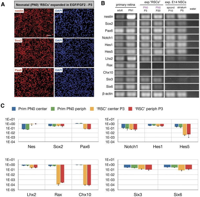
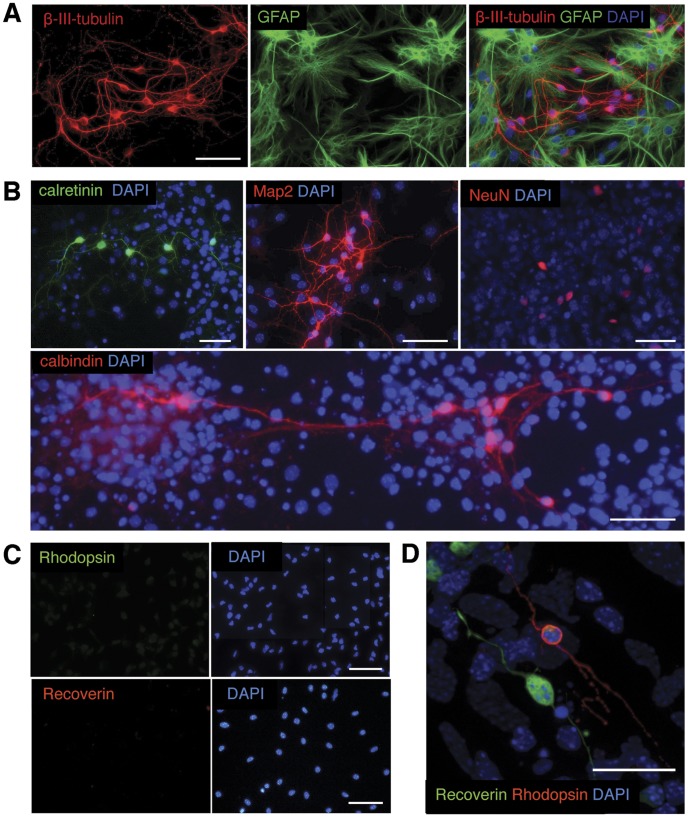
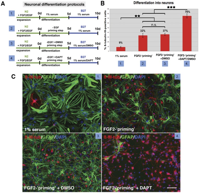
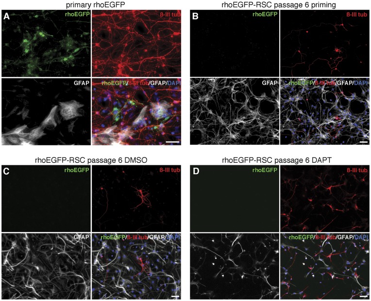
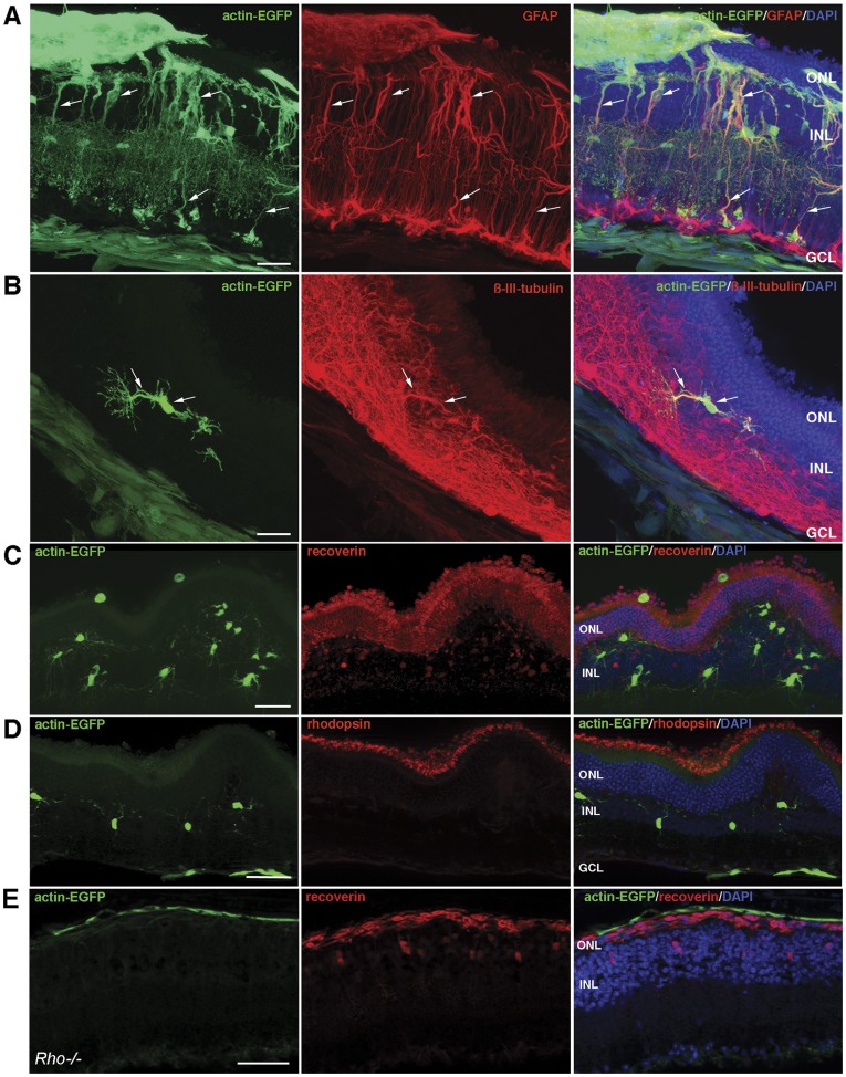
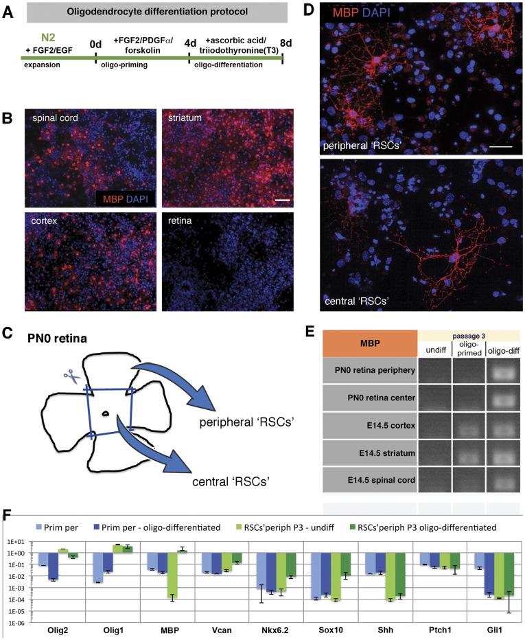
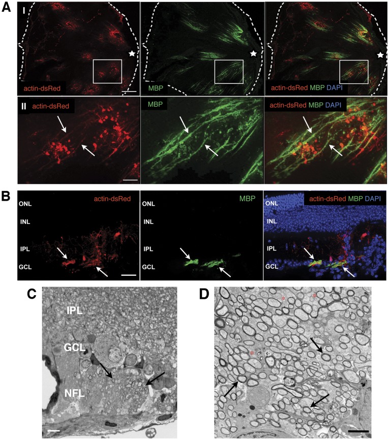
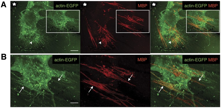
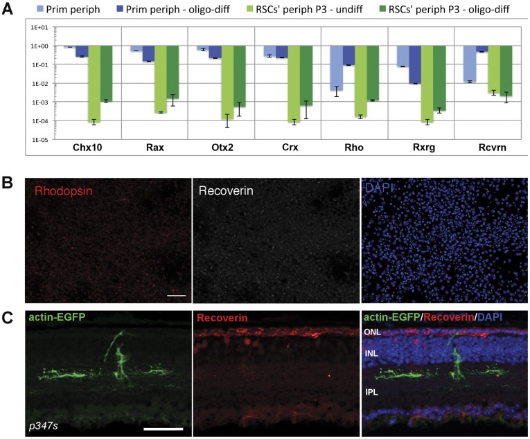
Cell transplantation to treat retinal degenerative diseases represents an option for the replacement of lost photoreceptor cells. In vitro expandable cells isolated from the developing mammalian retina have been suggested as a potential source for the generation of high numbers of donor photoreceptors. In this study we used standardized culture conditions based on the presence of the mitogens FGF-2 and EGF to generate high numbers of cells in vitro from the developing mouse retina. These presumptive 'retinal stem cells' ('RSCs') can be propagated as monolayer cultures over multiple passages, express markers of undifferentiated neural cells, and generate neuronal and glial cell types upon withdrawal of mitogens in vitro or following transplantation into the adult mouse retina. The proportion of neuronal differentiation can be significantly increased by stepwise removal of mitogens and inhibition of the notch signaling pathway. However, 'RSCs', by contrast to their primary counterparts in vivo, i.e. retinal progenitor cells, loose the expression of retina-specific progenitor markers like Rax and Chx10 after passaging and fail to differentiate into photoreceptors both in vitro or after intraretinal transplantation. Notably, 'RSCs' can be induced to differentiate into myelinating oligodendrocytes, a cell type not generated by primary retinal progenitor cells. Based on these findings we conclude that 'RSCs' expanded in high concentrations of FGF-2 and EGF loose their retinal identity and acquire features of in vitro expandable neural stem-like cells making them an inappropriate cell source for strategies aimed at replacing photoreceptor cells in the degenerated retina.


Conflict of interest statement

Competing Interests: The authors have declared that no competing interests exist.

Figures

Figure 1
Figure 1. Immunocytochemistry, RT-PCR and Q-PCR analyses of neonatal ‘RSCs’ during in vitro cultivation.
Following expansion in the presence of growth factors ‘RSCs’ show immunoreactivity for nestin, Sox2 and Pax6 at the protein (A) and mRNA levels (B, C). Further gene expression examination using semi-quantitative RT-PCR revealed that throughout all passages analyzed ‘RSCs’ express components of the notch signaling pathway (Notch1 receptor, Hes1 and Hes5). Levels of transcription factors crucial for eye and retina development (primary retina, PN1) were highly variable: Six3 expression was stable up to P20, Lhx2 and Six6 levels decreased with increasing passage number, and Rax and Chx10 were no longer detectable beginning with passage 3 (B). In comparison, NSCs isolated from E14.5 spinal cord or striatum and cultured for 10 or 5 passages, respectively, showed expression of nestin, Sox2, Pax6, and notch pathway components, but were negative for Rax, Chx10, Six3, and Six6. Adult and PN1 primary retina served as a positive or negative control (B). Q-PCR analysis performed on peripheral ‘RSCs’ from P3 and on their primary counterparts (peripheral retinal cells from PN0) confirmed the above RT-PCR results: expanded cells from low passages expressed Nes, Sox2, Pax6, Notch1, Hes1, Hes5, Six3 and Six6 (C). Although Lhx2 level in P3 ‘RSCs’ was as high as in primary retinal cells, Rax and Chx10 genes were undetectable (C). Gene expression levels are related to the mean expression levels of housekeeping genes. Scale bar: 50 µm. Abbreviations: E, embryonic day; exp, expanded; NSC, neural stem cells; P, passage; PN, postnatal day; ‘RSCs’, retinal stem cells; spcord, spinal cord.
Figure 2
Figure 2. Differentiation of ‘RSCs’ in vitro.
Following differentiation in 1% newborn calf serum expanded ‘RSCs’ showed immunoreactivity for the pan-neuronal markers β-III-tubulin (A, red) and MAP2 (B, red) or the glial marker GFAP (A, green). A subfraction of cells expressed the interneuron markers calretinin (B, green) or calbindin (B, red) or, after prolonged maintenance in differentiation conditions, the mature neuron marker NeuN (B, red). In contrast to primary neonatal retinal cells (D) subjected to the same differentiation conditions, expanded ‘RSC’ cultures are devoid of recoverin (red) or rhodopsin (green) expressing photoreceptors (C). Scale bars: 50 µm. Abbreviations: DAPI, 4,6-diamidino-2-phenylindole; GFAP, glial fibrillary acidic protein; MAP2, microtubule-associated protein 2; NeuN, neuron-specific nuclear antigen.
Figure 3
Figure 3. Increased neuronal differentiation of ‘RSCs’ by priming and notch inhibition.
Generation efficiency of neuronal cell types by expanded ’RSCs’ is dependent on differentiation conditions. ’RSC’ cultures subjected to different differentiation conditions detailed in (A) and immunolabeled with antibodies directed against GFAP (C, green) and β-III-tubulin (C, red) contain different percentages of neurons in dependence of the applied protocol (B, C). The percentage of β-III-tubulin -positive neurons is significantly increased by ’neuronal-priming’ from <17% to <32% when compared to differentiation conditions where mitogens are replaced by NCS and can be further increased to <76% by inhibition of notch-signaling using DAPT (B). DMSO represents the DAPT control experiment with neuron numbers (<37%) corresponding to ’priming’ (B, C). Scale bars: 50 µm. Abbreviations: d, days; DAPI, 4,6-diamidino-2-phenylindole; DAPT, [N-(3,5-difluorophenacetyl)-L-alanyl]-S-phenylglycine t-butylester; DMSO, dimethyl sulfoxide; GFAP, glial fibrillary acidic protein. **p<0.01, ***p<0.001.
Figure 4
Figure 4. Differentiation of primary retinal cells and in vitro expanded ‘RSCs’ isolated from rhoEGFP reporter mice.
Primary cells isolated at PN2 from retinas of rhoEGFP transgenic mice and cultured for 7 days in vitro (A) showed expression of GFP (green - identifying rod photoreceptors), β-III-tubulin (red - identifying neurons), or GFAP (white - identifying glial cells; merged images additionally contain nuclear DAPI staining). ‘RSCs’ generated from rhoEGFP transgenic mice and propagated in vitro for 6 passages were subjected to priming (B) or Notch inhibition with DAPT (D; C represents the DMSO control) differentiation conditions for 10 days. ‘RSCs’ differentiated into β-III-tubulin-positive neurons (red) and GFAP-positive glia (white), but did not show GFP expression indicative for photoreceptor differentiation (B, C, D). Scale bars: 20 um.
Figure 5
Figure 5. Transplantation of ‘RSCs’ into adult mouse retina.
Immunohistochemical analysis on adult wild-type retinas transplanted with GFP-positive ‘RSCs’ (green in A, B, C, and D) revealed that the majority of integrated donor cells expressed GFAP, indicative for extensive glial differentiation (A, arrows). While some transplanted ‘RSCs’ differentiated along the neuronal lineage and expressed β-III-tubulin (B, arrows), grafted cells did not show recoverin (C) or rhodopsin (D) immunoreactivity. Also following transplantation into the degenerative retina of rho−/− mice (E) donor ‘RSCs’ (green) did not show immunoreactivity for recoverin (red). Scale bars: 50 µm. Abbreviations: DAPI, 4,6-diamidino-2-phenylindole; GCL, ganglion cell layer; GFAP, glial fibrillary acidic protein; INL, inner nuclear layer; IPL, inner plexiform layer; ONL, outer nuclear layer.
Figure 6
Figure 6. Oligodendrocyte differentiation of ‘RSCs’ in vitro.
Whereas primary cells isolated at E14.5 from different regions of the CNS, i.e. spinal cord, striatum, and cortex, showed strong MBP expression (B) when subjected to the oligodendroglial differentiation protocol detailed in (A), primary retinal cells remained MBP-negative (B). However, after in vitro expansion central and peripheral ‘RSC’ (P3) cultures generated by appropriate dissection procedures (C) responded to the oligodendrocyte differentiation protocol with expression of MBP as shown by immunocytochemistry (D) and RT-PCR performed on RNA isolated from undifferentiated, oligo-primed and oligo-differentiated ‘RSCs’ and NSCs from P3 (E). Expanded peripheral ‘RSCs’ (P3) as well as their primary counterparts (peripheral retinal cells from PN0) were subjected to the oligo-differentiation protocol in vitro and their gene expression profile was investigated in detail using Q-PCR array for oligodendrocyte-related gene expression (F). Primary cells expressed moderate levels of Olig1/2 genes, whereas expanded ‘RSCs’ contained very high levels of these transcripts. While primary cells subjected to oligo-differentiation conditions did not respond to the treatment with an increase in the expression of oligodendrocyte-related genes, expanded ‘RSCs’ exhibited a significant increase in MBP transcript levels along with other oligodendrocyte-related genes that were analyzed (Vcan, Nkx6.2, Sox10). Generation of oligodendrocytes from expanded ‘RSCs’ was Shh-independent since we could not detect endogenous Shh nor its downstream target Gli1 (F). Scale bars: 100 µm (B) and 50 µm (D). Abbreviations: DAPI, 4,6-diamidino-2-phenylindole; E, embryonic day; MBP, myelin basic protein; Oligo1/1+2, oligodendroglial differentiation step 1/1+2; PN, postnatal day; T3, 3,3,5-triodothyronine; Rho, rhodopsin; Rxrg, retinoid X receptor gamma; Rcvrn - recoverin.
Figure 7
Figure 7. In vivo myelin-formation by pre-differentiated ‘RSCs’.
Analysis of retinas four weeks after intraretinal transplantation of oligo-primed actin-dsRed-‘RSCs’ (red) into adult mice. Many donor cells identified by dsRed expression (A) were located on the vitreal side of the retina (the edges of a flat mounted retina are marked by the dashed white line) and some formed elongated structures radiating towards the optic disc (white star)(some are labeled by arrows in AII; AII is an enlarged view of the boxed area in AI) that are positive for MBP (green, A). Following transplantation of oligo-primed actin-dsRed expressing ‘RSCs’ (red, B), donor cells integrated into the GCL and IPL (red, B) of the host retina. Co-localization of MBP immunoreactivity (green, B) and dsRed fluorescence was restricted to the GCL (B, nuclear DAPI staining (blue) is additionally present in the merged image). Histological analysis of a semi-thin section revealed myelinated axons in the nerve fiber layer of an experimental retina (C; arrows). Transmission electron microscopy confirmed the presence of compact myelin around many RGC axons (some labeled by arrows) in retinas transplanted with ‘RSCs’ (D). Note the increased diameter of myelinated in comparison to unmyelinated axons (D; some labeled by red stars). Scale bars: 200 µm (AI), 50 µm (AII, B), 10 µm (C), 2500 nm (D). Abbreviations: DAPI, 4,6-diamidino-2-phenylindole; GCL, ganglion cell layer; INL, inner nuclear layer; IPL, inner plexiform layer; ONL, outer nuclear layer.
Figure 8
Figure 8. In vivo myelin-formation by pre-differentiated ‘RSCs’ derived from peripheral regions of the developing retina.
Analysis of wild-type retinas four weeks after transplantation of oligo-primed actin-EGFP-‘RSCs’ (green) from P3 into adult mice. Many GFP-expressing donor cells form MBP-positive elongated structures on the vitreal side of the retina (red; some are labeled by arrows in B; B is an enlarged view of the boxed area in A) showing MBP-positive fibers radiating towards the optic disc (white star). Scale bars: 100 µm (A), 50 µm (B).
Figure 9
Figure 9. Oligo-differentiated ‘RSCs’ show slight elevated levels of photoreceptor-specific genes, but fail to generate photoreceptors.
Cultivated peripheral ‘RSCs’ subjected to the full oligodendrocyte differentiation protocol showed a slight increase in the expression of the retina-specific genes Chx10, Rax, Otx2, Crx, rhodopsin and RXRgamma as analysed by Q-PCR albeit at very low absolute levels (A). Immunocytochemical analysis of ‘RSCs’ subjected to oligodendrocyte differentiation in vitro did not reveal positive signals for rhodopsin (red) or recoverin (white) proteins and therefore no evidence for generation of photoreceptors (B). Also following transplantation of oligo-primed ‘RSCs’ (C; green) into the subretinal space of degenerative P347S mice (C) donor cells (green) did not show immunopositivity for the photoreceptor marker recoverin (red). Nuclear DAPI staining is shown in blue, rhodopsin localization in red, recoverin localization in white (B). Scale bars: 50 µm (B). Abbreviations: DAPI, 4,6-diamidino-2-phenylindole.

Similar articles

Cited by

References

    1. Delyfer MN, Leveillard T, Mohand-Said S, Hicks D, Picaud S, et al. Inherited retinal degenerations: therapeutic prospects. Biology of the cell/under the auspices of the European Cell Biology Organization. 2004;96:261–269. - PubMed
    1. Berson EL, Rosner B, Sandberg MA, Weigel-DiFranco C, Moser A, et al. Clinical trial of docosahexaenoic acid in patients with retinitis pigmentosa receiving vitamin A treatment. Archives of ophthalmology. 2004;122:1297–1305. - PubMed
    1. Maeda T, Cideciyan AV, Maeda A, Golczak M, Aleman TS, et al. Loss of cone photoreceptors caused by chromophore depletion is partially prevented by the artificial chromophore pro-drug, 9-cis-retinyl acetate. Human molecular genetics. 2009;18:2277–2287. - PMC - PubMed
    1. Zrenner E, Bartz-Schmidt KU, Benav H, Besch D, Bruckmann A, et al. Subretinal electronic chips allow blind patients to read letters and combine them to words. Proceedings Biological sciences/The Royal Society. 2011;278:1489–1497. - PMC - PubMed
    1. Millington-Ward S, Chadderton N, O’Reilly M, Palfi A, Goldmann T, et al. Suppression and Replacement Gene Therapy for Autosomal Dominant Disease in a Murine Model of Dominant Retinitis Pigmentosa. Molecular Therapy. 2011;19:642–649. - PMC - PubMed

Publication types

MeSH terms