

Minocycline inhibits alkali burn-induced corneal neovascularization in mice
- PMID: 22848638
- PMCID: PMC3405025
- DOI: 10.1371/journal.pone.0041858
Minocycline inhibits alkali burn-induced corneal neovascularization in mice
Abstract
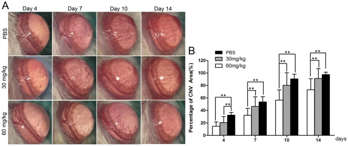
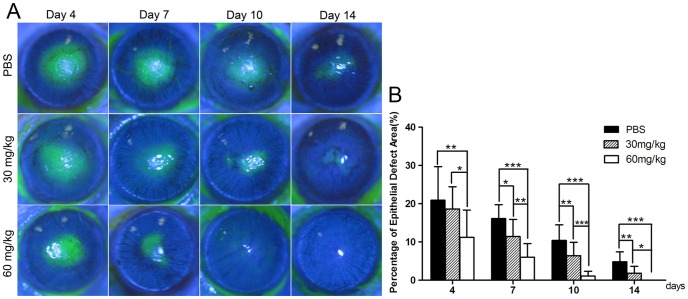
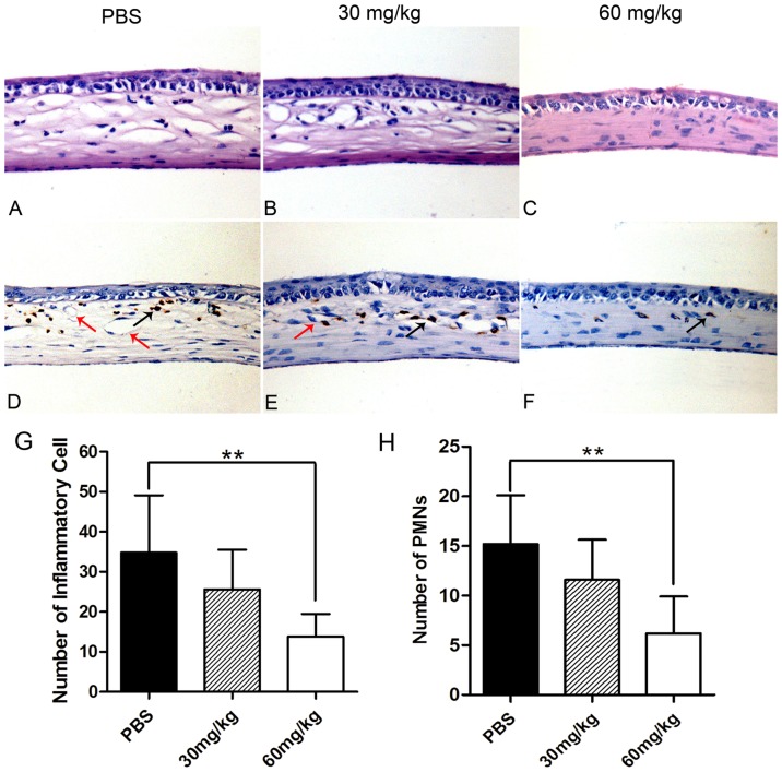
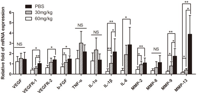
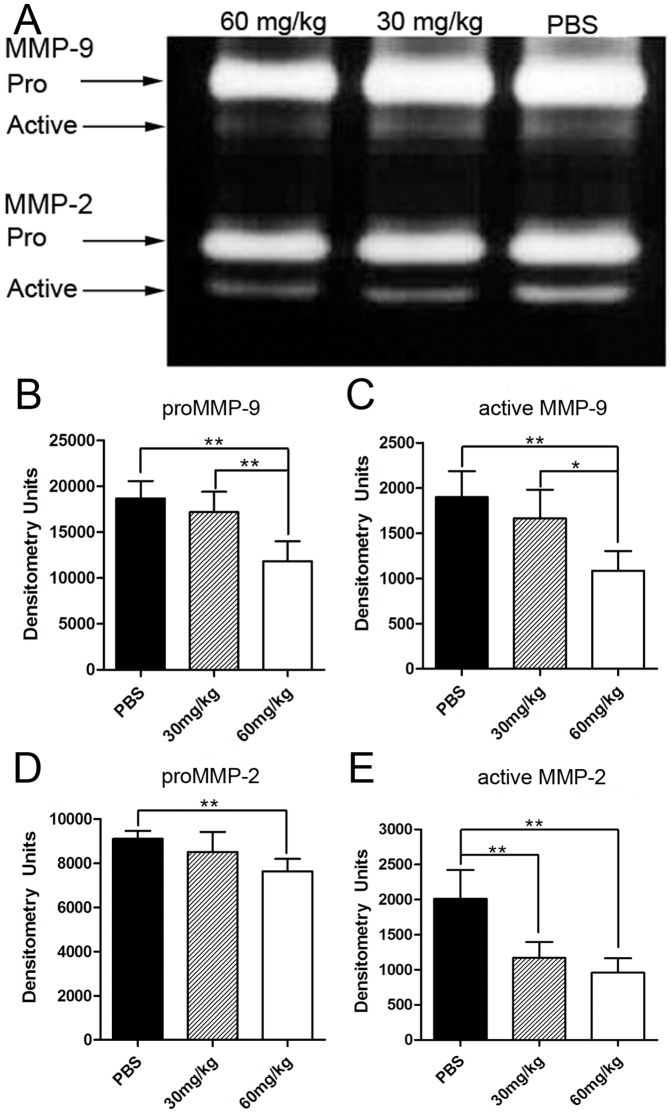
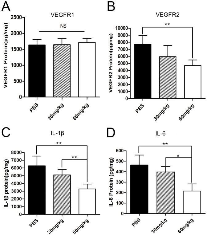
The purpose of this study was to investigate the effects of minocycline on alkali burn-induced corneal neovascularization (CNV). A total of 105 mice treated with alkali burns were randomly divided into three groups to receive intraperitoneal injections of either phosphate buffered saline (PBS) or minocycline twice a day (60 mg/kg or 30 mg/kg) for 14 consecutive days. The area of CNV and corneal epithelial defects was measured on day 4, 7, 10, and14 after alkali burns. On day 14, a histopathological examination was performed to assess morphological change and the infiltration of polymorphonuclear neutrophils (PMNs). The mRNA expression levels of vascular endothelial growth factor (VEGF) and its receptors (VEGFRs), basic fibroblast growth factor (bFGF), matrix metalloproteinases (MMPs), interleukin-1α, 1β, 6 (IL-1α, IL-1β, IL-6) were analyzed using real-time quantitative polymerase chain reaction. The expression of MMP-2 and MMP-9 proteins was determined by gelatin zymography. In addition, enzyme-linked immunosorbent assay was used to analyze the protein levels of VEGFR1, VEGFR2, IL-1β and IL-6. Minocycline at a dose of 60 mg/kg or 30 mg/kg significantly enhanced the recovery of the corneal epithelial defects more than PBS did. There were significant decreases of corneal neovascularization in the group of high-dosage minocycline compared with the control group at all checkpoints. On day 14, the infiltrated PMNs was reduced, and the mRNA expression of VEGFR1, VEGFR2, bFGF, IL-1β, IL-6, MMP-2, MMP-9, -13 as well as the protein expression of VEGFR2, MMP-2, -9, IL-1β, IL-6 in the corneas were down-regulated with the use of 60 mg/kg minocycline twice a day. Our results showed that the intraperitoneal injection of minocycline (60 mg/kg b.i.d.) can significantly inhibit alkali burn-induced corneal neovascularization in mice, possibly by accelerating corneal wound healing and by reducing the production of angiogenic factors, inflammatory cytokines and MMPs.
Conflict of interest statement
Figures
Similar articles
-
Doxycycline enhances the inhibitory effects of bevacizumab on corneal neovascularization and prevents its side effects.Invest Ophthalmol Vis Sci. 2011 Nov 25;52(12):9108-15. doi: 10.1167/iovs.11-7255. Invest Ophthalmol Vis Sci. 2011. PMID: 22039247
-
The effect of TC14012 on alkali burn-induced corneal neovascularization in mice.Ophthalmic Res. 2014;52(1):17-24. doi: 10.1159/000358201. Epub 2014 May 14. Ophthalmic Res. 2014. PMID: 24853648
-
Inhibition of mouse alkali burn induced-corneal neovascularization by recombinant adenovirus encoding human vasohibin-1.Mol Vis. 2010 Jul 26;16:1389-98. Mol Vis. 2010. PMID: 20680097 Free PMC article.
-
Fasudil hydrochloride, a potent ROCK inhibitor, inhibits corneal neovascularization after alkali burns in mice.Mol Vis. 2015 Jun 12;21:688-98. eCollection 2015. Mol Vis. 2015. PMID: 26120273 Free PMC article.
-
Emerging Strategies in Treating Corneal Alkali Burns: A Narrative Review.Cureus. 2023 Oct 25;15(10):e47662. doi: 10.7759/cureus.47662. eCollection 2023 Oct. Cureus. 2023. PMID: 38021904 Free PMC article. Review.
Cited by
-
Inhibiting the effect of (90)Sr-(90)Y ophthalmic applicators on rat corneal neovascularization induced by sutures.Int J Ophthalmol. 2016 Sep 18;9(9):1251-4. doi: 10.18240/ijo.2016.09.02. eCollection 2016. Int J Ophthalmol. 2016. PMID: 27672586 Free PMC article.
-
Pharmacological Potential of Small Molecules for Treating Corneal Neovascularization.Molecules. 2020 Jul 30;25(15):3468. doi: 10.3390/molecules25153468. Molecules. 2020. PMID: 32751576 Free PMC article. Review.
-
WIP1 regulates the proliferation and invasion of nasopharyngeal carcinoma in vitro.Tumour Biol. 2014 Aug;35(8):7651-7. doi: 10.1007/s13277-014-2034-6. Epub 2014 May 7. Tumour Biol. 2014. PMID: 24801909
-
Sorafenib-loaded nanostructured lipid carriers for topical ocular therapy of corneal neovascularization: development, in-vitro and in vivo study.Drug Deliv. 2022 Dec;29(1):837-855. doi: 10.1080/10717544.2022.2048134. Drug Deliv. 2022. PMID: 35277107 Free PMC article.
-
Comparison of the Effects of Subconjunctival Injections of Bevacizumab and Interferon Alpha-2a on Corneal Angiogenesis in a Rat Model.Medicina (Kaunas). 2018 Apr 16;54(2):16. doi: 10.3390/medicina54020016. Medicina (Kaunas). 2018. PMID: 30344247 Free PMC article.
References
-
- Wagoner MD. Chemical injuries of the eye: current concepts in pathophysiology and therapy. Surv Ophthalmol. 1997;41:275–313. - PubMed
-
- Bashshur ZF, Bazarbachi A, Schakal A, Haddad ZA, El Haibi CP, et al. Intravitreal bevacizumab for the management of choroidal neovascularization in age-related macular degeneration. Am J Ophthalmol. 2006;142:1–9. - PubMed
-
- Dana MR, Zhu SN, Yamada J. Topical modulation of interleukin-1 activity in corneal neovascularization. Cornea. 1998;17:403–409. - PubMed
Publication types
MeSH terms
Substances
LinkOut - more resources
Full Text Sources
Miscellaneous
