. 2012;7(7):e41917.
doi: 10.1371/journal.pone.0041917. Epub 2012 Jul 25.

Haploinsufficiency for translation elongation factor eEF1A2 in aged mouse muscle and neurons is compatible with normal function

Affiliations

Haploinsufficiency for translation elongation factor eEF1A2 in aged mouse muscle and neurons is compatible with normal function

Lowri A Griffiths et al. PLoS One. 2012.

Abstract

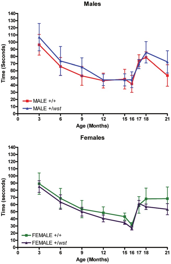
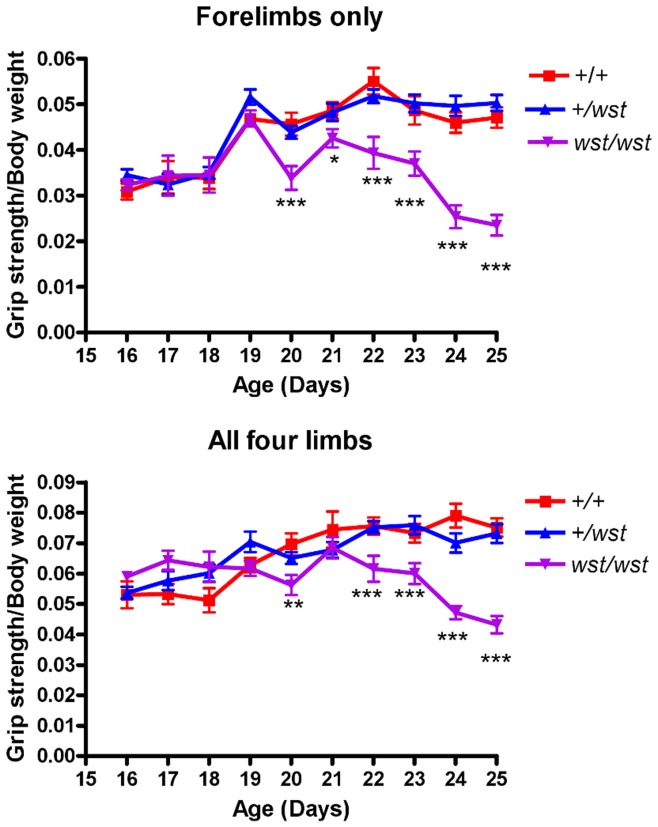
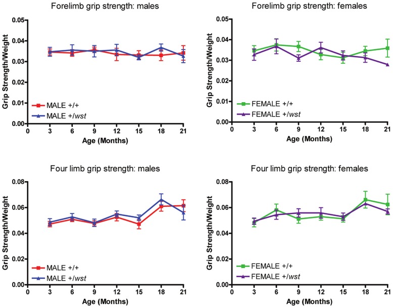
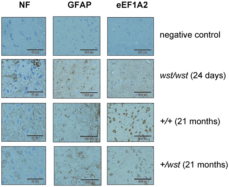
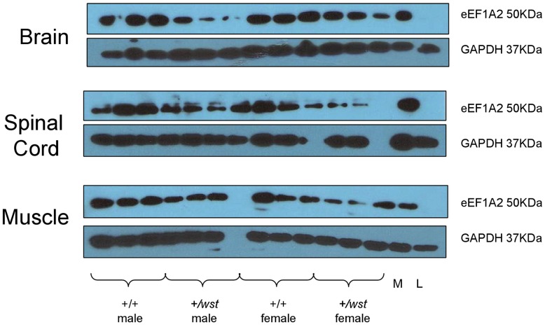
Translation elongation factor isoform eEF1A2 is expressed in muscle and neurons. Deletion of eEF1A2 in mice gives rise to the neurodegenerative phenotype "wasted" (wst). Mice homozygous for the wasted mutation die of muscle wasting and neurodegeneration at four weeks post-natal. Although the mutation is said to be recessive, aged heterozygous mice have never been examined in detail; a number of other mouse models of motor neuron degeneration have recently been shown to have similar, albeit less severe, phenotypic abnormalities in the heterozygous state. We therefore examined the effects of ageing on a cohort of heterozygous +/wst mice and control mice, in order to establish whether a presumed 50% reduction in eEF1A2 expression was compatible with normal function. We evaluated the grip strength assay as a way of distinguishing between wasted and wild-type mice at 3-4 weeks, and then performed the same assay in older +/wst and wild-type mice. We also used rotarod performance and immunohistochemistry of spinal cord sections to evaluate the phenotype of aged heterozygous mice. Heterozygous mutant mice showed no deficit in neuromuscular function or signs of spinal cord pathology, in spite of the low levels of eEF1A2.


Conflict of interest statement

Competing Interests: The authors have declared that no competing interests exist.

Figures

Figure 1
Figure 1. Rotarod analysis of aged mice.
Mean times of latency to fall from the rotarod over the 21 months of the ageing study. WT indicates wild-type animals, HET indicates heterozygote animals. The upper graph shows data from the male groups and the lower graph shows data from the female groups. Error bars represent the standard error of the mean.
Figure 2
Figure 2. Grip strength analysis of young wasted mice.
Forelimb (top panel) and all four limbs (bottom panel) grip strength analysis of wasted mice. The daily grip strength reading of 3 tests (measured in Newtons) were normalised to body weight (measured in grams). P values were calculated comparing wasted mice with controls (+/+ and +/wst combined). * indicates a P value<0.05, ** indicates a P value<0.01, and *** indicates a P value <0.001.
Figure 3
Figure 3. Grip strength analysis of aged mice.
Grip strength data for the entire ageing study. Graphs on the left display male data and graphs on the right display female data. Top graphs display forelimb data only and the bottom 2 graphs display data from all four limbs. WT indicates wild-type animals, HET indicates heterozygote animals. Error bars represent the standard error of the mean.
Figure 4
Figure 4. Immunohistochemistry in aged spinal cord sections.
Expression of phosphorylated neurofilaments, GFAP, and eEF1A2 in cervical spinal cord sections from 21 month old mice. The top panel in each case shows a section with primary antibody omitted from the protocol, the second panel from the top shows sections from a 24 day old wasted homozygote as a control, and the bottom two panels show sections from an aged matched wild-type and heterozygous male. The NF staining clearly shows perikaryal accumulation of NF staining in the wasted mouse section but not in the aged +/wst mouse. The GFAP staining shows a characteristic pattern of reactive astrocytes throughout the grey matter of the spinal cord, even in the aged wild-type mouse. The eEF1A2 shows no staining at all in the section from the wst/wst mouse as expected, and fainter but easily detectable, albeit reduced, staining in the aged +/wst sample.
Figure 5
Figure 5. Protein expression in aged mice.
Western blots showing protein expression of eEF1A2 and GAPDH (as a loading control) of animals from the ageing cohort. Three 21month old animals from each group are shown. WT indicates wild-type animals, HET indicates heterozygous animals, M indicates a muscle sample (as an eEF1A2 positive control) and L indicates a liver sample (an eEF1A2 negative control), both from wild-type mice.

References

    1. Lutsep HL, Rodriguez M. Ultrastructural, morphometric, and immunocytochemical study of anterior horn cells in mice with “wasted” mutation. J Neuropathol Exp Neurol. 1989;48:519–533. - PubMed
    1. Newbery HJ, Gillingwater TH, Dharmasaroja P, Peters J, Wharton SB, et al. Progressive loss of motor neuron function in wasted mice: effects of a spontaneous null mutation in the gene for the eEF1 A2 translation factor. J Neuropathol Exp Neurol. 2005;64:295–303. - PubMed
    1. Shultz LD, Sweet HO, Davisson MT, Coman DR. 'Wasted', a new mutant of the mouse with abnormalities characteristic to ataxia telangiectasia. Nature. 1982;297:402–404. - PubMed
    1. Chambers DM, Peters J, Abbott CM. The lethal mutation of the mouse wasted (wst) is a deletion that abolishes expression of a tissue-specific isoform of translation elongation factor 1alpha, encoded by the Eef1a2 gene. Proc Natl Acad Sci U S A. 1998;95:4463–4468. - PMC - PubMed
    1. Ann DK, Lin HH, Lee S, Tu ZJ, Wang E. Characterization of the statin-like S1 and rat elongation factor 1 alpha as two distinctly expressed messages in rat. J Biol Chem. 1992;267:699–702. - PubMed

Publication types

Substances