. 2012;7(7):e42107.
doi: 10.1371/journal.pone.0042107. Epub 2012 Jul 25.

Analysis of monensin sensitivity in Toxoplasma gondii reveals autophagy as a mechanism for drug induced death

Affiliations

Analysis of monensin sensitivity in Toxoplasma gondii reveals autophagy as a mechanism for drug induced death

Mark D Lavine et al. PLoS One. 2012.

Abstract

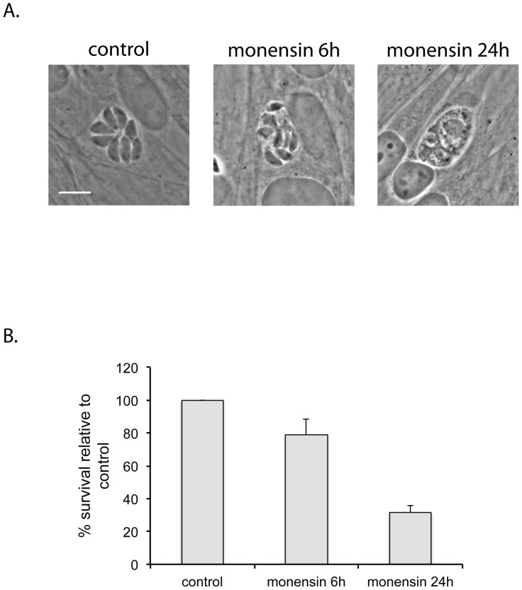
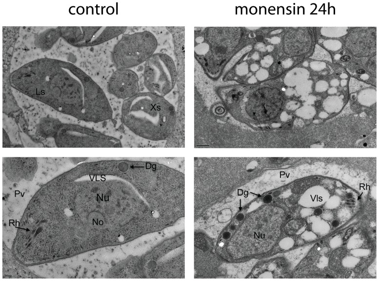
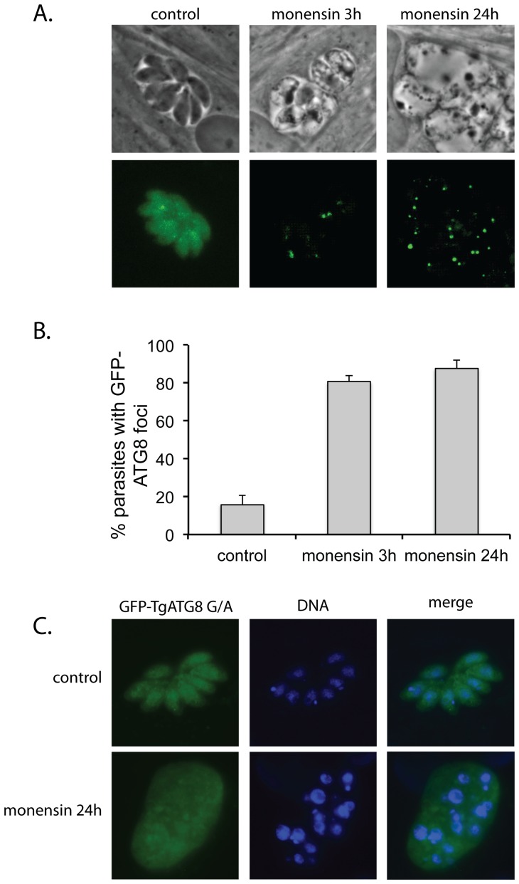
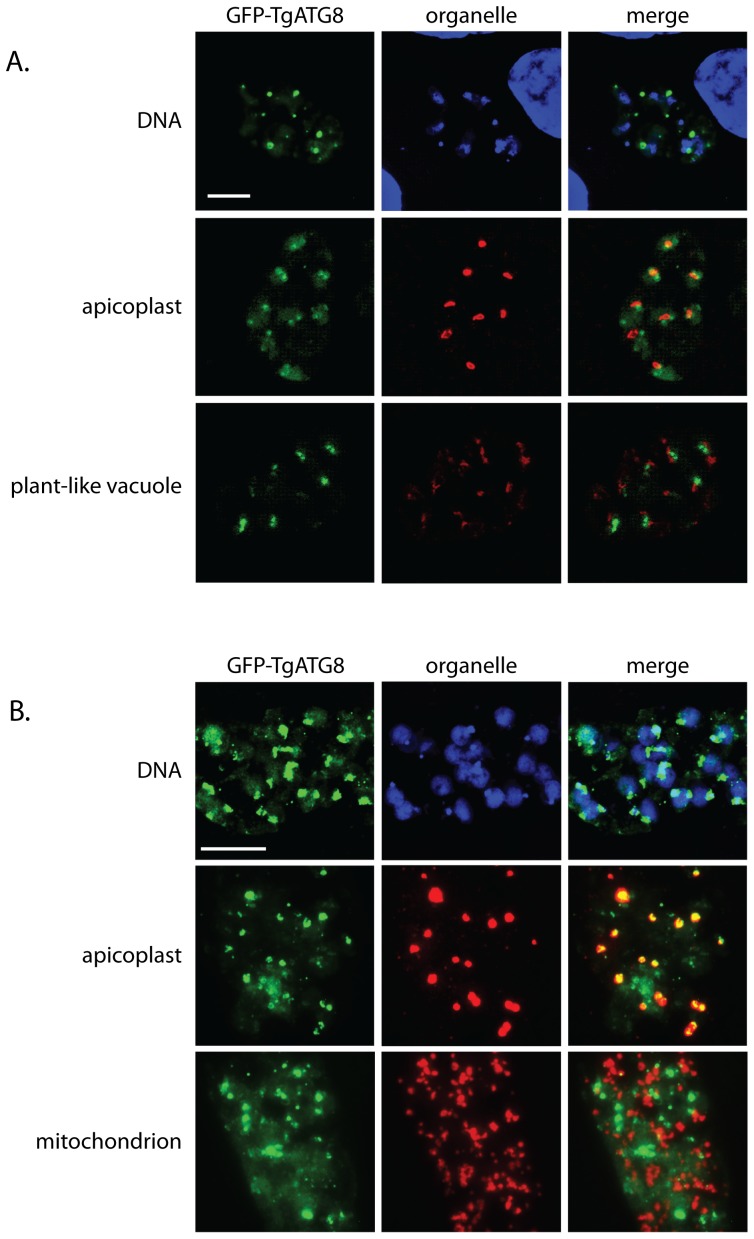
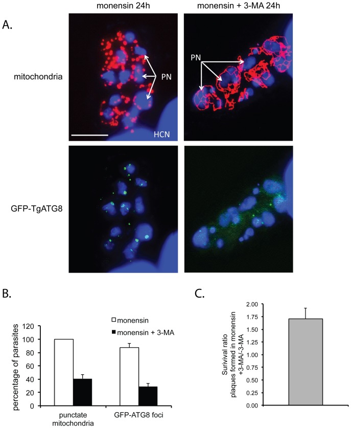
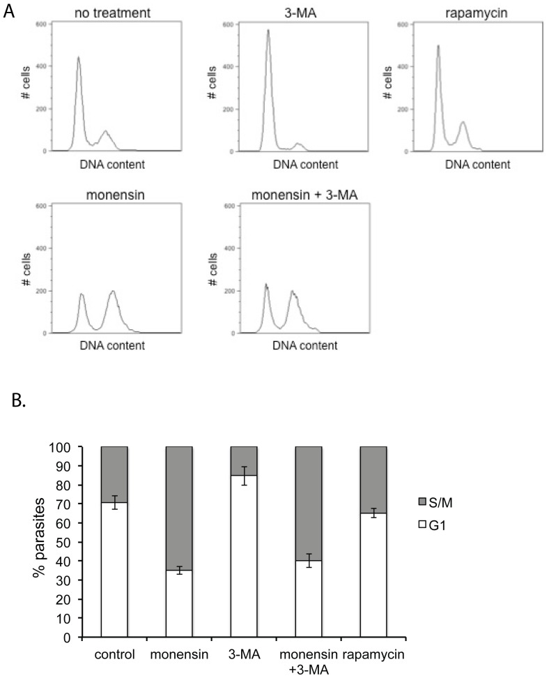
Understanding the mechanisms by which anti-parasitic drugs alter the physiology and ultimately kill is an important area of investigation. Development of novel parasitic drugs, as well as the continued utilization of existing drugs in the face of resistant parasite populations, requires such knowledge. Here we show that the anti-coccidial drug monensin kills Toxoplasma gondii by inducing autophagy in the parasites, a novel mechanism of cell death in response to an antimicrobial drug. Monensin treatment results autophagy, as shown by translocation of ATG8 to autophagosomes, as well as causing marked morphological changes in the parasites' mitochondria. Use of the autophagy inhibitor 3-methyladenine blocks autophagy and mitochondrial alterations, and enhances parasite survival, in monensin-exposed parasites, although it does not block other monensin-induced effects on the parasites, such as late S-phase cell cycle arrest. Monensin does not induce autophagy in a parasite strain deficient in the mitochondrial DNA repair enzyme TgMSH-1 an enzyme that mediates monensin-induced late S-phase arrest. TgMSH-1 therefore either mediates cell cycle arrest and autophagy independently, or autophagy occurs downstream of cell cycle arrest in a manner analogous to apoptosis of cells arrested in G(2) of the cell cycle. Overall, our results point to autophagy as a potentially important mode of cell death of protozoan parasites in response to antimicrobial drugs and indicate that disruption of the autophagy pathway could result in drug resistance.


Conflict of interest statement

Competing Interests: The authors have declared that no competing interests exist.

Figures

Figure 1
Figure 1. Effect of monensin on T. gondii is reversible.
(A) Phase-contrast micrographs of intracellular T. gondii after exposure to 0.75 ng/ml monensin for 6 hours or 24 hours. By 24 hours 100% of parasites show the altered appearance pictured. Scale bar = 10 µm. (B) Survival of T. gondii after exposure to monensin. Parasites were exposed to 0.75 ng/ml monensin for 6 hours or 24 hours, after which the monensin was removed and parasites were allowed to form plaques. Data is expressed as % survival relative to control (no-monensin exposure) parasites. Control parasites are considered to have 100% survival. Each bar represents the mean value for three independent replicates. Error bar = 1 standard deviation.
Figure 2
Figure 2. Monensin induces formation of vacuole like structures in T. gondii.
Transmission electron micrographs of intracellular T. gondii after 24 hours exposure to 0.75 ng/ml monensin. Ls, longitudinal section through parasite; Xs, cross-section through parasite; Pv, parasitophorous vacuole; Rh, rhoptries; Nu, nucleus; No, nucleolus; Dg, dense granule; Vls, vacuole-like structure. Scale bar (bottom left) = 0.5 µM.
Figure 3
Figure 3. Monensin affects mitochondrial morphology.
Phase-contrast and deconvolved immunofluorescence micrographs showing effect of monensin (0.75 ng/ml, 24 hours) on several T. gondii intracellular structures. (A) Phase contrast and DAPI staining of DNA showing parasite nuclei (PN) and apicoplast DNA (ADNA). HCN = host cell nuclei. (B) Phase contrast and immunofluorescence staining showing Atrx1 protein in the apicoplasts. (C) Phase contrast and immunofluorescence staining of TgNHE3 showing parasite plant-like vacuoles. (D) Phase contrast and immunofluorescence showing parasite mitochondria. Scale bars = 10 µm.
Figure 4
Figure 4. Monensin induces formation of GFP-ATG8 foci in intracellular parasites.
(A) Phase contrast and deconvolved immmuofluorescent micrographs of T. gondii expressing GFP-tagged ATG8 after exposure to monensin (0.75 ng/ml) for 3 hours or 24 hours. (B) Percentage of parasites containing one or more GFP-ATG8 foci after exposure to monensin. Each bar represents the mean value for three independent replicates. Error bar = 1 standard deviation. (C) Deconvolved immmuofluorescent micrographs show that T. gondii expressing a GFP-tagged ATG8 in which the terminal glycine was replaced with an alanine (GFP-TgATG8-G/A) do not show formation of GFP-ATG8 foci even after 24 hours exposure to monensin (0.75 ng/ml).
Figure 5
Figure 5. ATG8 colocalizes with the apicoplasts after prolonged monensin exposure.
Deconvolved immunofluorescence micrographs showing relative localization of GFP-ATG8 and T. gondii intracellular structures after exposure to monensin (0.75 ng/ml). (A) 3 hours monensin exposure showing localization of GFP-ATG8 and DNA, apicoplasts, and plant-like vacuoles. (B) 24 hours monensin exposure showing localization of DNA, apicoplasts, and mitochondria. Scale bar = 10 µm.
Figure 6
Figure 6. The autophagy inhibitor 3-methyladenine (3-MA) blocks autophagy and mitochondrial alteration induced by monensin.
(A) Deconvolved fluorescence micrographs of intracellular T. gondii exposed to 0.75 ng/ml monensin or 0.75 ng/ml monensin plus 10 mM 3-MA for 24 hours. Red = mitochondria, green = GFP-ATG8, blue = DNA. PN, parasite nuclei; HCN, host cell nuclei. (B) Quantification of number of parasites positive for punctate mitochondria or GFP-ATG8 autophagosome foci after exposure to 0.75 ng/ml monensin for 24 hours (white bars) or 0.75 ng/ml monensin+10 mM 3-MA for 24 hours (black bars). Each bar represents the mean value for three independent replicates. Error bar equals the standard deviation. Scale bar = 10 µm. (C) The bar represents the ratio of number of plaques formed in 0.75 ng/ml monensin+10 mM 3-MA over that in only 0.75 ng/ml monensin. Error bar is the standard deviation.
Figure 7
Figure 7. Rapamycin alone does not induce cell cycle arrest, nor does 3-MA rescue monensin-induced cell cycle arrest.
Flow cytometry analysis of T. gondii cell cycle in response to rapamycin, monensin and 3-MA. Intracellular parasites were exposed to either normal culture medium or normal culture medium plus 0.75 ng/ml monensin, normal culture medium plus 10 mM 3-MA, normal culture medium plus 0.75 ng/ml monensin and 10 mM 3-MA, or normal culture medium plus 5 µM rapamycin. After 24 hours exposure, DNA content was measured by Sytox green staining. (A) Representative histograms are shown. Each histogram represents 10,000 total events. (B) Percentage ± standard deviation of parasites in G1 or S/M phases determined by gating for three separate experiments is indicated in the bar graphs.

References

    1. Muller J, Hemphill A (2011) Drug target identification in intracellular and extracellular protozoan parasites. Curr Top Med Chem 11: 2029–2038. - PubMed
    1. Haberkorn A (1996) Chemotherapy of human and animal coccidioses: state and perspectives. Parasitol Res 82: 193–199. - PubMed
    1. Mead JR (2002) Cryptosporidiosis and the challenges of chemotherapy. Drug Resist Updat 5: 47–57. - PubMed
    1. Meneceur P, Bouldouyre MA, Aubert D, Villena I, Menotti J, et al. (2008) In vitro susceptibility of various genotypic strains of Toxoplasma gondii to pyrimethamine, sulfadiazine, and atovaquone. Antimicrob Agents Chemother 52: 1269–1277. - PMC - PubMed
    1. Petersen I, Eastman R, Lanzer M (2011) Drug-resistant malaria: molecular mechanisms and implications for public health. FEBS Lett 585: 1551–1562. - PubMed

Publication types

MeSH terms