. 2012;7(7):e42114.
doi: 10.1371/journal.pone.0042114. Epub 2012 Jul 27.

The cellular immune response of the pea aphid to foreign intrusion and symbiotic challenge

Affiliations

The cellular immune response of the pea aphid to foreign intrusion and symbiotic challenge

Antonin Schmitz et al. PLoS One. 2012.

Abstract

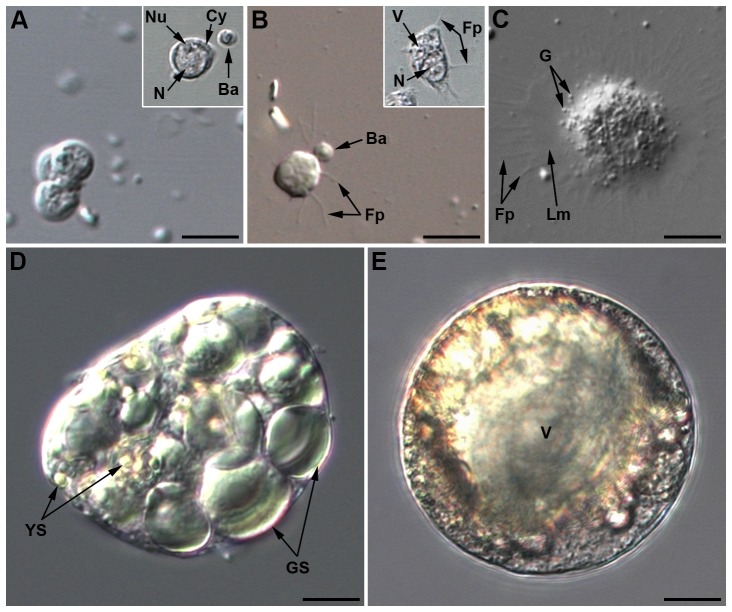
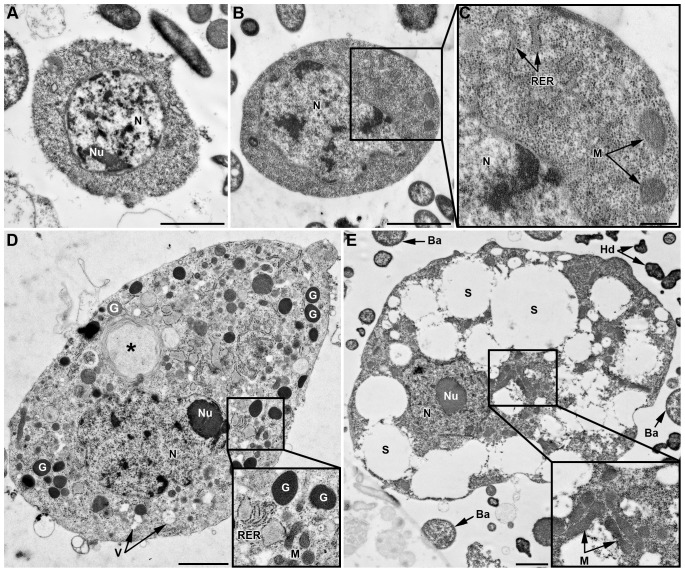
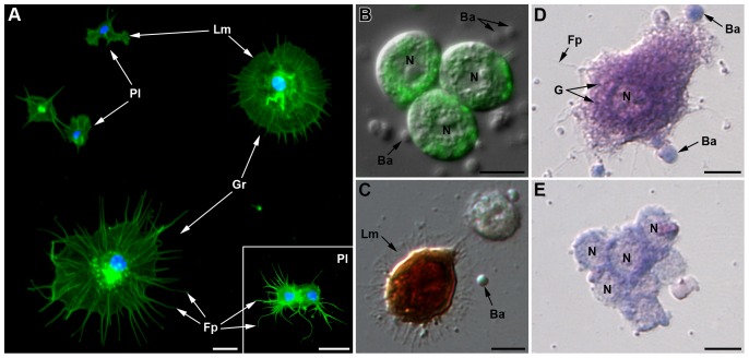
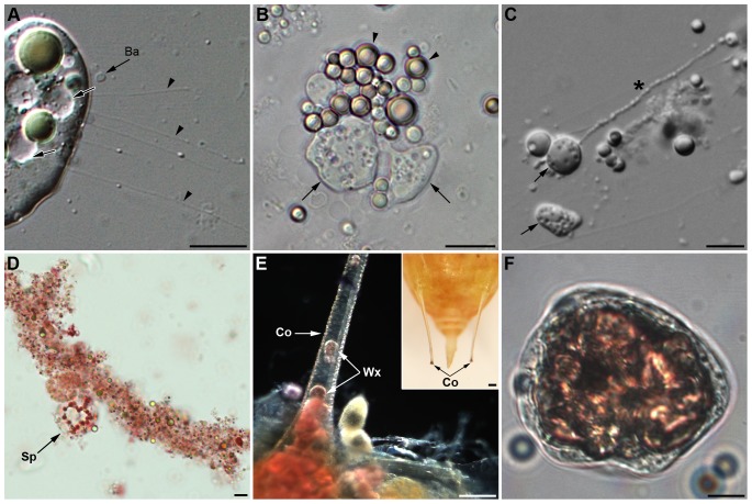
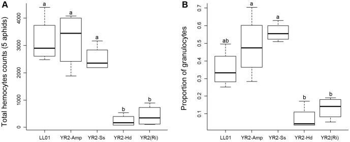
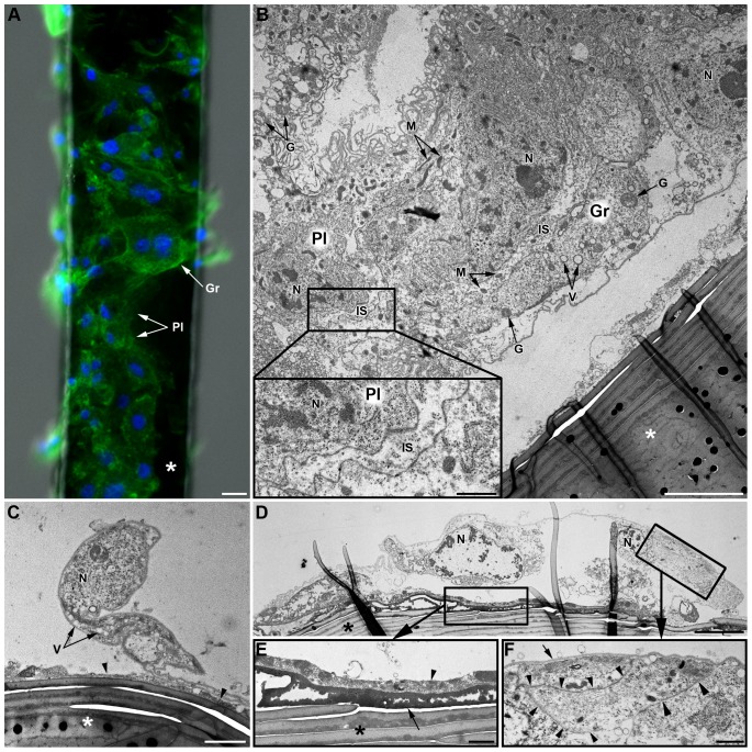
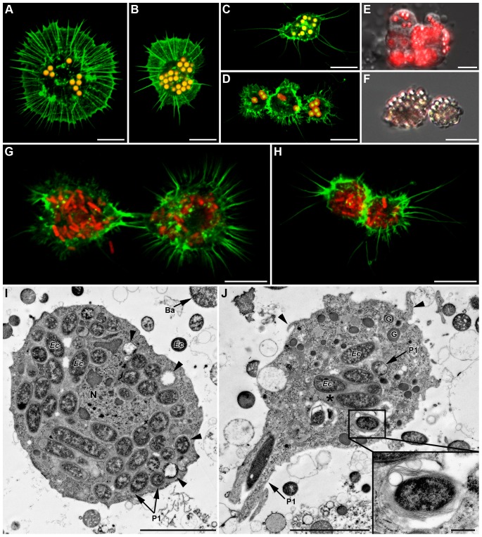
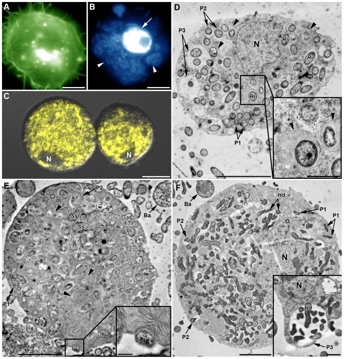
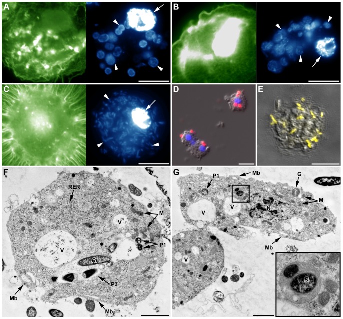
Recent studies suggest that the pea aphid (Acyrthosiphon pisum) has low immune defenses. However, its immune components are largely undescribed, and notably, extensive characterization of circulating cells has been missing. Here, we report characterization of five cell categories in hemolymph of adults of the LL01 pea aphid clone, devoid of secondary symbionts (SS): prohemocytes, plasmatocytes, granulocytes, spherulocytes and wax cells. Circulating lipid-filed wax cells are rare; they otherwise localize at the basis of the cornicles. Spherulocytes, that are likely sub-cuticular sessile cells, are involved in the coagulation process. Prohemocytes have features of precursor cells. Plasmatocytes and granulocytes, the only adherent cells, can form a layer in vivo around inserted foreign objects and phagocytize latex beads or Escherichia coli bacteria injected into aphid hemolymph. Using digital image analysis, we estimated that the hemolymph from one LL01 aphid contains about 600 adherent cells, 35% being granulocytes. Among aphid YR2 lines differing only in their SS content, similar results to LL01 were observed for YR2-Amp (without SS) and YR2-Ss (with Serratia symbiotica), while YR2-Hd (with Hamiltonella defensa) and YR2(Ri) (with Regiella insecticola) had strikingly lower adherent hemocyte numbers and granulocyte proportions. The effect of the presence of SS on A. pisum cellular immunity is thus symbiont-dependent. Interestingly, Buchnera aphidicola (the aphid primary symbiont) and all SS, whether naturally present, released during hemolymph collection, or artificially injected, were internalized by adherent hemocytes. Inside hemocytes, SS were observed in phagocytic vesicles, most often in phagolysosomes. Our results thus raise the question whether aphid symbionts in hemolymph are taken up and destroyed by hemocytes, or actively promote their own internalization, for instance as a way of being transmitted to the next generation. Altogether, we demonstrate here a strong interaction between aphid symbionts and immune cells, depending upon the symbiont, highlighting the link between immunity and symbiosis.


Conflict of interest statement

Competing Interests: The authors have declared that no competing interests exist.

Figures

Figure 1
Figure 1. Light microscopy pictures of Acyrthosiphon pisum hemocytes (LL01 clone).
(A) Three prohemocytes in cluster. Inset: phase contrast showing the large central nucleus and the nucleolus (Nu). Ba: B. aphidicola. (B) Plasmatocyte beginning to adhere, with filopodia (Fp) extension. Inset: phase contrast showing large cytoplasmic vacuolar formation. (C) Adherent granulocyte containing cytoplasmic granules (G) and filopodia (Fp) extending from a lamellipodium (Lm). (D) Spherulocyte with its large colored globular inclusions, small yellow spherules (YS) and large green spherules (GS). (E) Wax cell showing a large central vacuole (V) and colored globular inclusions that differ from those of spherulocytes. Same magnification for all micrographs; scale bar: 10 µm.
Figure 2
Figure 2. TEM characterization of hemocytes.
Cells from the LL01 clone (A–D) and the YR2-Hd line (E). (A) Prohemocyte characterized by a round nucleus (N), a high N/C ratio, and homogenous cytoplasm devoid of apparent organelles. (B) Plasmatocyte with homogeneous cytoplasm, a lobulated nucleus, a high N/C ratio and some cytoplasmic organelles clearly visible in box (C): Rough Endoplasmic Reticulum (RER), Mitochondria (M). (D) Granulocyte with a lobulated nucleus, a low N/C ratio, and granules (G). The cytoplasm contains numerous organelles (see box). A phagosome is observed that contains a large foreign particle (asterisk). (E) YR2-Hd spherulocyte with a round nucleus and a low N/C ratio. The large volume of cytoplasm is filled with spherules (S) of different sizes, and numerous mitochondria are found in a small region (enlarged box). Ba: B. aphidicola; Hd: H. defensa. Scale bar: 2 µm (A, B, D, E) and 0.5 µm (C).
Figure 3
Figure 3. Histological characterization of adherent hemocytes (LL01 clone).
(A) Fluorescent micrograph showing AHP after F-actin (green) and nucleus (blue) staining. Plasmatocytes (Pl) are the smaller cells; they display two adhesion profiles (i) lamellipodia (Lm) extension without apparent filopodia or (ii) filopodia extension (Fp) exclusively (inset). Granulocytes (Gr) have spread more than four times their original size; they display lamellipodia with radiates filopodia. (B) Merger of DIC and fluorescent micrographs showing three clumped ROS-producing granulocytes (green fluorescence of Rhodamine 123). (C) Intracellular PO staining on AHP using Dopamine hydrochloride showing a PO-positive hemocyte (brown staining). (D–E) Adherent hemocytes stained with May Grünwald Giemsa. (D) Granulocyte with an eosinophilic cytoplasm (pink staining) that contains basophilic granules (blue staining). (E) Clumped plasmatocytes with a limited layer of basophilic cytoplasm. Ba: B. aphidicola. Scale bars: 10 µm.
Figure 4
Figure 4. Functions of spherulocytes and wax cells.
(A–C) Early alterations of spherulocytes after hemolymph collection: (A) loss of spherules and fibrils' formation (arrowhead). Ba: B. aphidicola. (B) Unstable cytoplasmic blebs (arrow) derive from spherulocytes while spherules remain intact (arrowhead). (C) From blebs (arrow), long stable strands like strings of pearls extend (asterisk; see also movie 1). (D) Low magnification of a large coagulum stained by neutral red showing a granular aspect and the presence of an intact spherulocyte (Sp). (E–F) Wax cells (Wx) are localized at the base and inside the cornicles (Co), secretory appendices localized at the posterior end of aphids' bodies (inset in (E)). (E) Red lipid staining showing large accumulation of lipid-containing cells at the base of the cornicle. (F) Wax cell containing a large neutral lipidic inclusion (pink staining) that almost fills entirely the cytoplasm. Scale bars: 15 µm (A), 10 µm (B, C, D, F) and 100 µm (E and inset in (E)).
Figure 5
Figure 5. Total hemocyte counts (THC) and granulocyte proportions from AHPs of the different lines.
Box-plot representations of the THC (A) and the proportion of granulocytes (B), estimated on F-actin stained AHPs (pools of 5 adults per AHP), for aphid lines listed in Table 1. Box-plots with the same letter have means that are not significantly different (TukeyHSD, alpha = 5e-03, n = 4).
Figure 6
Figure 6. In vivo hemocyte adhesion assay (LL01 clone).
(A) Observations of a brush horsehair (asterisk) seven days after insertion in the hemocoele of an LL01 aphid: both plasmatocytes (Pl) and granulocytes (Gr) are observed (F-actin (green) and nuclei (blue) staining). Scale bar: 10 µm. (B–F) TEM micrographs of the “encapsulation” process. (B) Part of a multilayered hemocytic capsule containing both plasmatocytes (Pl) and granulocytes (Gr). The capsule is partly detached from the brush hair (asterisk). Intercellular spaces (IS) between hemocytes are relatively large (see box magnified bottom left) and they often contain some cytoplasmic organelles (mitochondria, granules…). Scale bar: 5 µm; box: 1 µm. (C) Plasmatocyte partly adhering to the brush hair (asterisk). An electron-dense matrix of granular aspect is observed at the interface between the hair and the hemocyte (arrowhead). Scale bar: 2 µm. (D) Granulocyte largely spread onto the hair. Two characteristic layers are observed at the interface between the horsehair and the hemocyte (box magnified in (E)). Scale bar: 5 µm. (E) The internal layer is highly electron-dense, compact and homogenous (arrow). The external layer is heterogeneous and composed of cell debris and coagulated hemolymph (arrowhead). Scale bar: 1 µm. This granulocyte is in tight contact with another “capsule”-forming hemocyte (box magnified in (F); arrowheads). An electron-dense thin layer covers the surface of the cell (arrow). Scale bar: 1 µm. N: Nucleus; M: Mitochondria; G: Granule; V: Vacuole.
Figure 7
Figure 7. In vivo phagocytosis of fluorescent latex beads and fluorescent E. coli 24 h post-injection in LL01 aphids.
(A–D) Confocal images of both granulocytes (A and B) and plasmatocytes (C and D) having ingested numerous latex beads (yellow-red fluorescence). (E) Merger of DIC and fluorescent micrographs showing an aggregate of hemocytes actively phagocytizing beads (red fluorescence). (F) Melanization of ingested latex beads (brown staining). (G–H) Granulocytes (in G) and plasmatocytes (in H) containing numerous red fluorescent bacteria (F-actin in green); Scale bar: 10 µm (A–H). (I–J) TEM micrographs showing uptake and degradation of ingested bacteria. (I) E. coli bacteria inside the cytoplasm of a granulocyte (arrowheads: bacteria at different degrees of degradation). (J) Granulocyte phagocytizing a bacterium in a zipper-like manner (P1 arrow). Phagolysosome-like structures are seen that contain a single bacterium (magnified in box) or several bacteria in an electron-dense matrix (asterisk). See also filopodial extensions (arrowhead). Scale bar: 5 µm (I–J) and 0.5 µm (magnifications box in (J)).
Figure 8
Figure 8. In vivo phagocytosis of symbionts by hemocytes from SS-containing YR2 lines.
(A–C) Fluorescent micrographs of YR2(Ri) hemocytes. (A) F-actin (green) and (B) DNA staining (blue), of an adherent granulocyte. The cytoplasm is filled with R. insecticola secondary symbionts as shown by bacterial DNA staining (arrowheads in B; arrow: cell nucleus), and (C) specific FISH detection (yellow, false color; DIC and confocal micrographs overlay). Nucleus (N) location is detectable by the absence of yellow coloration. (D–F) TEM micrographs showing phagocytosis of secondary symbionts by hemocytes of three YR2 lines. (D) YR2(Ri) granulocyte containing numerous R. insecticola inside the cytoplasm. Different symbiont-containing phagosomal structures are observed (P1: zipper-like, P2: trigger-like, P3: macropinocytosis), as well as phagolysosomes containing symbionts and membranous material (arrowheads and box). (E) YR2-Ss line granulocyte with numerous S. symbiotica inside cytoplasmic membranous phagolysosomes (arrowheads). Insert box: detail of S. symbiotica engulfment by trigger-like phagocytosis. (F) YR2-Hd granulocyte containing H. defensa symbionts in the three types of phagosomes. Inset: plasmatocyte with a large phagosome structure (macropinocytosis-like) enclosing several symbionts and extracellular fluid. Scale bar: 5 µm (A–B), 10 µm (C), 5 µm (D), 2 µm (F) and 1 µm (magnifications boxes in (D) and (E)).
Figure 9
Figure 9. In vivo phagocytosis of primary symbionts and injected secondary symbionts in LL01 and YR2-Amp aphids (devoid of SS).
(A–B) Fluorescent images of AHP from LL01 (A) and YR2-Amp (B) unchallenged lines. Hemocytes have taken up B. aphidicola (Ba) cells (arrowheads); F-actin in green, DNA in blue, arrows indicate cell nuclei. (C) Fluorescent images of AHP from the LL01 line, 24 h after injection of R. insecticola (Ri) secondary symbionts. Ri are observed in the cytoplasm of a granulocyte (arrowhead); F-actin in green, DNA in blue, arrows indicate cell nuclei. (D) Overlay of DIC and fluorescent images from AHP of unchallenged LL01 aphids, showing FISH detection of Ba cells (red fluorescence) in contact or within adherent hemocytes. Nuclei are counterstained with DAPI (blue fluorescence). (E) Overlay of DIC and confocal images showing FISH detection of Ri secondary symbionts (yellow fluorescence, false color) in a granulocyte of an LL01 aphid, 24 h post-injection. (F–G) TEM showing secondary symbionts Ri within LL01 hemocytes 24 h post-injection. Notice the high vacuolization (V: Vacuoles), the presence of membrane ruffles (Mb) and the phagosomes containing ingested Ri. (F) LL01 Plasmatocyte showing two zipper-like phagosomes containing individual bacteria (P1) and a macropinocytosis-like phagosome (P3) containing several symbionts, some in advanced state of degradation (high darkening). (G) LL01 granulocytes showing zipper-like phagosome (P1) and phagolysosomes containing symbionts (asterisk and box magnified). Scale bar: 10 µm (A–E), 1 µm (F) and 2 µm (G). N: Nucleus; M: Mitochondria; RER: Rough endoplasmic reticulum; G: Granule.

References

    1. Broderick NA, Welchman DP, Lemaitre B (2009) Recognition and response to microbial infection in Drosophila. In: Insect Infection and Immunity. Rolff J, Reynolds S, editors. Oxford University Press, USA 13–33.
    1. Cerenius L, Kawabata S-I, Lee BL, Nonaka M, Söderhäll K (2010) Proteolytic cascades and their involvement in invertebrate immunity. Trends Biochem Sci 35: 575–583. - PubMed
    1. Nappi A, Poirié M, Carton Y (2009) The Role of melanization and cytotoxic by-products in the cellular immune response of Drosophila against parasitic wasps. Adv Parasitol 70: 99–121. - PubMed
    1. Zou Z, Evans JD, Lu Z, Zhao P, Williams M, et al. (2007) Comparative genomic analysis of the Tribolium immune system. Genome Biol 8: R177. - PMC - PubMed
    1. Evans JD, Aronstein A, Chen YP, Hetru C, Imler J-L, et al. (2006) Immune pathways and defence mechanisms in honey bees Apis mellifera . Insect Mol Biol 15(5): 645–56. - PMC - PubMed

Publication types