. 2012:2012:931215.
doi: 10.1155/2012/931215. Epub 2012 Jul 15.

The phosphorylation-dependent regulation of mitochondrial proteins in stress responses

Affiliations

The phosphorylation-dependent regulation of mitochondrial proteins in stress responses

Yusuke Kanamaru et al. J Signal Transduct. 2012.

Abstract

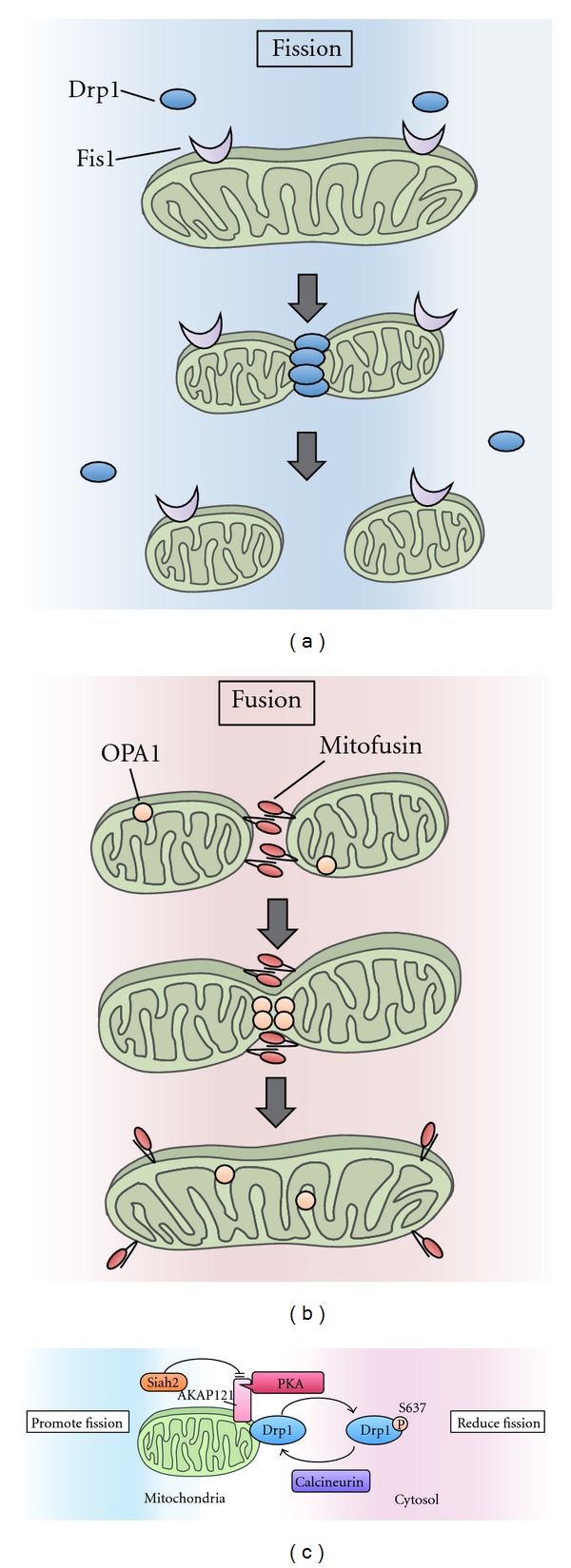
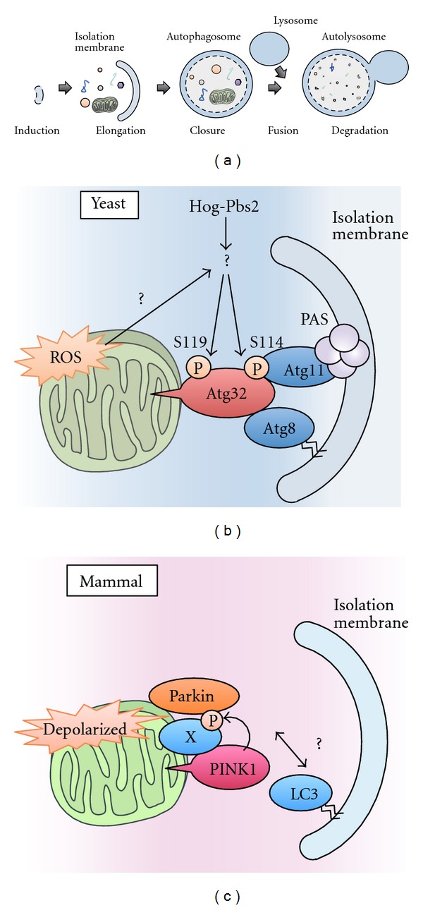
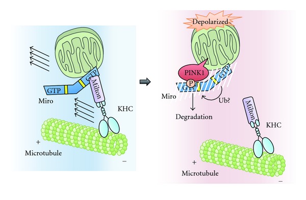
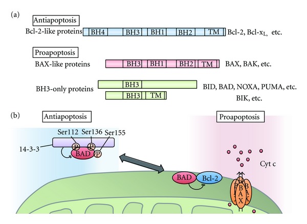
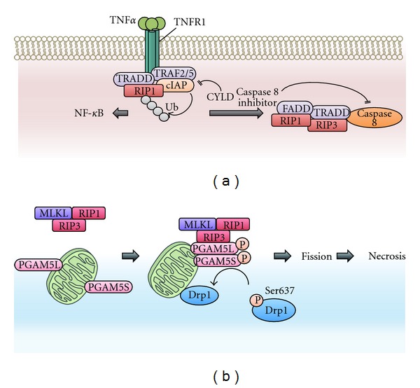
To maintain cellular homeostasis, cells are equipped with precise systems that trigger the appropriate stress responses. Mitochondria not only provide cellular energy but also integrate stress response signaling pathways, including those regulating cell death. Several lines of evidence suggest that the mitochondrial proteins that function in this process, such as Bcl-2 family proteins in apoptosis and phosphoglycerate mutase family member 5 (PGAM5) in necroptosis, are regulated by several kinases. It has also been suggested that the phosphorylation-dependent regulation of mitochondrial fission machinery, dynamin-related protein 1 (Drp1), facilitates appropriate cellular stress responses. However, mitochondria themselves are also damaged by various stresses. To avoid the deleterious effects exerted by damaged mitochondria, cells remove these mitochondria in a selective autophagic degradation process called mitophagy. Interestingly, several kinases, such as PTEN-induced putative kinase 1 (PINK1) in mammals and stress-responsive mitogen-activated protein (MAP) kinases in yeast, have recently been shown to be involved in mitophagy. In this paper, we focus on the phosphorylation-dependent regulation of mitochondrial proteins and discuss the roles of this regulation in the mitochondrial and cellular stress responses.


Figures

Figure 1
Figure 1
Mitochondrial fission and fusion. (a) Mitochondrial fission is driven by Drp1. Drp1 is recruited to the mitochondrial outer membrane at the fission sites and promotes mitochondrial fission. Fis1 resides on the mitochondrial outer membrane and is considered to function as a receptor for Drp1. (b) Fusion is driven by OPA1 and mitofusins. Interactions between these proteins tether two adjacent mitochondria. Mitofusins mediate mitochondrial outer membrane fusion, while Opa1 mediates mitochondrial inner membrane fusion. (c) The mitochondrial fission-promoting activity of Drp1 is controlled by the phosphorylation of Drp1 at Ser637. PKA phosphorylates Drp1 and inhibits the translocation of Drp1 to mitochondrial fission sites. Conversely, the calcineurin-mediated dephosphorylation of Drp1 results in the recruitment of Drp1 to the mitochondria and promotes mitochondrial fission. AKAP121 is a mitochondria-localized adaptor protein that regulates mitochondrial dynamics by facilitating Drp1 phosphorylation via PKA anchoring on mitochondria. Siah2 promotes the degradation of AKAP121, which decreases Drp1 phosphorylation, thus resulting in mitochondrial fission.
Figure 2
Figure 2
Mitophagy in yeast and mammals. (a) In autophagy, a double-membrane compartment, termed the isolation membrane, engulfs a portion of cytoplasm, including macromolecules and organelles, and forms autophagosomes. The late autophagosome fuses with the lysosome to form autolysosomes, which degrades the engulfed contents. (b) A model of mitophagy in yeast. Hog1 and Pbs2 regulate mitophagy by promoting the phosphorylation of Ser114 and Ser119 on Atg32. When Ser114 is phosphorylated, Atg32 binds to Atg11. Atg11 is a core component that facilitates the recruitment of specific cargoes to PAS, where the autophagosomes are generated. Atg32 also binds to Atg8, a factor that is essential for autophagosome formation. Autophagosomes envelop the mitochondria and fuse with lysosomes to degrade the mitochondria. (c) A model of mitophagy in mammalian cells. When mitochondria are depolarized, PINK1 accumulates on the mitochondrial outer membrane and recruits Parkin from the cytosol to the depolarized mitochondria in a manner dependent on the kinase activity of PINK1. The PINK1/Parkin complex triggers the autophagosome formation and the degradation of damaged mitochondria. Recent findings suggest that the proteasome is also involved in the destruction of mitochondria (see text for details).
Figure 3
Figure 3
The mechanism of PINK1/Parkin-dependent arrest of mitochondrial motility. Long-range mitochondrial transport relies on the action of the molecular motor complex, which includes KHC, Milton, and Miro. When mitochondria are depolarized, stabilized PINK1 phosphorylates Ser156 of Miro. Subsequent interaction of Parkin with Miro and likely ubiquitination by Parkin causes proteasomal degradation of Miro to arrest mitochondrial motility.
Figure 4
Figure 4
Bcl-2 family in apoptosis. (a) The domain structure of three classes of Bcl-2 family proteins. Antiapoptotic Bcl-2-like proteins have four Bcl-2 homology (BH) domains and a C-terminal transmembrane domain. Proapoptotic Bcl-2 family proteins are classified into two groups. Bax-like proteins have three BH domains (BH1-BH3) and a C-terminal transmembrane domain. Proapoptotic proteins, such as BID, BAD, NOXA, and PUMA, only have a BH3 domain and thus are called BH3-only proteins; some BH-3-only proteins, such as BIK, have a C-terminal transmembrane domain. (b) Survival signals suppress apoptosis by promoting the phosphorylation of BAD at Ser112, Ser136, and Ser155. When these sites are phosphorylated, BAD binds to 14-3-3 proteins, which suppresses the proapoptotic activity of BAD by inhibiting its association with antiapoptotic proteins such as Bcl-2. During apoptosis, dephosphorylated BAD dimerizes with Bcl-xL or Bcl-2 and promotes the release of inner mitochondrial proteins such as cytochrome c (Cyt c) through pores formed by BAX.
Figure 5
Figure 5
Necrosis signaling pathways. (a) Necrosis is induced downstream of death domain-containing receptors, including TNF receptors 1 (TNFR1) and TNFR2. When TNFα binds to TNFR1, RIP1 is recruited to the activated TNFR1. In this receptor-associated complex, E3 ligase cellular inhibitor of apoptosis proteins (cIAPs) mediate the Lys63-linked polyubiquitin of RIP1, activating the NF-κB pathway. Conversely, when the Lys63-linked polyubiquitin is removed by deubiquitinating enzymes, including CYLD, RIP1 functions as a cell death inducer rather than as a survival-promoting factor. When caspase 8 activity is blocked by inhibitors, such as a pan-caspase inhibitor or Z-VAD-fmk, or by the genetic ablation of caspase 8 itself, RIP1 interacts with RIP3 and forms a necrosis-inducing complex. RIP1 and RIP3 become phosphorylated and activated in this complex. Although the kinase activity of RIP1 is not required to activate the NF-κB pathway, its kinase activity is essential for necrosis induction. (b) The mitochondrial Ser/Thr protein phosphatase PGAM5 is a substrate of RIP3. The RIP3-mediated phosphorylation of PGAM5 enhances its protein phosphatase activity. Once activated by phosphorylation, PGAM5 dephosphorylates and activates the mitochondrial fission regulator Drp1, promoting mitochondrial fission and subsequent necrosis. PGAM5 has two splice variants in humans, PGAM5L and PGAM5S, each of which appears to have different functions in necrosis (see text for details).

References

    1. McBride HM, Neuspiel M, Wasiak S. Mitochondria: more than just a powerhouse. Current Biology. 2006;16(14):R551–R560. - PubMed
    1. Wang X. The expanding role of mitochondria in apoptosis. Genes and Development. 2001;15(22):2922–2933. - PubMed
    1. West AP, Shadel GS, Ghosh S. Mitochondria in innate immune responses. Nature Reviews Immunology. 2011;11(6):389–402. - PMC - PubMed
    1. Wallace DC. A mitochondrial paradigm of metabolic and degenerative diseases, aging, and cancer: a dawn for evolutionary medicine. Annual Review of Genetics. 2005;39:359–407. - PMC - PubMed
    1. Detmer SA, Chan DC. Functions and dysfunctions of mitochondrial dynamics. Nature Reviews Molecular Cell Biology. 2007;8(11):870–879. - PubMed