

doi: 10.1155/2012/508956.
Epub 2012 Jul 10.
Duplex ultrasound evaluation of hemodialysis access: a detailed protocol
Affiliations
- PMID: 22848824
- PMCID: PMC3400354
- DOI: 10.1155/2012/508956
Item in Clipboard
Duplex ultrasound evaluation of hemodialysis access: a detailed protocol
Int J Nephrol.
2012.
Abstract
A detailed protocol for the performance and interpretation of duplex ultrasound evaluation of hemodialysis access is described.
Figures
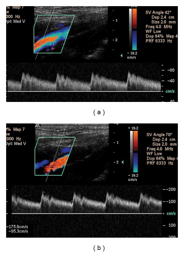
(a) The angle of insonation, noted by the phrase “SV Angle” in the upper left hand corner, has been set at 42°. The peak systolic velocity (PSV), the highest point at the top of the waveform, is nearly 80 cm/sec. (b) The same vessel is examined now at an angle of 70°. Marking the highest and lowest points along the waveforms instructs the machine to calculate PSV and the end diastolic velocity (EDV), shown in the lower left hand corner. Using this incorrect angle, the PSV has more than doubled to 175.9 cm/sec. Great care must be taken to avoid this error as PSV is widely used as a diagnostic measure. An improper angle of insonation may thus result in a false impression of stenosis where none actually exists.
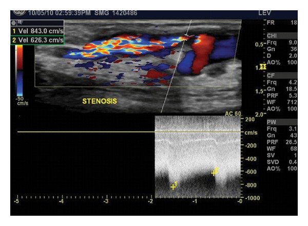
A high-grade stenosis is noted just distal to the take-off of a branch, where a marked elevation in both peak systolic (843.0 cm/sec) and end diastolic (626.3 cm/sec) velocities is found. Doppler color flow imaging demonstrates post-stenotic turbulence distal to the narrowest segment of the vein.
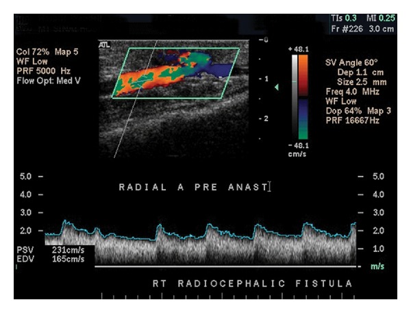
The radial artery just proximal to a Brescia-Cimino fistula demonstrates spectral broadening and diastolic flow seen characteristically in arterial beds with low resistance outflow in addition to elevation of both PSV and EDV. In the absence of dialysis access, a normal radial artery will exhibit triphasic waveforms with no spectral broadening and PSV >40 cm/sec.
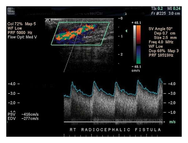
Normal arterio-venous fistula demonstrating marked spectral broadening and elevated velocities. The cephalic vein in this image is relatively superficial, sitting about a centimeter or less below the surface of the skin as denoted by the scale to the right of the color-flow image.
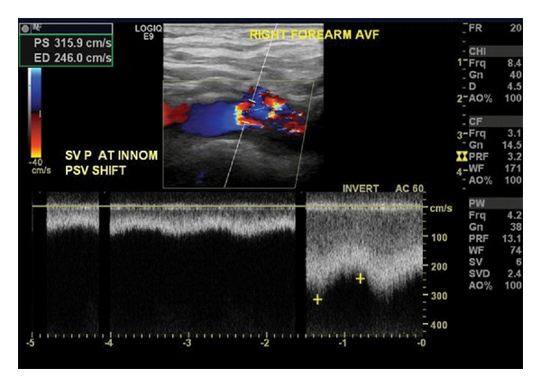
Marked turbulence and a velocity shift at the confluence of the subclavian and innominate veins indicates the presence of outflow stenosis.
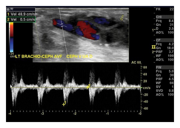
This brachiocephalic fistula has thrombosed. Waveforms demonstrate a to-and-from characteristic indicative of a vessel with no outflow. Low PSV, the absence of color flow throughout the access, and the presence of echogenic material within the fistula are other findings compatible with access thrombosis.
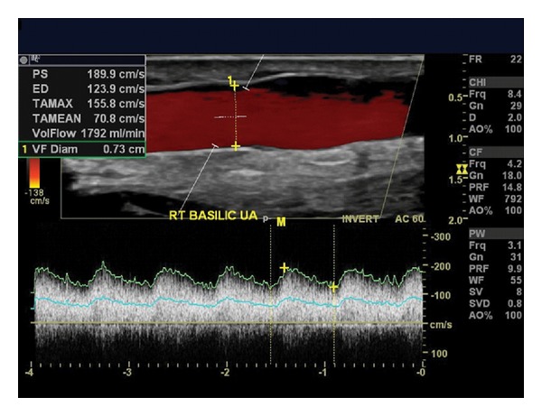
Ultrasound findings indicate this recently created transposed basilic fistula is maturing well. The scale to the right of the image confirms that a 5-cm length is superficial enough for easy cannulation, lying 0.5 cm or less from the surface of the skin. The diameter measures 0.73 cm. With PSV of 189.9 cm/sec and EDV of 123.9 cm/sec, a volume-flow of 1792 mL/min has been calculated by a software package incorporated into the ultrasound equipment. These details are shown in the upper left hand corner.
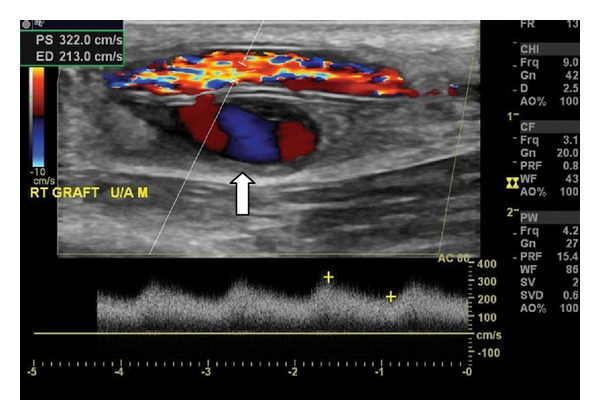
This ultrasound demonstrates a pseudoaneurysm (PSA), denoted by the white arrow, arising from the posterior wall of an access graft, presumably as a consequence of through-and-through puncture. Color Doppler shows the classic swirling “yin-yang” pattern of blood flow typically seen in PSAs.
References
-
- NKF-DOQI. NKF-DOQI clinical practice guidelines for vascular access. American Journal of Kidney Diseases . 1997;30(4, supplement):S150–S191. - PubMed
-
- KDOQI. Clinical practice guidelines for vascular access. American Journal of Kidney Diseases . 2006;48(supplement 1):S176–S247. - PubMed
-
- Silva MB, Jr., Hobson RW, II, Pappas PJ, et al. A strategy for increasing use of autogenous hemodialysis access procedures: impact of preoperative noninvasive evaluation. Journal of Vascular Surgery . 1998;27(2):302–308. - PubMed
-
- Ferring M, Henderson J, Wilmink A, Smith S. Vascular ultrasound for the pre-operative evaluation prior to arteriovenous fistula formation for haemodialysis: review of the evidence. Nephrology Dialysis Transplantation . 2008;23(6):1809–1815. - PubMed
LinkOut - more resources
Full Text Sources
Other Literature Sources
