. 2012:2012:640762.
doi: 10.1155/2012/640762. Epub 2012 Jul 15.

The phospholipid profile of mycoplasmas

Affiliations

The phospholipid profile of mycoplasmas

Jonathan D Kornspan et al. J Lipids. 2012.

Abstract

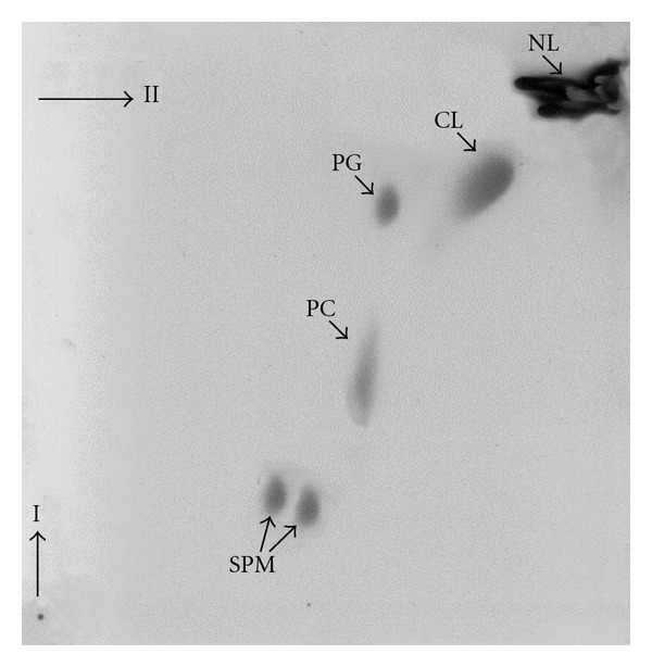
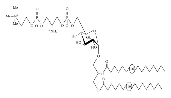
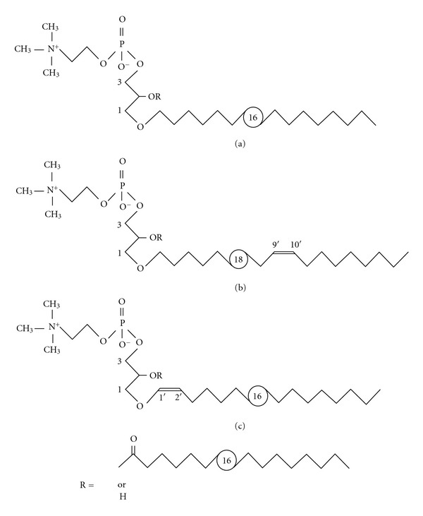
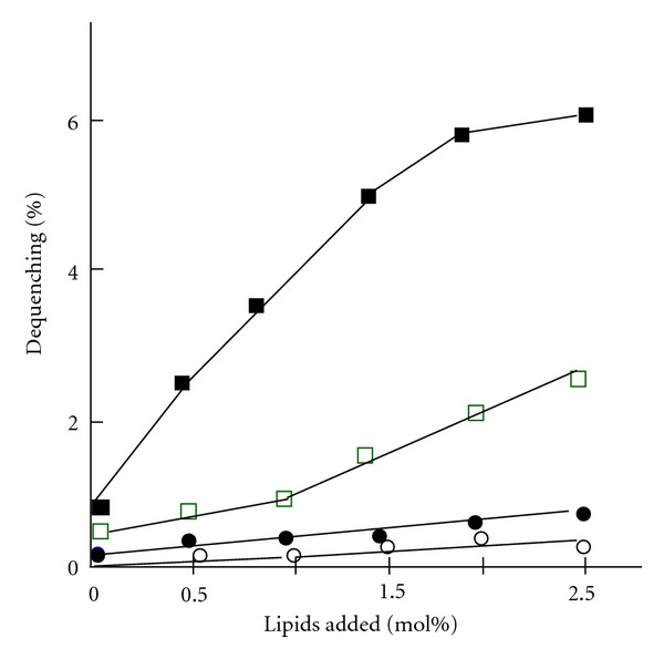
The de novo synthesized polar lipids of Mycoplasma species are rather simple, comprising primarily of the acidic glycerophospholipids PG and CL. In addition, when grown in a medium containing serum, significant amounts of PC and SPM are incorporated into the mycoplasma cell membrane although these lipids are very uncommon in wall-covered bacteria. The exogenous lipids are either incorporated unchanged or the PC incorporated is modified by a deacylation-acylation enzymatic cycle to form disaturated PC. Although their small genome, in some Mycoplasma species, other genes involved in lipid biosynthesis were detected, resulting in the synthesis of a variety of glycolipis, phosphoglycolipids and ether lipids. We suggest that analyses and comparisons of mycoplasma polar lipids may serve as a novel and useful tool for classification. Nonetheless, to evaluate the importance of polar lipids in mycoplasma, further systematic and extensive studies on more Mycoplasma species are needed. While studies are needed to elucidate the role of lipids in the mechanisms governing the interaction of mycoplasmas with host eukaryotic cells, the finding that a terminal phosphocholine containing glycolipids of M. fermentans serves both as a major immune determinants and as a trigger of the inflammatory responses, and the findings that the fusogenicity of M. fermentans with host cells is markedly stimulated by lyso-ether lipids, are important steps toward understanding the molecular mechanisms of M. fermentans pathogenicity.


Figures

Figure 1
Figure 1
The major polar lipids of M. hyorhinis. Lipids were extracted from membrane preparations by the method of Bligh and Dyer [18]. The lipids were separated by TLC on silica gel plates (Kiesel-gel 60 HR, Merck, Darmstadt, Germany) developed at room temperature by a two-dimensional system using chloroform-methanol-ammonia (65 : 35 : 4 by vol.) for the first dimension and chloroform-methanol-acetic acid-water (85 : 25 : 5 : 4 by vol.) for the second dimension. Lipid spots were detected by iodine vapor, while phospholipid spots were detected by the molybdate spray reagent [29]. The spots were tentatively identified based on their comigration with commercial standards as NL, neutral lipids; CL, cardiolipin; PG, phosphatidylglycerol; PC, phosphatidylcholine; SPM, sphingomyelin.
Figure 2
Figure 2
Structure of MfGL-II of M. fermentans. The chemical structure of the major polar lipid of M. fermentans, was investigated by GLC-MS, MALDI-TOF mass spectrometry, as well as one-and two-dimensional homo- and heteronuclear NMR spectroscopy and identified as 6′-O-[3-phosphocholine-2-amino-1-phospho-1,3-propanediol]-α-D-glucopyranosyl-(1′→3)-1,2-diacyl-glycerol (MfGL-II). Palmitate (16 : 0) and stearate (18 : 0), in a 3.6 : 1 molar ratio, constitute the major fatty acids present [32].
Figure 3
Figure 3
Structures of ether lipids of M. fermentans. Both MfEL (R=acyl) and lyso-MfEL (R=H) are mixtures of different molecular species with respect to the alkyl/alkenyl residues which are hexadecyl (a), (9Z)-octadec-9′-enyl (b), or (1Z)-alk-1′-enyl (c) [37].
Figure 4
Figure 4
The effect of M. fermentans lipids on the fusion of SUV with Molt-3 cells. Small unilamellar vesicles (SUVs) were prepared by sonicating a buffer solution containing egg-phosphatidylcholine (about 5 mg per mL) with various amounts of the glycophospholipid MfGL-II or the ether lipids MfEL or lyso-MfEL [37] as previously described [39]. The SUVs were fluorescently labeled with the octadecyl rhodamine B chloride (R18). Fusion of SUV with T-lymphocytes of the human CD4+ Molt-3 was monitored by following the fluorescence intensity of the Molt-3 cells, and the results were presented as % dequenching [40]. (○): SUV alone; (●): SUV + MFGL-II; (□): SUV + MfEL; (■): SUV + lyso-MfEL.

References

    1. Razin S, Yogev D, Naot Y. Molecular biology and pathogenicity of Mycoplasmas . Microbiology and Molecular Biology Reviews. 1998;62(4):1094–1156. - PMC - PubMed
    1. Herrmann R, Gohlmann NWH, Regula JT, et al. Mycoplasmas, the smallest known bacteria. In: Göbel UB, Reinbeck BRR, editors. Microbial Evolution and Infection. Hamburg, Germany: Einhorn-Presse; 1999. pp. 71–79.
    1. Maniloff J. The minimal cell genome: on being the right size. Proceedings of the National Academy of Sciences of the United States of America. 1996;93(19):10004–10006. - PMC - PubMed
    1. Rottem S. Membrane lipids of Mycoplasmas . Biochimica et Biophysica Acta. 1980;604(1):65–90. - PubMed
    1. McElhaney RN. Physical studies of lipid organization and dynamics in Mycoplasma membranes. Sub-Cellular Biochemistry. 1993;20:53–108. - PubMed

LinkOut - more resources