. 2012 Nov;32(11):1989-2001.
doi: 10.1038/jcbfm.2012.109. Epub 2012 Aug 1.

Transport of biogenic amine neurotransmitters at the mouse blood-retina and blood-brain barriers by uptake1 and uptake2

Affiliations

Transport of biogenic amine neurotransmitters at the mouse blood-retina and blood-brain barriers by uptake1 and uptake2

Pascal André et al. J Cereb Blood Flow Metab. 2012 Nov.

Abstract

Uptake1 and uptake2 transporters are involved in the extracellular clearance of biogenic amine neurotransmitters at synaptic clefts. We looked for them at the blood-brain barrier (BBB) and blood-retina barriers (BRB), where they could be involved in regulating the neurotransmitter concentration and modulate/terminate receptor-mediated effects within the neurovascular unit (NVU). Uptake2 (Oct1-3/Slc22a1-3, Pmat/Slc29a4) and Mate1/Slc47a1 transporters are also involved in the transport of xenobiotics. We used in situ carotid perfusion of prototypic substrates like [(3)H]-1-methyl-4-phenylpyridinium ([(3)H]-MPP(+)), [(3)H]-histamine, [(3)H]-serotonin, and [(3)H]-dopamine, changes in ionic composition and genetic deletion of Oct1-3 carriers to detect uptake1 and uptake2 at the BBB and BRB. We showed that uptake1 and uptake2 are involved in the transport of [(3)H]-dopamine and [(3)H]-MPP(+) at the blood luminal BRB, but not at the BBB. These functional studies, together with quantitative RT-PCR and confocal imaging, suggest that the mouse BBB lacks uptake1 (Net/Slc6a2, Dat/Slc6a3, Sert/Slc6a4), uptake2, and Mate1 on both the luminal and abluminal sides. However, we found evidence for functional Net and Oct1 transporters at the luminal BRB. These heterogeneous transport properties of the brain and retina NVUs suggest that the BBB helps protect the brain against biogenic amine neurotransmitters in the plasma while the BRB has more of a metabolic/endocrine role.


Figures

Figure 1
Figure 1
Modulation of [3H]-1-methyl-4-phenylpyridinium ([3H]-MPP+) ocular (BRB, solid bars) and cerebral (BBB, empty bars) transport by biogenic amine and aminergic reuptake inhibitors (A), and changes in vascular or intracellular pH (pHi) (B). Both panels show Kin (μL/s per gram) values obtained after in situ carotid perfusion of Swiss mice with 9 nmol/L [3H]-MPP+ for 120 seconds. (A) Effect of coperfusing unlabeled compounds in Krebs-carbonate perfusion fluid (pHe 7.40). (B) Effect of changing the pHe (5.4, 6.4, or 8.4) or increasing the pHi by acute exposure to NH4+ during perfusion. Controls were perfused with regular Krebs-bicarbonate fluid (pHe 7.40). Data are mean values±s.d. (n=4 to 6 mice). *P<0.05, **P<0.01, ***P<0.001 compared with the appropriate control Kin obtained in the same tissue. BRB, blood–retina barrier; BBB, blood–brain barrier; pHe, extracellular vascular pH.
Figure 2
Figure 2
Uptake1 transporters involved in the ocular (BRB, solid bars) and cerebral (BBB, empty bars) transport of [3H]-1-methyl-4-phenylpyridinium ([3H]-MPP+). Both panels show Kin (μL/s per gram) values obtained after in situ carotid perfusion of Swiss mice with 9 nmol/L [3H]-MPP+ for 120 seconds. (A) Krebs-carbonate perfusion fluid (pHe 7.40, control) lacking Cl (replaced by gluconate and nitrate, ‘Glu'), Na+ (replaced by N-methyl D-glucamine chloride ‘NMDG-Cl', potassium ‘K+' or lithium ‘Li+') or both Cl and Na+ (replaced by mannitol). (B) Perfusion fluid with or without (Control) 1 μmol/L GBR12909 (Dat inhibitor), 1 μmol/L citalopram (Sert inhibitor), or 1 μmol/L nisoxetine (Net inhibitor). Data are mean values±s.d. (n=4 to 6 mice). **P<0.01, ***P<0.001 compared with the appropriate control Kin obtained in the same tissue. BRB, blood–retina barrier; BBB, blood–brain barrier; pHe, extracellular vascular pH.
Figure 3
Figure 3
Effects of uptake2, Oct1,2 and Oct3 transporters and uptake2 inhibitors on [3H]-1-methyl-4-phenylpyridinium ([3H]-MPP+) (9 nmol/L) transport (Kin; μL/s per gram) measured by in situ carotid perfusion of Swiss, and Oct-deficient Fvb mice for 120 seconds. (A) [3H]-MPP+ transport (Kin; μL/s per gram) measured in Fvb (wild-type, control), Oct1,2(−/−) and Oct3(−/−) Fvb mice. (B) [3H]-MPP+ transport (Kin; μL/s per gram) at the eye (BRB, black column) and brain (BBB, empty column) measured in Swiss mice with or without (control) coperfusion of cold compound in Krebs-carbonate perfusion fluid (pHe 7.40). Data are mean values±s.d. (n=4 to 6 mice). *P<0.05, **P<0.01, ***P<0.001 compared with the appropriate control obtained in the same tissue. BRB, blood–retina barrier; BBB, blood–brain barrier; pHe, extracellular vascular pH.
Figure 4
Figure 4
Eye (BRB, solid bars) and brain (BBB, empty bars) transport of [3H]-histamine (A), [3H]-serotonin (B) and [3H]-dopamine (C, D). Kin values (μL/s per gram) measured after carotid perfusion of the radiolabeled compounds in Krebs-carbonate buffer (pHe 7.40) for 60 seconds. (A) [3H]-histamine (60 nmol/L) transport measured in Swiss mice with or without (control) coperfusion of unlabeled histamine (1 or 10 mmol/L) and in Oct1,2(−/−) Fvb mice. (B) [3H]-serotonin (40 nmol/L) transport measured in Swiss mice with or without (wild-type; control) coperfusion of unlabeled serotonin (10 mmol/L) or L-tryptophan (5 mmol/L) or with Na+and Cl depletions (mannitol), and in Oct1,2(−/−) Fvb mice. (C) [3H]-dopamine (40 nmol/L) transport measured in Swiss mice under the following conditions: normal Krebs-carbonate perfusion fluid (control), coperfusion of unlabeled dopamine (1 or 10 μmol/L), norepinephrine (1 or 100 μmol/L), or nisoxetine (0.1 or 1 μmol/L), depletion of chloride (Glu), sodium (NMDG-Cl) or both sodium and chloride (mannitol). (D) [3H]-dopamine transport measured in Fvb (control), Oct3(−/−) and double knock-out Oct1,2(−/−) Fvb mice with and without coperfusion of nisoxetine (1 μmol/L). Data are mean values±s.d. (n=4 to 6 mice). *P<0.05, **P<0.01, ***P<0.001 compared with the appropriate control Kin obtained in the same tissue. †P<0.05 comparing Oct1,2(−/−) mice with and without nisoxetine (1 μmol/L) in the same tissue. BRB, blood–retina barrier; BBB, blood–brain barrier; pHe, extracellular vascular pH.
Figure 5
Figure 5
Uptake1 (Dat, Sert, Net) and uptake2 (Oct3, Pmat) immunostaining in the Swiss mouse brain. (A) Immunostaining for Dat, Sert, and Net (yellow) along with P-glycoprotein (P-gp) or laminin (magenta). (B) Immunostaining for Oct3 and Pmat (yellow) along with P-gp or laminin (magenta). All images are of brain sections from Swiss mice. Images are 3D reconstructions obtained by maximum intensity projection after the acquisition of a z-stack of confocal images. Although some fibers are sometimes found running close to capillaries (arrowheads), the cerebral endothelium was not immunostained for Dat, Sert, Net, Oct3, or Pmat. Bar: 20 μm.
Figure 6
Figure 6
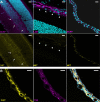
Cryosections immunostained for Net in the Swiss whole eye. Nuclei are stained with TOPRO-3 (cyan). (A) Immunostaining for Net (yellow) and Bcrp (magenta) or laminin (magenta). Net was not detected in retinal capillaries (arrows, left column) or the retinal pigmentary epithelium (RPE) (arrowheads, central column), but was intensely stained in the iris/ciliary bodies (only iris is shown, right column). (B) Net (yellow) and tyrosine hydroxylase (TH) (magenta) labeling in the iris. Net immunostaining was restricted to TH-positive varicose fibers. Bar: 20 μm.

References

    1. Abbott NJ, Patabendige AA, Dolman DE, Yusof SR, Begley DJ. Structure and function of the blood-brain barrier. Neurobiol Dis. 2010;37:13–25. - PubMed
    1. Amphoux A, Vialou V, Drescher E, Brüss M, Mannoury La Cour C, Rochat C, Millan MJ, Giros B, Bönisch H, Gautron S. Differential pharmacological in vitro properties of organic cation transporters and regional distribution in rat brain. Neuropharmacology. 2006;50:941–952. - PubMed
    1. Brust P, Friedrich A, Krizbai IA, Bergmann R, Roux F, Ganapathy V, Johannsen B. Functional expression of the serotonin transporter in immortalized rat brain microvessel endothelial cells. J Neurochem. 2000;74:1241–1248. - PubMed
    1. Buck KJ, Amara SG. Chimeric dopamine-norepinephrine transporters delineate structural domains influencing selectivity for catecholamines and 1-methyl-4-phenylpyridinium. Proc Natl Acad Sci USA. 1994;91:12584–12588. - PMC - PubMed
    1. Catravas JD, Gillis CN. Single-pass removal of [14C]-5-hydroxytryptamine and [3H]norepinephrine by rabbit lung, in vivo: kinetics and sites of removal. J Pharmacol Exp Ther. 1983;224:28–33. - PubMed

MeSH terms

LinkOut - more resources