. 2012 Oct 1;18(19):5329-40.
doi: 10.1158/1078-0432.CCR-12-1632. Epub 2012 Jul 31.

Oncogenic BRAF(V600E) promotes stromal cell-mediated immunosuppression via induction of interleukin-1 in melanoma

Affiliations

Oncogenic BRAF(V600E) promotes stromal cell-mediated immunosuppression via induction of interleukin-1 in melanoma

Jahan S Khalili et al. Clin Cancer Res. .

Abstract

Purpose: In this study, we assessed the specific role of BRAF(V600E) signaling in modulating the expression of immune regulatory genes in melanoma, in addition to analyzing downstream induction of immune suppression by primary human melanoma tumor-associated fibroblasts (TAF).

Experimental design: Primary human melanocytes and melanoma cell lines were transduced to express WT or V600E forms of BRAF, followed by gene expression analysis. The BRAF(V600E) inhibitor vemurafenib was used to confirm targets in BRAF(V600E)-positive melanoma cell lines and in tumors from melanoma patients undergoing inhibitor treatment. TAF lines generated from melanoma patient biopsies were tested for their ability to inhibit the function of tumor antigen-specific T cells, before and following treatment with BRAF(V600E)-upregulated immune modulators. Transcriptional analysis of treated TAFs was conducted to identify potential mediators of T-cell suppression.

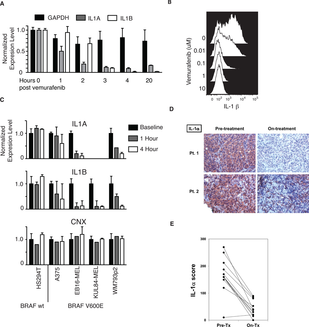
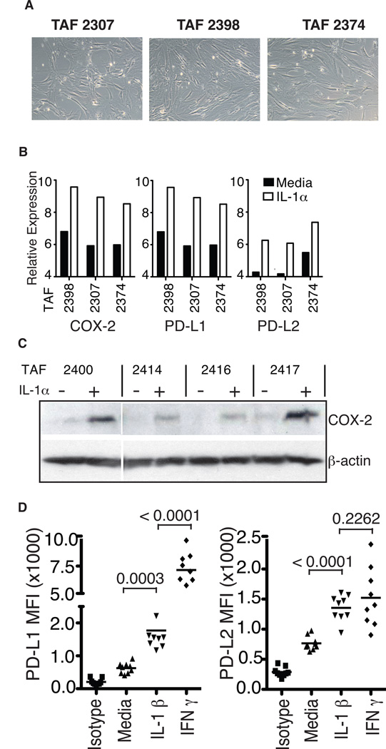
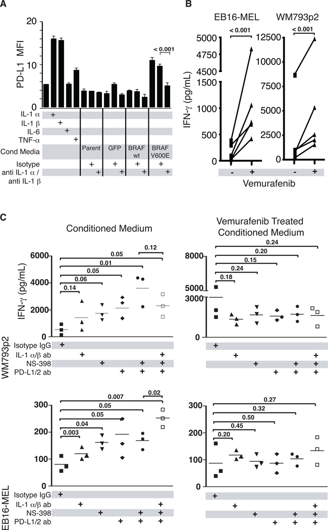
Results: Expression of BRAF(V600E) induced transcription of interleukin 1 alpha (IL-1α) and IL-1β in melanocytes and melanoma cell lines. Further, vemurafenib reduced the expression of IL-1 protein in melanoma cell lines and most notably in human tumor biopsies from 11 of 12 melanoma patients undergoing inhibitor treatment. Treatment of melanoma-patient-derived TAFs with IL-1α/β significantly enhanced their ability to suppress the proliferation and function of melanoma-specific cytotoxic T cells, and this inhibition was partially attributable to upregulation by IL-1 of COX-2 and the PD-1 ligands PD-L1 and PD-L2 in TAFs.

Conclusions: This study reveals a novel mechanism of immune suppression sensitive to BRAF(V600E) inhibition, and indicates that clinical blockade of IL-1 may benefit patients with BRAF wild-type tumors and potentially synergize with immunotherapeutic interventions.


Conflict of interest statement

Disclosure of Potential Conflicts of Interest: Michael A. Davies has served on advisory boards and received research funding from Roche-Genentech and GlaxoSmithKline.

Figures

Figure 1
Figure 1. Ectopic expression of BRAF(V600E) upregulates IL-1α/β expression in melanocytes and melanoma cells
(A and B) Flow cytometric analysis of green fluorescent protein (GFP) and BRAF expression in dermal melanocytes following transduction with lentiviral expression vectors BRAF(wt)-IRES-GFP, BRAF(V600E)-IRES-GFP, or empty-IRES-GFP. Gated GFP(dim) cells were flow sorted for use in subsequent studies. (C) Affymetrix gene expression profiling of selected genes classically implicated in immune modulation of the tumor microenvironment. Transduced and sorted dermal melanocytes (36 hours following transduction) or HS294T cells (24 hours post-transduction) were analyzed, and the heatmap shown represents color-coded expression levels for each sample compared to GFP-transduced controls. (D) Luminex assay showing cytokine profiles in supernatants of transduced dermal melanocyte preparations cultured for 5 days. Results are representative of 4 independent experiments. ND, not detected.
Figure 2
Figure 2. Inhibition of BRAF(V600E) abrogates IL-1 expression in melanoma cell lines and patient tumors
(A) RT-PCR analysis of IL-1α, IL-1β and GAPDH transcripts in BRAF(V600E)-positive WM793p2 cells at different time points following treatment with 1µM vemurafenib. (B) Flow cytometric analysis showing intracellular IL-1β protein expression in live cell-gated WM793p2 cells 48 hours following treatment with titrated doses of PLX4032. (C) RT-PCR analysis showing transcript levels of IL1α, IL1β and CNX in five vemurafenib-treated melanoma cell lines expressing either wt BRAF (HS294T) or V600E-mutated BRAF (A375, EB16-MEL, KUL84-MEL, WM793p2). Transcript levels were normalized to GAPDH expression and adjusted to corresponding baseline samples. (D) Immunohistochemical (IHC) analysis of IL-1α protein expression in tumor biopsies resected from two representative metastatic melanoma patients harboring the BRAF(V600E) mutation, both prior to and on vemurafenib treatment. (E) Summary of changes to IL-1α expression in response to vemurafenib treatment, as assessed by IHC analysis of 12 total melanoma patient tumor biopsy pairs analyzed.
Figure 3
Figure 3. IL-1α treated tumor-associated fibroblasts induce suppression of melanoma-specific CD8+ T-cells
(A) Tissue sections from two representative melanoma metastases labeled with anti-αSMA antibody and visualized with peroxidase immunostaining. Red-brown color shows staining of αSMA-positive tumor-associated fibroblasts (TAF), asterisks denote tumor cells, arrows indicate tumor infiltrating lymphocytes (TIL), and ‘V’ denotes tumor vasculature. (B) Interferon-gamma release by MART-1 reactive TIL stimulated with MART-1 peptide-pulsed T2 cells in the presence or absence of untreated or IL-1α treated melanoma TAFs, with or without the addition of IL-1 neutralizing antibodies. Data are representative of six different TAF lines analyzed and three experimental replicates. (C) Frequency of CD107a-positive TIL following co-culture with MART-1 peptide-pulsed dermal fibroblasts pretreated with or without IL-1α, as determined by flow cytometry. Data from 2 different melanoma patient TIL are shown, and are representative of 2 independent experiments. Asterisks indicate statistical significance (P < 0.05); ns, not significant.
Figure 4
Figure 4. IL-1α upregulates the expression of immunosuppressive genes in melanoma-derived TAFs
(A) Phase contrast images of three short-term cultured TAFs derived from melanoma patient biopsies (10X). (B) Normalized relative transcriptional expression levels of COX-2, PD-L1 and PD-L2 in 24 h IL-1α treated or untreated TAFs, as analyzed by Affymetrix gene expression array. (C) Western blot analysis showing COX-2 and β-actin protein expression in four additional patient-derived TAF lines, prior to and 24 hours following treatment with IL-1α. (D) Surface expression of PD-1 ligands PD-L1 and PD-L2 on TAFs 24 hours after treatment with IL-1α or IFN-γ, as determined by flow cytometry. Data from 9 different melanoma-derived TAF lines are shown. Asterisks indicate statistical significance (P < 0.05); ns, not significant.
Figure 5
Figure 5. BRAF(V600E) can induce T-cell suppression through IL-1 mediated upregulation of PD-1 ligands and COX-2 on TAFs
(A) Flow cytometric analysis of surface PD-L1 expression on TAFs cultured overnight with IL-1α, IL-1β, IL-6, TNF-α, or conditioned medium from cultured primary melanocytes transduced with lentiviral vectors expressing BRAF(wt)-GFP, BRAF(V600E)-GFP, or GFP alone. As indicated, conditioned media experiments were also performed in the presence of isotype control or anti-IL1α and anti-IL1β blocking antibodies. (B) Interferon-gamma release by T2-stimulated MART-1 reactive TIL in the presence of melanoma patient-derived TAFs previously exposed to conditioned media from BRAF(V600E) mutant-expressing melanoma cell lines that were either untreated or treated with the BRAF(V600E) inhibitor vemurafenib. Results from five different TAF lines are shown. (C) To assess the relative contributions of IL-1α/β, COX-2, and PD-1 ligands in the induction of T-cell suppression, three melanoma TAF lines were pre-treated with conditioned media from untreated or vemurafenib-treated melanoma cell lines (WM793p2 and EB16-MEL), in the presence of either IL-1α/β blocking antibodies or the COX-2 inhibitor NS398. Pre-conditioned TAFs were then incubated with MART-1-reactive TIL and MART-1 peptide-pulsed T2 cells in the presence of isotype control antibody or antibodies specific for PD-L1 and PD-L2. Asterisks indicate statistical significance (P < 0.05); ns, not significant.
Figure 6
Figure 6. Model of BRAF(V600E)-mediated immune suppression by TAFs in the melanoma tumor microenvironment
Illustration of proposed mechanistic model showing how constitutively activated BRAF(V600E) in melanoma tumor cells may initiate and sustain T-cell suppression in vivo. In this model, T-cell suppression is manifested by tumor-associated fibroblasts (TAFs) that upregulate COX-2 and PD-1 ligands PD-L1 and PD-L2 in response to BRAF(V600E)-induced IL-1α/β production by melanoma cells. Targeted therapies that inhibit BRAF(V600E) could abrogate IL-1α/β production by tumor cells, thus interfering with the cellular crosstalk leading to TAF-mediated immune suppression.

References

    1. Ostrand-Rosenberg S. Myeloid-derived suppressor cells: more mechanisms for inhibiting antitumor immunity. Cancer Immunol Immunother. 2010;59:1593–1600. - PMC - PubMed
    1. Poschke I, Mougiakakos D, Kiessling R. Camouflage and sabotage: tumor escape from the immune system. Cancer Immunol Immunother. 2011 - PMC - PubMed
    1. Lizee G, Cantu MA, Hwu P. Less yin, more yang: confronting the barriers to cancer immunotherapy. Clinical cancer research. 2007;13:5250–5255. - PubMed
    1. Lizee G, Radvanyi LG, Overwijk WW, Hwu P. Improving antitumor immune responses by circumventing immunoregulatory cells and mechanisms. Clin Cancer Res. 2006;12:4794–4803. - PubMed
    1. Lizee G, Radvanyi LG, Overwijk WW, Hwu P. Immunosuppression in melanoma immunotherapy: potential opportunities for intervention. Clin Cancer Res. 2006;12:2359s–2365s. - PubMed

Publication types

MeSH terms