. 2012 Aug 29;31(17):3588-95.
doi: 10.1038/emboj.2012.204. Epub 2012 Jul 31.

Arabidopsis Argonaute MID domains use their nucleotide specificity loop to sort small RNAs

Affiliations

Arabidopsis Argonaute MID domains use their nucleotide specificity loop to sort small RNAs

Filipp Frank et al. EMBO J. .

Abstract

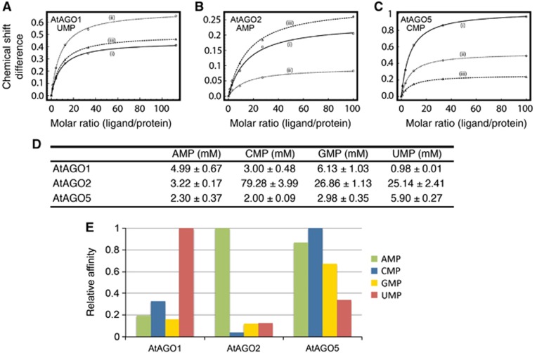
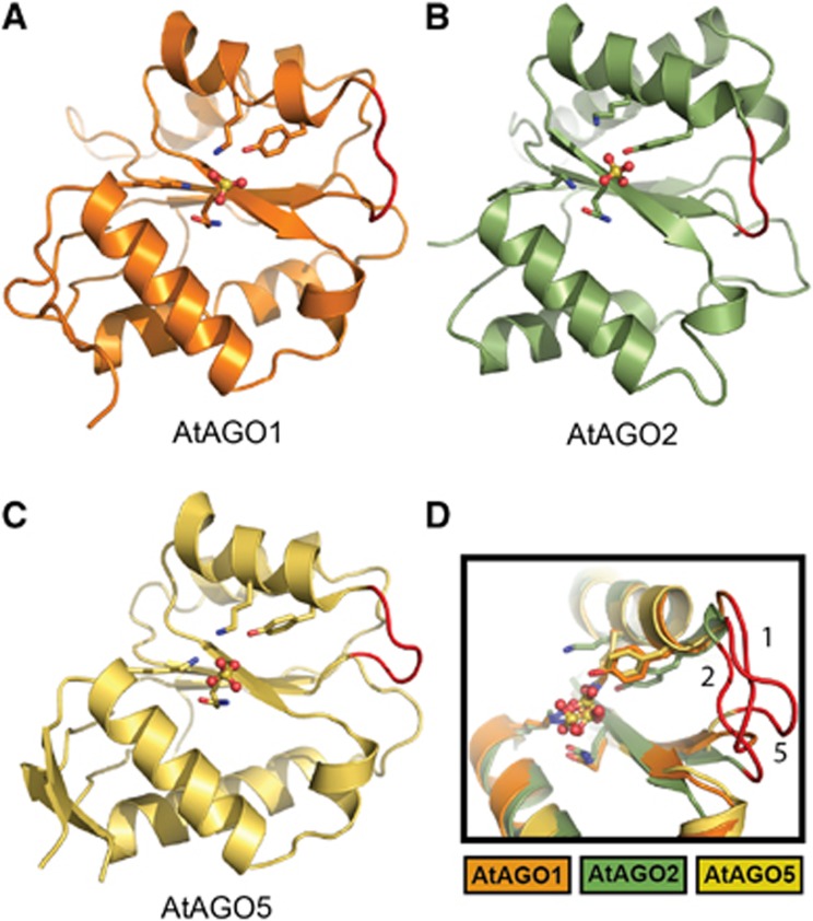
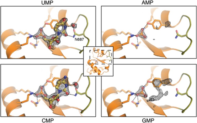
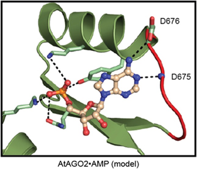
The 5'-nucleotide of small RNAs associates directly with the MID domain of Argonaute (AGO) proteins. In humans, the identity of the 5'-base is sensed by the MID domain nucleotide specificity loop and regulates the integrity of miRNAs. In Arabidopsis thaliana, the 5'-nucleotide also controls sorting of small RNAs into the appropriate member of the AGO family; however, the structural basis for this mechanism is unknown. Here, we present crystal structures of the MID domain from three Arabidopsis AGOs, AtAGO1, AtAGO2 and AtAGO5, and characterize their interactions with nucleoside monophosphates (NMPs). In AtAGOs, the nucleotide specificity loop also senses the identity of the 5'-nucleotide but uses more diverse modes of recognition owing to the greater complexity of small RNAs found in plants. Binding analyses of these interactions reveal a strong correlation between their affinities and evolutionary conservation.


Conflict of interest statement

The authors declare that they have no conflict of interest.

Figures

Figure 1
Figure 1
Binding affinities of NMPs with the MID domains of AtAGO1, AtAGO2 and AtAGO5 probed by NMR titration spectroscopy. (AC) Chemical shift differences were calculated from 1H-15N HSQC NMR titration experiments and plotted as a function of the molar ratio (nucleotide/protein). Representative binding curves for AtAGO1, AtAGO2 and AtAGO5, with their preferred 5′-nucleotides, UMP, AMP and CMP, respectively, are shown. The data for multiple peaks were fit using the maximum shift and dissociation constant (KD) as adjustable parameters. (D) Summary of dissociation constants of NMPs with MID domains from AtAGO1, AtAGO2 and AtAGO5. Values are means of curve fits from three different peaks with indicated standard deviations. The complete set of NMR spectra and curve fits for all interactions are shown in Supplementary Figures 3–5. (E) Graphical representation of data shown in (D). Vertical bars represent the association constant of the interactions (KA=KD−1), so that a higher bar represents stronger binding. Data were normalized to one for the highest affinity association constant for each protein. This representation makes apparent the stringency of binding selectivity for each AtAGO.
Figure 2
Figure 2
Crystal structures of the MID domains of AtAGO1 (A), AtAGO2 (B) and AtAGO5 (C). Structures are represented as ribbon diagrams and the sulphate ions bound to the 5′-phosphate-binding sites are shown as balls and sticks. Highly conserved phosphate interacting residues (Tyr, Lys, Gln, Lys) are shown as sticks. The loop region corresponding to the nucleotide specificity loop is coloured red. (D) Structural alignment of MID domains from AtAGO1, AtAGO2 and AtAGO5 highlighting the structural variability of the nucleotide specificity loops. Loops are coloured red and numbered according to the AtAGO from which they originate.
Figure 3
Figure 3
AtAGO1 MID domain in complex with NMPs. UMP and CMP are modelled and shown in ball and stick representation. The nucleotide specificity loop is coloured green. Only the phosphate groups of GMP and AMP were modelled (not shown). Protein side chains interacting with the 5′-phosphate or the base portion of the NMPs are shown as sticks with carbon, oxygen and nitrogen atoms coloured orange, red and blue, respectively. A water molecule that indirectly hydrogen bonds N687 to the NMP is shown as a red sphere. Difference electron density contoured at 2.5σ is shown before inclusion of any nucleotide in the model. Dotted black lines indicate hydrogen bonds.
Figure 4
Figure 4
Modelling of AMP into the structure of the AtAGO2 MID domain. The MID domain is shown in ribbon representation coloured green with the nucleotide specificity loop highlighted in red. AMP is shown in ball and stick representation. Shown as sticks, are conserved phosphate-binding residues as well as Asp676, which could potentially hydrogen bond with AMP. The amide backbone nitrogen atom of Asp675, which could also hydrogen bond with AMP, is represented as a sphere.

References

    1. Adams PD, Grosse-Kunstleve RW, Hung LW, Ioerger TR, McCoy AJ, Moriarty NW, Read RJ, Sacchettini JC, Sauter NK, Terwilliger TC (2002) PHENIX: building new software for automated crystallographic structure determination. Acta Crystallogr D Biol Crystallogr 58: 1948–1954 - PubMed
    1. Boland A, Tritschler F, Heimstädt S, Izaurralde E, Weichenrieder O (2010) Crystal structure and ligand binding of the MID domain of a eukaryotic Argonaute protein. EMBO Rep 11: 522–527 - PMC - PubMed
    1. Brennecke J, Aravin AA, Stark A, Dus M, Kellis M, Sachidanandam R, Hannon GJ (2007) Discrete small RNA-generating loci as master regulators of transposon activity in Drosophila. Cell 128: 1089–1103 - PubMed
    1. Brünger AT, Adams PD, Clore GM, DeLano WL, Gros P, Grosse-Kunstleve RW, Jiang JS, Kuszewski J, Nilges M, Pannu NS, Read RJ, Rice LM, Simonson T, Warren GL (1998) Crystallography & NMR system: a new software suite for macromolecular structure determination. Acta Crystallogr D Biol Crystallogr 54: 905–921 - PubMed
    1. Czech B, Hannon GJ (2011) Small RNA sorting: matchmaking for Argonautes. Nat Rev Genet 12: 19–31 - PMC - PubMed

Publication types