. 2012 Sep 12;31(18):3667-77.
doi: 10.1038/emboj.2012.180. Epub 2012 Jul 31.

Rif1 regulates the replication timing domains on the human genome

Affiliations

Rif1 regulates the replication timing domains on the human genome

Satoshi Yamazaki et al. EMBO J. .

Abstract

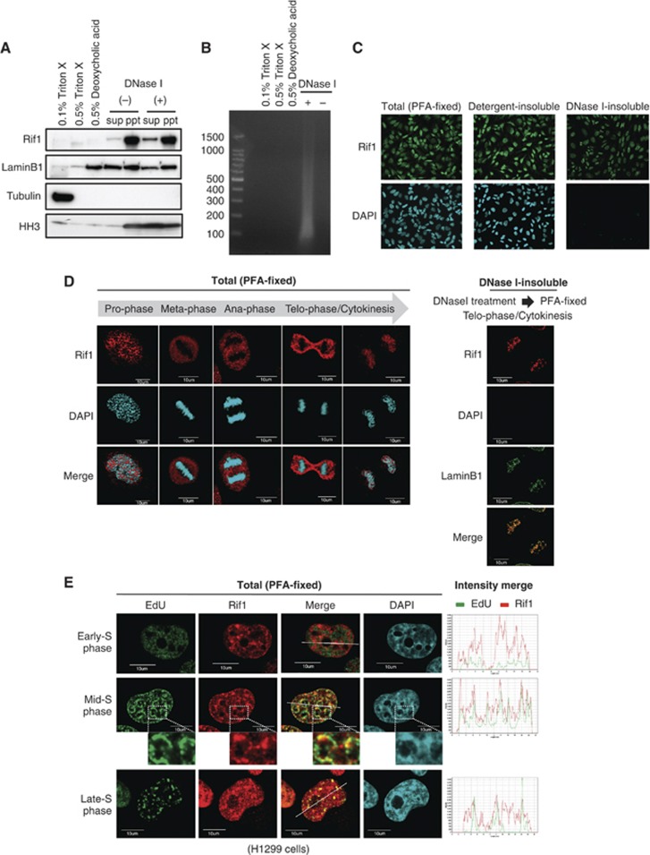
DNA replication is spatially and temporally regulated during S-phase. DNA replication timing is established in early-G1-phase at a point referred to as timing decision point. However, how the genome-wide replication timing domains are established is unknown. Here, we show that Rif1 (Rap1-interacting-factor-1), originally identified as a telomere-binding factor in yeast, is a critical determinant of the replication timing programme in human cells. Depletion of Rif1 results in specific loss of mid-S replication foci profiles, stimulation of initiation events in early-S-phase and changes in long-range replication timing domain structures. Analyses of replication timing show replication of sequences normally replicating early is delayed, whereas that normally replicating late is advanced, suggesting that replication timing regulation is abrogated in the absence of Rif1. Rif1 tightly binds to nuclear-insoluble structures at late-M-to-early-G1 and regulates chromatin-loop sizes. Furthermore, Rif1 colocalizes specifically with the mid-S replication foci. Thus, Rif1 establishes the mid-S replication domains that are restrained from being activated at early-S-phase. Our results indicate that Rif1 plays crucial roles in determining the replication timing domain structures in human cells through regulating higher-order chromatin architecture.


Conflict of interest statement

The authors declare that they have no conflict of interest.

Figures

Figure 1
Figure 1
Depletion of Rif1 protein enhances initiation events in early-S-phase. (A) HeLa cells, transfected with Rif1 siRNA for 48 and 72 h, were harvested and the whole-cell extracts were immunoblotted with antibodies against Rif1. The Rif1 protein level was reduced by >90% at 48 h compared with control. Tubulin is shown as a loading control (C). (B) Asynchronous cells transfected with control or Rif1 siRNA for 48 h were stained with PI and DNA contents were examined by FACS. (C) HeLa cells treated with control or Rif1 siRNA were synchronized at the G1/S-phase by double-thymidine block. At indicated times after release, cell-cycle progression was examined by FACS. (D) HeLa and NHDF cells were harvested at 48 h after control (−) or Rif1 (+) siRNA transfection, and the cells were fractionated into Triton-soluble and -insoluble fractions by CSK buffer (containing 0.1% Triton X-100). The latter fractions (enriched in chromatin-bound proteins) were immunoblotted with the antibodies indicated. (E) HeLa cells treated with control (−) or Rif1 (+) siRNA were synchronized at the G1/S boundary, released into cell cycle and harvested at indicated times after release. The whole-cell extracts were immunoblotted with the antibodies indicated. (F) The chromatin-enriched fractions (insoluble fractions in CSK containing 0.1% Triton X-100) from HeLa cells, transfected with Rif1 and/or Cdc7 siRNAs for 48 h, were immunoblotted with the antibodies indicated. (G) HeLa cells treated with control or Rif1 siRNA were synchronized at the G1/S boundary by double-thymidine block. At the times indicated after release, cells were harvested and fractionated into Triton-soluble and Triton-insoluble chromatin-enriched fractions in CSK buffer (containing 0.1% Triton X-100). The latter fractions were immunoblotted with the antibodies indicated. In all the experiments, Rif1 siRNA #4 was used.
Figure 2
Figure 2
Mid-S-phase replication foci pattern is selectively lost in Rif1-depleted cells. (A) HeLa cells, transfected with control or Rif1 siRNA, were arrested at the G1/S boundary by double-thymidine block and were released into cell cycle. After treatment with BrdU for 30 min at the indicated times, cells were fixed, treated with 2.0 N HCl to denature DNA, and then stained with anti-BrdU antibody to detect DNA replication sites. Representative BrdU images of nuclei in cells at each stage of S-phase are shown. (B) Cells were analysed under fluorescence microscopy (Olympus FV1000 microscopy) and the numbers of the cells with each foci pattern (representing early-S-, mid-S- or late-S-phase cells) were counted at each time point after release. The averages of three independent experiments (N=100 for each) are presented. (C) Enlarged foci patterns of the cells from Supplementary Figure S7A. (D) The fractions of the two replication foci patterns were counted in control and Rif1-depleted cells under microscopy. The averages of three independent experiments (N=100 for each) are presented. (E) The cell-cycle progression of the cells used in (C) monitored by FACS. In all the experiments, Rif1 siRNA #4 was used.
Figure 3
Figure 3
Rif1 depletion causes major changes in replication timing domains on a 42-Mb segment of the human chromosome 5. (A) Asynchronously growing HeLa cells, treated with control or Rif1 siRNA #4, were pulse labelled for 90 min with BrdU and were fractionated into early- and late-S-phase fractions by FACS cell sorter (see Supplementary Figure S15). Nascent BrdU-substituted DNA, immunoprecipitated with anti-BrdU antibody, in each fraction was hybridized with oligonucleotide tiling arrays containing one probe every 1.0 kb, as described in ‘Materials and methods’. Data, processed in Genomic workbench software, generated the timing pattern on the 42-Mb segment on the chromosome 5. In the scatter plots, red and green indicate early- and late-replicating regions, respectively. The smoothed curve shows replication timing domains. The R/G band pattern of this segment is shown below. (B) Overlay of the results of the control (red) and Rif1-depleted (blue) cells. (C) Validation of the timing array results by quantitative PCR. Asynchronously growing HeLa cells, treated with control or Rif1 siRNA #2, were pulse labelled for 90 min with BrdU and were fractionated into early- and late-S-phase fractions by FACS cell sorter. BrdU-labelled nascent DNA was immunoprecipitated in each fraction. The nascent DNAs at the indicated loci (A–G; left) on the chromosome 5 were amplified by quantitative PCR to determine replication timing. The values presented are the ratios to the level of amplified mtDNA (right). Data means were calculated from at least three independent experiments. The data are in excellent agreement with those of tiling array analyses.
Figure 4
Figure 4
Rif1 tightly binds to DNase I-insoluble nuclear structures in nuclei at late M/early G1 and colocalizes with mid-S replication foci. (A, B) Asynchronously growing HeLa cells were fractionated as described in ‘Materials and methods’. (A) Each fraction was immunoblotted with indicated antibodies. (B) The soluble fractions were run on 2% agarose gel to examine the presence of DNA. (C) Immunostaining of Rif1 protein (green) after various treatment. Total, fixed with 4% paraformaldehyde; detergent-insoluble, pretreated with 0.5% Triton X-100 before fixation; DNase I-insoluble, pretreated with DNase I before fixation. (D) (Left) Mitotic HeLa cells as shown were fixed with 4% paraformaldehyde and immunostained with anti-Rif1 antibody. Rif1 dissociates from DNA in prophase and is not present on mitotic chromosomes. Rif1 binds to chromatin at late telophase. (Right) Rif1 staining on late telophase chromosome is resistant to prior DNase I treatment. (E) H1299 cells in early-, mid- and late-S-phase were labelled with EdU. The sites of the EdU signals (green) and Rif1 localization (red) were compared in each stage of S-phase. The insets show the enlarged images of the segments indicated. The intensities of both green and red signals on the white line are shown in the right graph (Olympus FV1000 microscopy). Blue, DNA.
Figure 5
Figure 5
Rif1 depletion causes increase of chromatin-loop sizes. (A, B) Control and Rif1-depleted HeLa cells were synchronized at G1/S-phase and released into cell cycle for 1 h. Cells were harvested and DNA halo assays were conducted (N=50). (A) Phase-contrast images of representative cells. (B) Chromatin-loop sizes were calculated according to the formula described in ‘Materials and methods’ (Guillou et al, 2010). The data with two representative siRNAs (#2 and #4) are presented.
Figure 6
Figure 6
A model for Rif1-mediated determination of replication timing domains. (Left) Normally, Rif1 binds to nuclear-insoluble structures at late-M to early-G1, generating mid-S replication domains some of which are clustered at nuclear periphery as well as around nucleoli. This could be related to TDP known to occur at early-G1. The origins associated with the mid-S domains are sequestered from activation until mid-S-phase (shown with dotted grey arrow emanating from Cdc7). (Right) In Rif1-depleted cells, mid-S replication domains are not generated and the origins normally associated with mid-S domains are scattered throughout the nuclei. This permits access of Cdc7 (shown with solid arrow) and other replication factors to mid-S origins throughout early- to mid-S-phase, resulting in stimulation of initiation events (Cdc7-mediated phosphorylation of MCM and chromatin loading of Cdc45 and PCNA, etc.) at early-S-phase. Portions of this figure were adopted from Gilbert (2010).

Comment in

References

    1. Aparicio JG, Viggiani CJ, Gibson DG, Aparicio OM (2004) The Rpd3-Sin3 histone deacetylase regulates replication timing and enables intra-S origin control in Saccharomyces cerevisiae. Mol Cell Biol 24: 4769–4780 - PMC - PubMed
    1. Arias EE, Walter JC (2007) Strength in numbers: preventing rereplication via multiple mechanisms in eukaryotic cells. Genes Dev 21: 497–518 - PubMed
    1. Buongiorno-Nardelli M, Micheli G, Carri MT, Marilley M (1982) A relationship between replicon size and supercoiled loop domains in the eukaryotic genome. Nature 298: 100–102 - PubMed
    1. Buonomo SB, Wu Y, Ferguson D, de Lange T (2009) Mammalian Rif1 contributes to replication stress survival and homology-directed repair. J Cell Biol 187: 385–398 - PMC - PubMed
    1. Courbet S, Gay S, Arnoult N, Wronka G, Anglana M, Brison O, Debatisse M (2008) Replication fork movement sets chromatin loop size and origin choice in mammalian cells. Nature 455: 557–560 - PubMed

Publication types

LinkOut - more resources