Review
. 2012;88(7):345-67.
doi: 10.2183/pjab.88.345.

Unnatural base pair systems toward the expansion of the genetic alphabet in the central dogma

Affiliations
Review

Unnatural base pair systems toward the expansion of the genetic alphabet in the central dogma

Ichiro Hirao et al. Proc Jpn Acad Ser B Phys Biol Sci. 2012.

Abstract

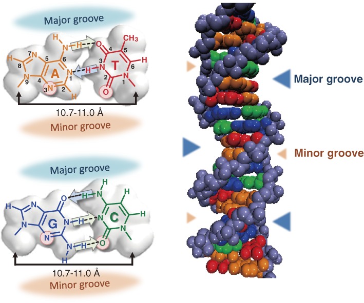
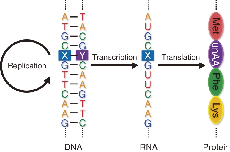
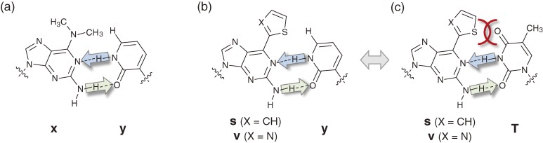
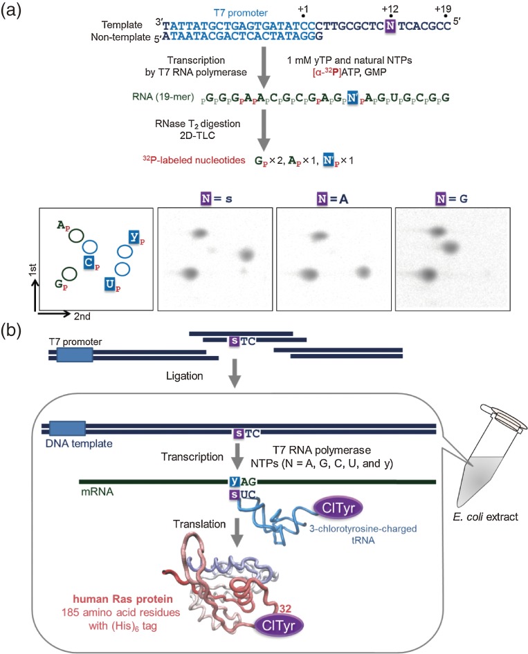
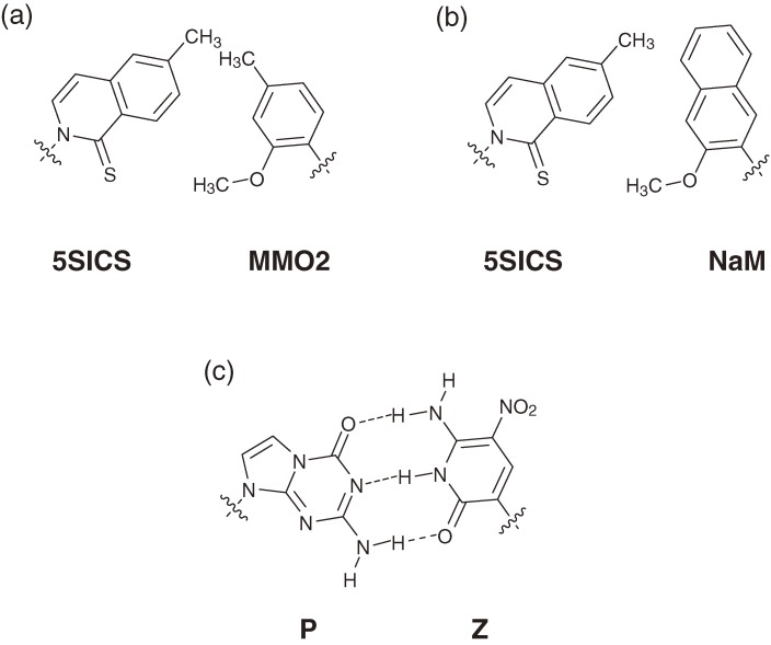
Toward the expansion of the genetic alphabet of DNA, several artificial third base pairs (unnatural base pairs) have been created. Synthetic DNAs containing the unnatural base pairs can be amplified faithfully by PCR, along with the natural A-T and G-C pairs, and transcribed into RNA. The unnatural base pair systems now have high potential to open the door to next generation biotechnology. The creation of unnatural base pairs is a consequence of repeating "proof of concept" experiments. In the process, initially designed base pairs were modified to address their weak points. Some of them were artificially evolved to ones with higher efficiency and selectivity in polymerase reactions, while others were eliminated from the analysis. Here, we describe the process of unnatural base pair development, as well as the tests of their applications.


Figures

Figure 1.
Figure 1.
Structures of the natural A–T and G–C pairs and the DNA duplex. The common properties of the natural base pairs, including the proton acceptor residues on the minor groove side (circles) for recognition by polymerases, the hydrogen-bonding patterns (arrows, from a proton donor to a proton acceptor residue), and the distances between the glycosidic bond positions of each base pair are indicated (left). The major and minor grooves in the DNA duplex are indicated by triangles (right).
Figure 2.
Figure 2.
Chemical structures of Benner’s base pairs. (a) The unnatural isoGisoC pair. (b) The unnatural Xκ pair. (c) The non-cognate isoG (enol)–T pair. (d) The non-cognate isoG (enol)–TS pair. (e) The cognate A–TS pair.
Figure 3.
Figure 3.
Unnatural base pair systems toward the expansion of the genetic alphabet and code. The unnatural base pair (X–Y), which works together with the natural A–T and G–C base pairs in the central dogma, allows the site-specific incorporation of extra unnatural nucleotides (X and Y) into DNA and RNA, and unnatural amino acids (unAA) into proteins.
Figure 4.
Figure 4.
Chemical structures of Kool’s base pairs. (a) The hydrophobic unnatural ZF pair. (b) The natural A–T pair. (c) The hydrophobic unnatural QF pair.
Figure 5.
Figure 5.
Hydrogen-bonded, unnatural base pairs developed by our group. (a) The unnatural xy pair. (b) The unnatural sy or vy pair. (c) The non-cognate pairing of s or v with T.
Figure 6.
Figure 6.
Transcription using the unnatural sy pair. (a) Scheme of the transcription experiments and 2D-TLC for nucleotide composition analyses of transcripts. (b) The coupled transcription–translation system involving the sy pair. The DNA template containing a CTs sequence and the 3-chlorotyrosine-charged tRNA were prepared separately and added to the transcription-coupled translation system. The yAG codon–CUs anticodon interaction was used for the site-specific incorporation of 3-chlorotyrosine at position 32 of the human Ras protein.
Figure 7.
Figure 7.
Preparation of 3-chlorotyrosyl tRNACUs. The tRNA was prepared by ligation of the 5′-half fragment derived from the native S. cerevisiae tyrosine tRNA and the chemically synthesized 3′-half fragment containing the CUs anticodon. The aminoacylation with 3-chlorotyrosine was performed by using a mutated S. cerevisiae tyrosyl-tRNA synthetase.
Figure 8.
Figure 8.
Site-specific modification of RNA molecules using the unnatural sy and vy pairs. (a) Scheme for the site-specific modification of RNA molecules at the 3′ terminal region. The DNA templates containing s or v at specific positions can be prepared by PCR, using a 5′ primer including the T7 promoter and a 3′ primer containing s or v. Functional substrates of modified y bases are shown in the yellow box. (b) Site-specific fluorescent labeling of an anti-theophylline aptamer. FAM-y was introduced into position 6, in place of U6, in the aptamer.
Figure 9.
Figure 9.
Structures of the hydrogen-bonded, unnatural base pairs designed by our group. (a) The cognate sy pair. (b) The non-cognate A–y pair. (c) The cognate sz pair. (d) The non-cognate A–z pair. Space filling models of the base alone (with a methyl group in place of the ribose) are shown.
Figure 10.
Figure 10.
Hydrophobic, unnatural base pairs. (a) The hypothetical self-pair between 4-methylpyridin-2-one (4MP) bases. (b) The self-pair between 6-propynylcarbostyril (PICS) bases. (c) The QF pair. The clash of the hydrogen atoms in the center of the QF pairing surface is indicated in red. (d) The QPa pair.
Figure 11.
Figure 11.
Site-specific fluorescent labeling of RNA molecules using the unnatural sPa pair. (a) The structure of the sPa pair. (b) Scheme for the site-specific incorporation of the fluorescent s base (excitation: 352 nm; emission: 434 nm) into RNA opposite Pa in the DNA template by T7 transcription. (c) The tertiary structure of yeast tRNAPhe. The positions substituted with s, U47 and G57, are indicated in blue and red, respectively. The yellow sphere represents Mg2+. (d) The fluorescent spectra of tRNA containing s at position 47 at different MgCl2 concentrations with excitation at 352 nm. (e) The fluorescent intensity profiles of tRNA containing s at position 47 (blue) or 57 (red), in the presence of 0.1 mM EDTA (thin line) and 2 mM MgCl2 (bold line), at different temperatures.
Figure 12.
Figure 12.
Hydrophobic, unnatural base pairs designed by our group. (a) The unnatural DsPa pair. (b) The usual triphosphate (R=OH) and γ-amidotriphosphate (R=NH2) used as substrates in the unnatural DsPa pair system for PCR. (c) The unnatural DsPn pair. (d) The non-cognate A–Pn pair. (e) The unnatural DsPx pair. (f) The unnatural Ds–Diol-Px pair.
Figure 13.
Figure 13.
Chemical syntheses of the usual 2′-deoxyribonucleoside triphosphate of Ds (dDsTP, 4) and its γ-amidotriphosphate (dDsTPNH2, 5).
Figure 14.
Figure 14.
Sequencing of DNA fragments containing Ds using the DsPa′ pair. In Method 1, using Ds-containing DNA fragments supplemented with dPa′TP, the unnatural base position of DNA fragments is identified as a gap on the sequencing peak pattern. In Method 2, using Ds-containing DNA fragments in the absence of dPa′TP, all sequencing peaks after the unnatural base positions disappear, due to the termination of sequencing at the unnatural base position.
Figure 15.
Figure 15.
Single-nucleotide incorporation efficiencies and selectivities of our unnatural base pairs by KF. A* corresponds to the γ-amidotriphosphate of A.
Figure 16.
Figure 16.
Unnatural base pairs that function in PCR with more than 99% selectivity.
Figure 17.
Figure 17.
The unnatural base pair between fluorophore and quencher analogs. (a) The unnatural Dss (fluorophore)–Pn/Px (quencher) pair. (b) Fluorescence quenching of Dss by Pn in double-stranded 12-mer DNA fragments. The fluorescence of the DNA fragments was detected upon irradiation at 365 nm. (c) Scheme for target DNA detection using molecular beacons containing the DssPn pair. The Dss fluorescence of the molecular beacon was observed in the presence of target DNA upon irradiation at 365 nm.

References

    1. Watson J.D., Crick F.H. (1953) Molecular structure of nucleic acids; a structure for deoxyribose nucleic acid. Nature 171, 737–738 - PubMed
    1. Nirenberg M.W., Matthaei J.H. (1961) The dependence of cell-free protein synthesis in E. coli upon naturally occurring or synthetic polyribonucleotides. Proc. Natl. Acad. Sci. U.S.A. 47, 1588–1602 - PMC - PubMed
    1. Nirenberg M., Leder P. (1964) RNA codewords and protein synthesis. The effect of trinucleotides upon the binding of sRNA to ribosomes. Science 145, 1399–1407 - PubMed
    1. Nishimura S., Jones D.S., Khorana H.G. (1965) Studies on polynucleotides. 48. The in vitro synthesis of a co-polypeptide containing two amino acids in alternating sequence dependent upon a DNA-like polymer containing two nucleotides in alternating sequence. J. Mol. Biol. 13, 302–324 - PubMed
    1. Rich, A. (1962) Problems of evolution and biochemical information transfer. In Horizons in Biochemistry (eds. Kasha, M. and Pullman, B.). Academic Press, New York, pp. 103–126.

Publication types