. 2012 Sep;122(9):3239-47.
doi: 10.1172/JCI62949. Epub 2012 Aug 1.

Human RHOH deficiency causes T cell defects and susceptibility to EV-HPV infections

Affiliations

Human RHOH deficiency causes T cell defects and susceptibility to EV-HPV infections

Amandine Crequer et al. J Clin Invest. 2012 Sep.

Abstract

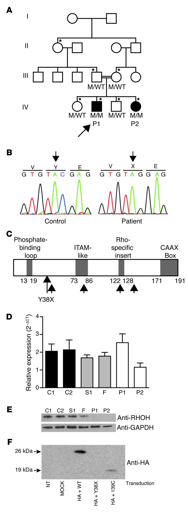
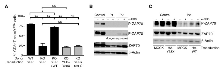
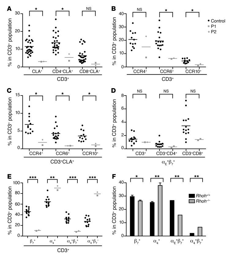
Epidermodysplasia verruciformis (EV) is a rare genetic disorder characterized by increased susceptibility to specific human papillomaviruses, the betapapillomaviruses. These EV-HPVs cause warts and increase the risk of skin carcinomas in otherwise healthy individuals. Inactivating mutations in epidermodysplasia verruciformis 1 (EVER1) or EVER2 have been identified in most, but not all, patients with autosomal recessive EV. We found that 2 young adult siblings presenting with T cell deficiency and various infectious diseases, including persistent EV-HPV infections, were homozygous for a mutation creating a stop codon in the ras homolog gene family member H (RHOH) gene. RHOH encodes an atypical Rho GTPase expressed predominantly in hematopoietic cells. Patients' circulating T cells contained predominantly effector memory T cells, which displayed impaired TCR signaling. Additionally, very few circulating T cells expressed the β7 integrin subunit, which homes T cells to specific tissues. Similarly, Rhoh-null mice exhibited a severe overall T cell defect and abnormally small numbers of circulating β7-positive cells. Expression of the WT, but not of the mutated RHOH, allele in Rhoh-/- hematopoietic stem cells corrected the T cell lymphopenia in mice after bone marrow transplantation. We conclude that RHOH deficiency leads to T cell defects and persistent EV-HPV infections, suggesting that T cells play a role in the pathogenesis of chronic EV-HPV infections.


Figures

Figure 1
Figure 1. Homozygous RHOH loss-of-expression mutation in 2 patients with persistent EV-HPV infections.
(A) Pedigree of the family with susceptibility to EV-HPV infections and other infectious manifestations. Generations are designated by a Roman numeral (I, II, III, IV, and V). P1 and P2 are represented by black symbols. The proband is indicated by an arrow. The asterisk indicates the individuals genotyped with the Affymetrix Genome-Wide SNP 6.0 array. (B) Automated sequencing profile showing the Y38X RHOH mutation in genomic DNA (gDNA) extracted from EBV-B cells from the patients and comparison with the sequence obtained from a healthy control. (C) Schematic representation of the structure of the RHOH protein adapted from the work of Fueller et al. (17). Y38X is situated between the phosphate-binding loop and the ITAM-like domain. The 4 possible reinitiation sites downstream from the mutation are indicated by small black arrows. (D) RHOH mRNA production, as assessed by qRT-PCR on total RNA isolated from saimiri T cells from the 2 patients, 1 healthy sibling (S1), and the father (F), both heterozygous for the mutant allele, and 2 healthy controls (C1 and C2). Mean + SD for 3 experiments is presented for all samples except for P1 (2 experiments). (E) Immunoblot analyses of 30 μg of total protein extracted from the saimiri T cells of P1, P2, S1, F, C1, and C2, with an antibody directed against RHOH and an antibody against GAPDH as a protein-loading control. (F) Immunoblot analysis of NIH/3T3 cells transfected with WT RHOH, Y38X RHOH, or 139-C RHOH. NT, not transduced.
Figure 2
Figure 2. The Y38X RHOH allele is loss of function in the mouse model and is associated with an impaired phosphorylation of ZAP70 upon TCR stimulation in the patients’ saimiri T cells.
(A) Bone marrow cells from Rhoh–/– mice (KO) of mixed background were transduced with an empty retroviral vector encoding YFP or a retroviral vector coexpressing the YFP gene and WT RHOH, Y38X RHOH, or 139-C RHOH genes and transplanted into sublethally irradiated Rag2–/– recipient mice. Normal bone marrow cells transduced with an empty vector were included as a control. The percentage of CD3+ cells within the YFP+-transduced T cell group was assessed in the blood of recipient mice 3 months after transplantation by flow cytometry (mean ± SEM, n = 4 recipients per construct). *P < 0.05; **P < 0.005. (B) Representative immunoblots of cell lysates extracted from saimiri T cells of a healthy control and the 2 patients (P1 and P2) following OKT3 stimulation, probed with anti-ZAP70 and anti–phospho-ZAP70 antibodies. β-Actin was used as an additional protein-loading control. (C) Representative immunoblot analysis of cell lysates extracted from YPF+ sorted saimiri T cells (control and patient P2) transduced with a vector encoding YFP and the HA-tagged mutant Y38X RHOH (HA-Y38X), YFP and the HA-tagged WT RHOH (HA-WT), or YFP alone (MOCK), subsequently stimulated with OKT3. Lysates were blotted with anti-ZAP70 and anti-phospho-ZAP70 antibodies. β-Actin was used as an additional protein-loading control.
Figure 3
Figure 3. Human and mouse RHOH deficiencies lead to an abnormal integrin expression pattern.
(AE) Tissue-homing T cell subsets were assessed on live CD3+-, CD3+CD4+-, and CD3+CD8+-gated cryopreserved PBMCs from the 2 patients (P1 values indicated by gray squares, P2 values indicated by gray diamonds) and healthy controls (indicated by black circles) by flow cytometry. (A) Skin-homing CLA+ subsets were assessed for both patients and 28 healthy controls. (B) CCR4+, CCR6+, and CCR10+ subsets were assessed for both patients and 12, 17, and 12 healthy controls, respectively. (C) Skin-homing CLA+CCR4+, CLA+CCR6+, and CLA+CCR10+ subsets were assessed for both patients and for 12, 17, and 12 healthy controls, respectively. (D) αE+β7+ cells were assessed for both patients and 14 healthy controls. (E) β7+, α4+, α4+β7+, and α4+β7 subsets were assessed for both patients and 12 healthy controls. The frequencies of the various subsets are expressed in percentages of CD3+ cells. Viability rates were about 95% for all PBMC preparations. Patients’ samples were tested at least twice, except for the chemokine receptors, for which assessments were carried out only once. Mean values are indicated by horizontal bars. The values obtained in all experiments were similar. (F) The frequencies within the CD3+ population of β7+, α4+, αE+β7+, and α4+β7+ cells were assessed by flow cytometry on CD3+-gated peripheral blood cells from Rhoh+/+ (n = 5) and Rhoh–/– mice (mean ± SEM, n = 5 mice of mixed background). *P < 0.05; **P < 0.005; ***P < 0.0005.

References

    1. Lutzner MA. Epidermodysplasia verruciformis. An autosomal recessive disease characterized by viral warts and skin cancer. A model for viral oncogenesis. Bull Cancer. 1978;65(2):169–182. - PubMed
    1. Orth G. Host defenses against human papillomaviruses: lessons from epidermodysplasia verruciformis. Curr Top Microbiol Immunol. 2008;321:59–83. - PubMed
    1. Cockayne E. Inherited Abnormalities Of The Skin And Its Appendages. London, United Kingdom: Oxford University Press; 1933.
    1. Ramoz N, Rueda LA, Bouadjar B, Montoya LS, Orth G, Favre M. Mutations in two adjacent novel genes are associated with epidermodysplasia verruciformis. Nat Genet. 2002;32(4):579–581. doi: 10.1038/ng1044. - DOI - PubMed
    1. Alcais A, et al. Life-threatening infectious diseases of childhood: single-gene inborn errors of immunity? Ann N Y Acad Sci. 2010;1214:18–33. doi: 10.1111/j.1749-6632.2010.05834.x. - DOI - PubMed

Publication types

MeSH terms