. 2012 Sep;122(9):3227-38.
doi: 10.1172/JCI62182. Epub 2012 Aug 1.

Targets of antibodies against Plasmodium falciparum-infected erythrocytes in malaria immunity

Affiliations

Targets of antibodies against Plasmodium falciparum-infected erythrocytes in malaria immunity

Jo-Anne Chan et al. J Clin Invest. 2012 Sep.

Abstract

Plasmodium falciparum is the major cause of malaria globally and is transmitted by mosquitoes. During parasitic development, P. falciparum-infected erythrocytes (P. falciparum-IEs) express multiple polymorphic proteins known as variant surface antigens (VSAs), including the P. falciparum erythrocyte membrane protein 1 (PfEMP1). VSA-specific antibodies are associated with protection from symptomatic and severe malaria. However, the importance of the different VSA targets of immunity to malaria remains unclear, which has impeded an understanding of malaria immunity and vaccine development. In this study, we developed assays using transgenic P. falciparum with modified PfEMP1 expression to quantify serum antibodies to VSAs among individuals exposed to malaria. We found that the majority of the human antibody response to the IE targets PfEMP1. Furthermore, our longitudinal studies showed that individuals with PfEMP1-specific antibodies had a significantly reduced risk of developing symptomatic malaria, whereas antibodies to other surface antigens were not associated with protective immunity. Using assays that measure antibody-mediated phagocytosis of IEs, an important mechanism in parasite clearance, we identified PfEMP1 as the major target of these functional antibodies. Taken together, these data demonstrate that PfEMP1 is a key target of humoral immunity. These findings advance our understanding of the targets and mediators of human immunity to malaria and have major implications for malaria vaccine development.


Figures

Figure 1
Figure 1. Phenotypic analyses of var promoter knockdown parasites.
(A) Northern blot of var gene transcription by hybridization with a specific var exon 2 sequence. Compared with that in 3D7 parental parasites, var transcripts (arrow) are markedly reduced or absent in 3D7vpkd parasites. RNA was extracted from highly synchronous ring-stage IEs at approximately 10 hours after invasion. The position of molecular weight standards (kb) is indicated on the left. Ethidium bromide–stained gel prior to blotting was used as the loading control (Supplemental Figure 1B). (B) Western blot analyses of membrane extracts from mature trophozoite-IEs probed with anti-PfEMP1 and anti-RIF29 antibodies. In E8B parental parasites, full-length PfEMP1, which was absent in E8Bvpkd parasites, was detected at approximately 300 kDa (arrows). The double band represents different PfEMP1 variants expressed by E8B parental parasites. This anti-PfEMP1 antibody cross-reacts with erythrocyte spectrin (asterisk), as shown by comparison with extracts from uninfected erythrocytes (uRBC). The anti-RIF29 antibodies detected a protein at approximately 40 kDa, representing RIFIN in both E8B parental and E8Bvpkd parasites (bottom). ATS, acidic terminal sequence of PfEMP1. Adhesion of IEs to immobilized (C) ICAM-1 and (D) CD36 was significantly reduced in E8Bvpkd parasites compared with that in E8B parental parasites. (E) Adhesion of IEs to immobilized CD36 was significantly reduced in 3D7vpkd parasites compared with that in 3D7 parental parasites. However, adhesion to CD36 was partially retained in E8Bvpkd and 3D7vpkd parasites. Values are expressed as a percentage of parental parasites binding to each receptor. Assays were performed twice independently; bars represent median and interquartile ranges of samples tested in triplicate.
Figure 2
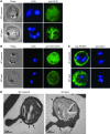
Figure 2. Exported proteins remained expressed by 3D7 parental and 3D7vpkd transgenic parasites.
Immunofluorescence assays demonstrate the expression of (A) RIFIN and (B) STEVOR proteins by mature trophozoite-stage parasites (green). Despite the lack of PfEMP1 expression, RIFIN and STEVOR proteins were detectable in the transfected 3D7vpkd parasites similar to 3D7 parental parasites. (C) Mid-trophozoite-stage parasites from 3D7 parental and 3D7vpkd lines were probed with anti-PfEMP3 antibodies as a positive control and anti-AMA1 antibodies as a negative control (green). As expected, the pattern of staining by anti-PfEMP3 antibodies was consistent with labeling of PfEMP3 in the IE membrane, and there was no apparent labeling of AMA1. In all assays, cells were fixed with a mixture of acetone (90%) and methanol (10%), and DAPI was used to stain nuclear DNA (blue). (AC) All images were taken with equal exposure for both parasite lines (original magnification, ×1000). (D) Electron-dense knobs in the erythrocyte membrane (arrows) were observed for IEs of 3D7 parental and 3D7vpkd parasites by transmission electron microscopy. Scale bar: 1 εm.
Figure 3
Figure 3. Antibodies among sera from Kenyan adults to surface antigens expressed by P. falciparum–IEs.
(A and C) IgG binding to the surface of erythrocytes infected with 3D7vpkd and E8Bvpkd parasites was significantly reduced compared with that to (A) 3D7 parental and (C) E8B parental parasites. Assays were performed twice independently; bars represent median and interquartile ranges of samples tested in duplicate (n = 26 for 3D7; n = 22 for E8B). P values were calculated using a paired Wilcoxon signed-rank test. (B and D) A representative selection of serum samples tested for (B) antibodies to 3D7 parental and 3D7vpkd parasites and (D) antibodies to E8B parental and E8Bvpkd parasites. Samples tested were from adults (K1–K7) exposed to malaria residing in the Kilifi district, Kenya, and nonexposed Melbourne residents (Cont). IgG binding to 3D7vpkd and E8Bvpkd parasites was substantially reduced in most individuals. There was minimal reactivity observed among sera from Melbourne residents. Assays were performed twice independently; bars represent mean and range of samples tested in duplicate. IgG binding levels are expressed as geometric MFI for all graphs.
Figure 4
Figure 4. Antibodies among sera from Kenyan children and adults to surface antigens expressed by P. falciparum–IEs.
(A) IgG binding to the surface of erythrocytes infected with 3D7vpkd parasites was markedly reduced compared with that of 3D7 parental parasites. Assays were performed twice independently; bars represent median and interquartile ranges of samples tested in duplicate (n = 296). The P value was calculated using a paired Wilcoxon signed-rank test. (B) A representative selection of samples tested for antibodies to 3D7 parental and 3D7vpkd parasites. Samples tested were from residents (C1–C7) exposed to malaria in the Chonyi cohort, Kenya, and nonexposed United Kingdom residents (Cont). IgG binding to 3D7vpkd parasites was substantially reduced in most individuals. There was minimal reactivity observed among sera from nonexposed United Kingdom residents. Assays were performed twice independently; bars represent mean and range of samples tested in duplicate. (C) Antibody responses among children of different age groups from the Chonyi cohort. Antibody acquisition was age-dependent as older children had higher levels of IgG binding to 3D7 parental parasites. Children from all age groups had very low IgG binding levels to 3D7vpkd parasites. Bars represent median and interquartile ranges. (AC) IgG binding levels are expressed as geometric MFI for all graphs. (D) Antibodies to IE surface proteins measured by agglutination assays among a selection of sera from children (n = 20). A representative selection is shown (C8–C14); most individuals have antibodies that agglutinated 3D7 parental parasites to a much greater extent than 3D7vpkd parasites. Bars represent mean and range of samples tested in duplicate.
Figure 5
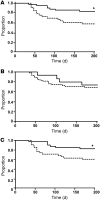
Figure 5. Antibodies to IEs and the risk of symptomatic P. falciparum episode during follow-up.
Kaplan-Meier survival curves show the proportion of children who remained free of malaria episodes over time for IgG responses to (A) surface antigens of 3D7 parental parasites, (B) surface antigens of 3D7vpkd parasites, and (C) 3D7-PfEMP1 (defined as IgG binding to 3D7 parental minus 3D7vpkd). Solid lines represent children who are positive for IgG binding; dashed lines represent children who are negative for IgG binding. *P < 0.05, comparing antibody-positive and antibody-negative individuals (log-rank test). Unadjusted data are shown. Symptomatic P. falciparum infection was defined as fever plus a parasite load of 2,500 parasites per εl. Survival analysis was restricted to children between 1 and 10 years of age who were parasite positive at baseline (n = 112).
Figure 6
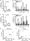
Figure 6. Opsonic phagocytosis of P. falciparum–IEs by undifferentiated THP-1 monocytes using sera from adults from Kilifi.
(A and C) Opsonic phagocytosis activity of sera was significantly reduced in 3D7vpkd and E8Bvpkd parasites compared with that in (A) 3D7 parental and (C) E8B parental parasites. The level of phagocytosis is expressed as a percentage of the positive control for all graphs. Assays were performed twice independently; bars represent median and interquartile ranges (n = 24 for 3D7; n = 31 for E8B). P values were calculated using a paired Wilcoxon signed-rank test. (B and D) A representative selection of sera tested for phagocytosis activity with (B) 3D7 and (D) E8B parasites is shown. Samples were from malaria-exposed adults (K1–K7) residing in the Kilifi district, Kenya, and nonexposed Melbourne residents (Cont). In most samples, opsonic phagocytosis activity to 3D7vpkd and E8Bvpkd parasites was substantially reduced compared with that to (B) 3D7 parental and (D) E8B parental parasites. Assays were performed twice independently; bars represent mean and range, with samples tested in duplicate. The correlation between antibody levels measured as IgG binding and as opsonic phagocytosis activity is shown for (E) E8B parental and (F) E8Bvpkd parasites. Symbols represent results for individual serum samples.

References

    1. World Health Organization . Global Malaria Programme. World Malaria Report 2010. Geneva, Switzerland: World Health Organization; 2010.
    1. Elliott SR, Beeson JG. Estimating the burden of global mortality in children aged <5 years by pathogen-specific causes. Clin Infect Dis. 2008;46(11):1794–1795. doi: 10.1086/588049. - DOI - PubMed
    1. Dondorp AM, et al. Artemisinin resistance in Plasmodium falciparum malaria. N Engl J Med. 2009;361(5):455–467. doi: 10.1056/NEJMoa0808859. - DOI - PMC - PubMed
    1. Trape JF, et al. Malaria morbidity and pyrethroid resistance after the introduction of insecticide-treated bednets and artemisinin-based combination therapies: a longitudinal study. Lancet Infect Dis. 2011;11(12):925–932. doi: 10.1016/S1473-3099(11)70194-3. - DOI - PubMed
    1. Bull PC, et al. Parasite antigens on the infected red cell surface are targets for naturally acquired immunity to malaria. Nat Med. 1998;4(3):358–360. doi: 10.1038/nm0398-358. - DOI - PMC - PubMed

Publication types

MeSH terms

Substances