. 2012 Sep 21;287(39):32770-9.
doi: 10.1074/jbc.M112.377614. Epub 2012 Jul 31.

Enzymatic engineering of polysialic acid on cells in vitro and in vivo using a purified bacterial polysialyltransferase

Affiliations

Enzymatic engineering of polysialic acid on cells in vitro and in vivo using a purified bacterial polysialyltransferase

Abderrahman El Maarouf et al. J Biol Chem. .

Abstract

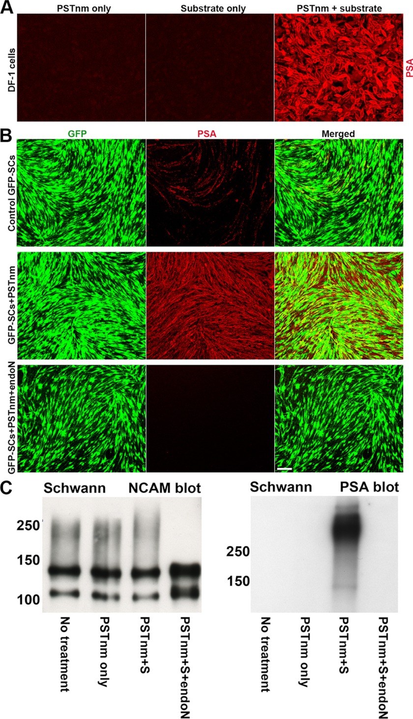
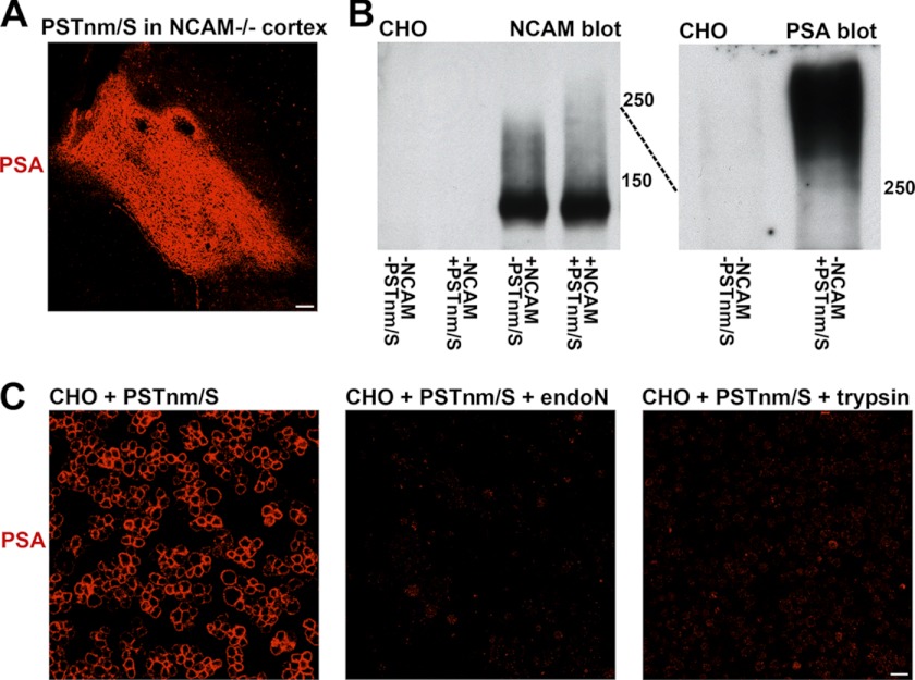
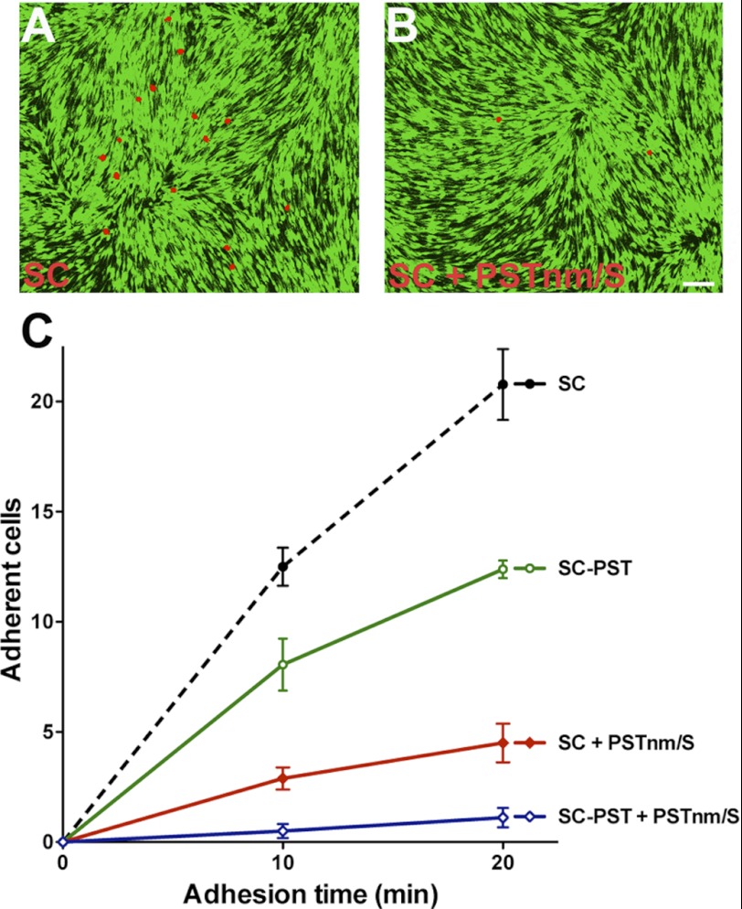
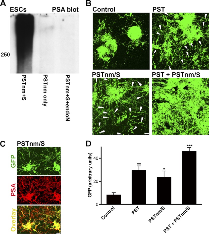
In vertebrates, polysialic acid (PSA) is typically added to the neural cell adhesion molecule (NCAM) in the Golgi by PST or STX polysialyltransferase. PSA promotes plasticity, and its enhanced expression by viral delivery of the PST or STX gene has been shown to promote cellular processes that are useful for repair of the injured adult nervous system. Here we demonstrate a new strategy for PSA induction on cells involving addition of a purified polysialyltransferase from Neisseria meningitidis (PST(Nm)) to the extracellular environment. In the presence of its donor substrate (CMP-Neu5Ac), PST(Nm) synthesized PSA directly on surfaces of various cell types in culture, including Chinese hamster ovary cells, chicken DF1 fibroblasts, primary rat Schwann cells, and mouse embryonic stem cells. Similarly, injection of PST(Nm) and donor in vivo was able to produce PSA in different adult brain regions, including the cerebral cortex, striatum, and spinal cord. PSA synthesis by PST(Nm) requires the presence of the donor CMP-Neu5Ac, and the product could be degraded by the PSA-specific endoneuraminidase-N. Although PST(Nm) was able to add PSA to NCAM, most of its product was attached to other cell surface proteins. Nevertheless, the PST(Nm)-induced PSA displayed the ability to attenuate cell adhesion, promote neurite outgrowth, and enhance cell migration as has been reported for endogenous PSA-NCAM. Polysialylation by PST(Nm) occurred in vivo in less than 2.5 h, persisted in tissues, and then decreased within a few weeks. Together these characteristics suggest that a PST(Nm)-based approach may provide a valuable alternative to PST gene therapy.


Figures

FIGURE 1.
FIGURE 1.
Addition of PSTNm/substrate induces high levels of PSA on DF-1 and Schwann cells in culture. PSA was detected using the highly specific 5A5 monoclonal antibody. A, chicken DF-1 fibroblast cells are known to have no detectable PSA expression, and incubation either with PSTNm or donor substrate alone produced no PSA. However, simultaneous addition of PSTNm and CMP-Neu5Ac produced high levels of PSA expression on cell surfaces (red). B, rat primary GFP-SC (green) cultures were treated with PSTNm/substrate and then fixed and immunostained for PSA. Control cultures express insignificant amounts of PSA (red). However, with PSTNm/substrate treatment, very high levels of PSA expression are observed on both the cell bodies and processes of GFP-SCs. Incubation with the PSA-specific endo-N completely abolished PSTNm-induced PSA staining. Scale bar, 100 μm. C, SC cultures, with or without PSTNm treatment, were processed for electrophoresis. Immunoblotting for NCAM revealed the presence of the 120- and 140-NCAM isoforms in untreated cells, with a light polysialylation smear extending to about 250 kDa. The same pattern was observed with addition of PSTNm alone. Incubation of cells with both enzyme and donor substrate shifted the smear to higher molecular masses. Addition of endo-N completely eliminated all the PSA-associated smears. Immunoblotting for PSA revealed that PSTNm and donor substrate produced a heavy band above 250 kDa, which was absent in control cells or samples treated with enzyme alone. This band disappeared completely after endo-N treatment.
FIGURE 2.
FIGURE 2.
In vivo administration of PSTNm and donor substrate induces high levels of PSA expression in brain tissues. PSTNm and CMP-Neu5Ac were co-injected into the cerebral cortex, striatum, or spinal cord. Animals were perfused 24 h later and immunostained for PSA. Injection of enzyme alone produced no PSA in all three regions (A, D, and F; arrow, needle track). With co-administration of PSTNm and substrate, large amounts of PSA were detected in the cerebral cortex (B), and treatment with endo-N (C) completely abolished this staining (transverse sections). Although large portions of the striatum (transverse section) were polysialylated following injection of PSTNm and donor substrate (E), the dense bundles of myelinated fibers that traverse the striatum (stars) showed little or no PSA staining. High PSA expression was also observed in the spinal cord (G) (sagittal section), with good rostro-caudal as well as dorso-ventral extension of staining (SG, endogenous PSA in substantia gelatinosa). Scale bars, 100 μm in A–G. An example from the spinal gray matter (H) shows PSA staining in the neuropil (star) as well as on cell surfaces (arrows), with the cytoplasm of cells remaining devoid of PSA (dark). Arrowhead indicates a capillary. Scale bar, 50 μm. In the white matter, such as the corticospinal tract (I), PSA was readily visible on axons (arrows). Scale bar, 50 μm. J, view of a sagittal spinal section shows an example with good rostro-caudal spread of PSA staining, which sometimes exceeded 3 mm. Note the irregular shape of the stained area (double arrow indicates the extent of PSA staining. Dotted line, injection track. Scale bar, 100 μm). The bottom drawings are overlays of coronal maps (red, blue, green, and yellow) from four representative samples with cortical injection (K) and four samples with striatal injection (L) showing the variability in size and shape of the area of PSA expression among PSTNm/S-injected animals. Each color represents one sample. Cx, cerebral cortex; S, striatum; CC, corpus callosum; V, lateral ventricle.
FIGURE 3.
FIGURE 3.
In vivo production of PSA by PSTNm is rapid and persistent but not permanent. PSA expression by PSTNm in the cerebral cortex was detected within 2 h 30 min after injection, was still intense after 2 weeks, and began to decrease in amount after 3 weeks. In the striatum, this reduction was already more pronounced by 2 weeks. Arrow, needle track; arrowheads, diffusion and polysialylation along the corpus callosum (cc); stars, bundles of myelinated fibers. Transverse sections are shown. Scale bar, 100 μm.
FIGURE 4.
FIGURE 4.
PSTNm adds PSA to NCAM as well as to other surface proteins. A, injection of PSTNm and donor substrate in the cerebral cortex of NCAM-null mice (lacking all NCAM isoforms) produced high levels of PSA expression as shown by immunostaining 2 days later (sagittal section shown; scale bar, 100 μm). B, PSTNm treatment and NCAM immunoblotting of CHO cells showed no band in control cells. An NCAM-140 band was detected when cells were transfected with this isoform. This NCAM was lightly polysialylated (smear below 250 kDa), and the PSA smear was extended above 250 kDa when cells were incubated with PSTNm and substrate. PSA immunoblotting of preparations of CHO cells (with no NCAM construct) showed that PSTNm produced a strong band above 250 kDa, which was absent in control samples. C, suspensions of CHO cells were incubated with PSTNm and donor substrate and immunostained for PSA. Staining was clearly present on their surfaces, and it was completely removed following treatment of the live cells with either endo-N or trypsin. Scale bar, 20 μm.
FIGURE 5.
FIGURE 5.
PSA produced by PSTNm attenuates cell-cell adhesion. Suspensions of red-labeled SCs were seeded onto a confluent monolayer of GFP-expressing SCs, and the red cells that adhered to the monolayer were examined 10 or 20 min later. A, example shows red-labeled SCs that adhered after 20-min attachment time. B, there were much fewer adherent cells following polysialylation by PSTNm. Scale bar, 100 μm. C, quantification revealed that PSTNm-produced PSA inhibits cell-cell adhesion even more effectively (SC + PSTNm/S) than PSA produced by introduction of the PST gene (SC-PST). The effects of PSTNm and PST are additive (SC-PST + PSTNm/S) (two-way ANOVA: p < 0.0001, F = 128.7, n = 6 for each data point, error bars: S.E.).
FIGURE 6.
FIGURE 6.
PSTNm-induced PSA promotes neurite outgrowth and cell migration. Theses studies utilized mESCs expressing GFP under the HB9 promoter. A, undifferentiated cultures of mESCs were processed for PSA immunoblotting. These cells have undetectable levels of PSA as shown in control samples treated with the enzyme without donor substrate. However, incubation with both PSTNm and substrate produced a large PSA band above 250 kDa, which was removed with endo-N treatment. B, EBs of these mESCs were induced to differentiate toward a motoneuronal phenotype, using retinoic acid and SHH analog. At the end of incubation on day 6, control cells had extended GFP-positive neurites. With cells that were modified to stably express mouse PST (PST), there was substantially more neurite outgrowth, and GFP-positive cell bodies were observed migrating out of the EBs (arrowheads), which started to flatten and disperse. Treatment of the EB cultures with PSTNm and donor substrate produced an effect similar to mouse PST, enhancing both neurite outgrowth and cell migration. When cells expressing mouse PST were also treated with PSTNm and substrate, the effects on neurite outgrowth and cell migration were enhanced, with EBs appearing to completely disintegrate and neurites and cells covering most of the dish floor. Scale bar, 100 μm. C, immunostaining of 6-day cultures that were pretreated with PSTNm and substrate showed PSA on the GFP-positive neurites of motoneurons as well as on the GFP-negative neurites (see overlay) of cells that differentiated into other neuronal types. Scale bar, 100 μm. D, the PSA-induced spreading of neurites and cells on the dish floor was estimated by measuring the density of GFP fluorescence/surface area (NIH ImageJ 1.45). Like the PST gene, PSTNm/substrate increased the spreading of neurites and cells, and the effect was much higher with the use of both enzymes (one-way ANOVA: p < 0.0001, F = 23.33; Tukey's post-test: *, p < 0.05; **, p < 0.01; ***, p < 0.001; error bars: S.E.).

Similar articles

Cited by

References

    1. Eckhardt M., Mühlenhoff M., Bethe A., Koopman J., Frosch M., Gerardy-Schahn R. (1995) Molecular characterization of eukaryotic polysialyltransferase-1. Nature 373, 715–718 - PubMed
    1. Johnson C. P., Fujimoto I., Rutishauser U., Leckband D. E. (2005) Direct evidence that neural cell adhesion molecule (NCAM) polysialylation increases intermembrane repulsion and abrogates adhesion. J. Biol. Chem. 280, 137–145 - PubMed
    1. Fujimoto I., Bruses J. L., Rutishauser U. (2001) Regulation of cell adhesion by polysialic acid: effects on cadherin, immunoglobulin cell adhesion molecule, and integrin function and independence from neural cell adhesion molecule binding or signaling activity. J. Biol. Chem. 276, 31745–31751 - PubMed
    1. Rutishauser U. (2008) Polysialic acid in the plasticity of the developing and adult vertebrate nervous system. Nat. Rev. Neurosci. 9, 26–35 - PubMed
    1. Angata K., Huckaby V., Ranscht B., Terskikh A., Marth J. D., Fukuda M. (2007) Polysialic acid-directed migration and differentiation of neural precursors are essential for mouse brain development. Mol. Cell. Biol. 27, 6659–6668 - PMC - PubMed

Publication types