Review
. 2012 Oct 22;51(43):10724-38.
doi: 10.1002/anie.201200408. Epub 2012 Jul 31.

Red fluorescent proteins: advanced imaging applications and future design

Affiliations
Review

Red fluorescent proteins: advanced imaging applications and future design

Daria M Shcherbakova et al. Angew Chem Int Ed Engl. .

Abstract

In the past few years a large series of the advanced red-shifted fluorescent proteins (RFPs) has been developed. These enhanced RFPs provide new possibilities to study biological processes at the levels ranging from single molecules to whole organisms. Herein the relationship between the properties of the RFPs of different phenotypes and their applications to various imaging techniques are described. Existing and emerging imaging approaches are discussed for conventional RFPs, far-red FPs, RFPs with a large Stokes shift, fluorescent timers, irreversibly photoactivatable and reversibly photoswitchable RFPs. Advantages and limitations of specific RFPs for each technique are presented. Recent progress in understanding the chemical transformations of red chromophores allows the future RFP phenotypes and their respective novel imaging applications to be foreseen.


Figures

Figure 1
Figure 1
Applications of permanently fluorescent RFPs of different phenotypes. Cylinders represent FPs, which are colored corresponding to their emission spectral range. Colored arrows denote the respective excitation or emission light. a) Multiple FRET pairs. Several spectrally distinguishable FRET-biosensors may be used for simultaneous imaging of multiple FRET pairs. A combined orange FP–far-red FP caspase biosensor (top panel) and CFP–YFP calcium biosensor (bottom panel) are shown as examples; b) Single-FP biosensors. A shift in excitation/emission maxima (top panel, pH sensor) or a change in intensity (bottom panel, calcium sensor) under specific stimuli is the readout of these biosensors; c) Multicolor BiFC. Several independent pairs of interacting proteins may be identified; d) Deep-tissue imaging. This application requires far-red FPs exhibiting excitation and emission maxima within the NIRW (650–690 nm) to obtain the highest light transmission and the lowest autofluorescence; e) Super-resolution imaging: Most commercially available STED microscopes use a 638 nm excitation beam (red circle), which is superimposed with a doughnut-shaped STED beam (750 nm). The STED beam instantly quenches excited FPs at the periphery of the excitation area, thus confining the fluorescence emission to a spot of only a few tens of nanometers (red dot). To date, using FPs a resolution of 50 nm can be achieved;[29] f) Multicolor single-laser FCCS. A single excitation wavelength is used for a set of LSSFPs fluorescing at different wavelengths; g) Multicolor two-photon microscopy. With this technique, a subcellular resolution in deep tissues can be achieved with 2P single wavelength excitation of a set of LSSFPs fluorescing at different wavelengths; h) Long-term particle tracking with FTs. A slow change of FT color allows prolonged tracking of the particle inside the cell.
Figure 2
Figure 2
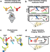
Applications of irreversibly and reversibly photoswitchable RFPs. Cylinders represent FPs, which are colored corresponding to their emission spectral range. Colored arrows denote the excitation, emission, and photo-activation light. a) Intravital imaging with selective photolabeling. PSFPs may be photoswitched inside an animal, and the photolabeled cells can be tracked; b) Super-resolution imaging with PALM-based techniques: multicolor PALM, PALMIRA (with rsFPs), and sptPALM. PALM microscopy is based on the accurate localization (up to ca. 20 nm)[65] of individual PAFP molecules (colored dots). The imaging cycle includes the stochastic photoactivation of the PAFPs molecules, imaging, and subsequent photobleaching of the photo-activated molecules. The cycle is repeated many times to reconstruct a super-resolution image. sptPALM allows tracking of localized single molecules in live cells; c) Photochromic FRET. rsFPs that change their absorbance upon photoswitching are used in photochromic FRET. The process of FRET occurs when rsFP is in the ON state, and does not occur when rsFP is in the OFF state. Thus, pcFRET allows a control image of the same sample to obtained without FRET by temporarily switching it the FRET OFF; d) Super-resolution imaging: RESOLFT microscopy utilizes an ON beam (blue arrow) and a super-imposed doughnut-shaped OFF beam (yellow arrow) to confine a diffraction-limited spot of switched ON rsFP molecules to a subdiffraction-sized spot (red dot). The resolution achieved to date using FPs is 40 nm.[66]
Figure 3
Figure 3
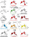
Mechanisms of chromophore formation for the DsRed-like, Kaede-like, mOrange-like, zFP538-like, asFP595-like, far-red DsRed-like, PSmOrange-like, LSS-red, and LSS-orange chromophores: Red, dark-red, orange, blue, green, and yellow colors of the chromophores correspond to the spectral range of the chromophore fluorescence. Gray denotes the non-fluorescent state. Transitions that can be photo-induced with 405 nm or 488 nm light are indicated by hν1 and hν2, respectively. [O] and [H] denote the oxidizing and reducing agents, respectively. Chromophores are shown in the cis form. Possible structures of trans chromophores and cistrans isomerizations are not shown.
Figure 4
Figure 4
Possible applications of future RFPs with combined phenotypes. Different FP phenotypes are shown in the bottom row. Two phenotypes that can be combined in a single FP are connected with an arrow pointing to the imaging applications of the new resulting FP. Possible combinations of LSSFPs, PAFPs/PSFPs, rsFPs, and FTs are presented. The colors of the boxes correspond to different FP phenotypes and show the spectral range of chromophore fluorescence.

References

    1. Lavis LD, Raines RT. ACS Chem. Biol. 2008;3:142–155. - PMC - PubMed
    2. Johnson I, Spence MTZ. The Molecular Probes Handbook: A Guide to Fluorescent Probes and Labeling Technologies. 11th ed. USA: Life Technologies Corp.; 2010.
    1. Subach FV, Malashkevich VN, Zencheck WD, Xiao H, Filonov GS, Almo SC, Verkhusha VV. Proc. Natl. Acad. Sci. USA. 2009;106:21097–21102. - PMC - PubMed
    2. Subach OM, Malashkevich VN, Zencheck WD, Morozova KS, Piatkevich KD, Almo SC, Verkhusha VV. Chem. Biol. 2010;17:333–341. - PMC - PubMed
    3. Mizuno H, Mal TK, Tong KI, Ando R, Furuta T, Ikura M, Miyawaki A. Mol. Cell. 2003;12:1051–1058. - PubMed
    4. Pakhomov AA, Martynov VI. Biochemistry. 2007;46:11528–11535. - PubMed
    5. Pletnev S, Subach FV, Dauter Z, Wlodawer A, Verkhusha VV. J. Am. Chem. Soc. 2010;132:2243–2253. - PMC - PubMed
    6. Remington SJ, Wachter RM, Yarbrough DK, Branchaud B, Anderson DC, Kallio K, Lukyanov KA. Biochemistry. 2005;44:202–212. - PubMed
    7. Shu X, Shaner NC, Yarbrough CA, Tsien RY, Remington SJ. Biochemistry. 2006;45:9639–9647. - PubMed
    8. Yampolsky IV, Remington SJ, Martynov VI, Potapov VK, Lukyanov S, Lukyanov KA. Biochemistry. 2005;44:5788–5793. - PubMed
    9. Piatkevich KD, Malashkevich VN, Almo SC, Verkhusha VV. J. Am. Chem. Soc. 2010;132:10762–10770. - PMC - PubMed
    1. Strack RL, Strongin DE, Bhattacharyya D, Tao W, Berman A, Broxmeyer HE, Keenan RJ, Glick BS. Nat. Methods. 2008;5:955–957. - PMC - PubMed
    2. Tao W, Evans BG, Yao J, Cooper S, Cornetta K, Ballas CB, Hangoc G, Broxmeyer HE. Stem Cells. 2007;25:670–678. - PubMed
    1. Strack RL, Hein B, Bhattacharyya D, Hell SW, Keenan RJ, Glick BS. Biochemistry. 2009;48:8279–8281. - PMC - PubMed
    1. Strack RL, Bhattacharyya D, Glick BS, Keenan RJ. BMC Biotechnol. 2009;9:32. - PMC - PubMed

Publication types

Substances

LinkOut - more resources