. 2012 Sep 14;111(7):894-906.
doi: 10.1161/CIRCRESAHA.112.273649. Epub 2012 Jul 31.

Tracking chromatid segregation to identify human cardiac stem cells that regenerate extensively the infarcted myocardium

Affiliations

Tracking chromatid segregation to identify human cardiac stem cells that regenerate extensively the infarcted myocardium

Jan Kajstura et al. Circ Res. .

Erratum in

  • Correction.
    [No authors listed] [No authors listed] Circ Res. 2015 Dec 4;117(12):e131. doi: 10.1161/RES.0000000000000086. Circ Res. 2015. PMID: 26635385 No abstract available.

Retraction in

Expression of concern in

  • Expression of Concern.
    [No authors listed] [No authors listed] Circ Res. 2019 Jan 18;124(2):e4-e5. doi: 10.1161/RES.0000000000000241. Circ Res. 2019. PMID: 30582460 No abstract available.
  • Expression of Concern.
    [No authors listed] [No authors listed] Circulation. 2019 Jan 15;139(3):e5-e6. doi: 10.1161/CIR.0000000000000639. Circulation. 2019. PMID: 30615475 No abstract available.

Abstract

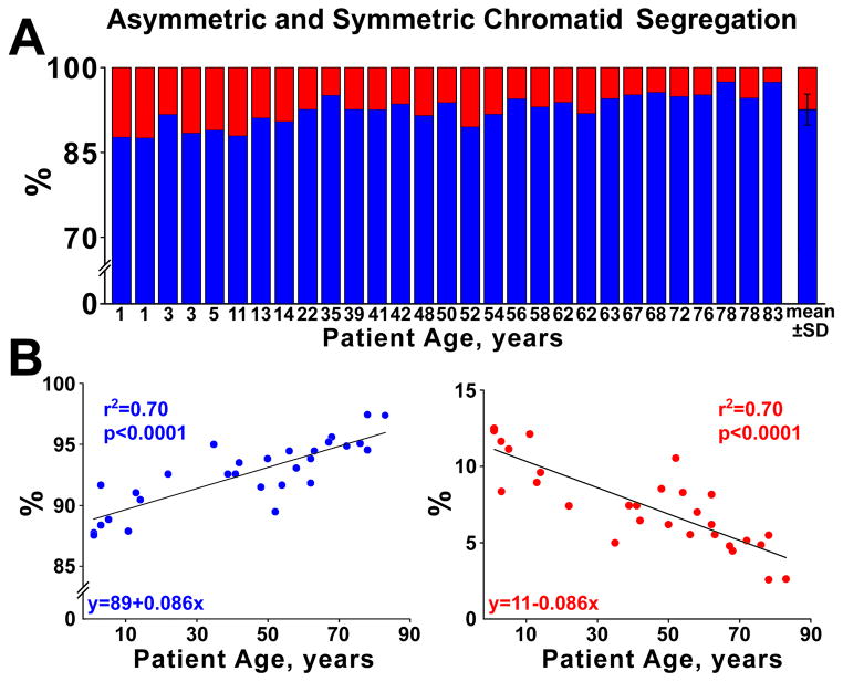
Rationale: According to the immortal DNA strand hypothesis, dividing stem cells selectively segregate chromosomes carrying the old template DNA, opposing accumulation of mutations resulting from nonrepaired replication errors and attenuating telomere shortening.

Objective: Based on the premise of the immortal DNA strand hypothesis, we propose that stem cells retaining the old DNA would represent the most powerful cells for myocardial regeneration.

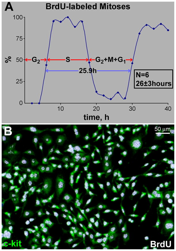
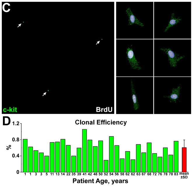
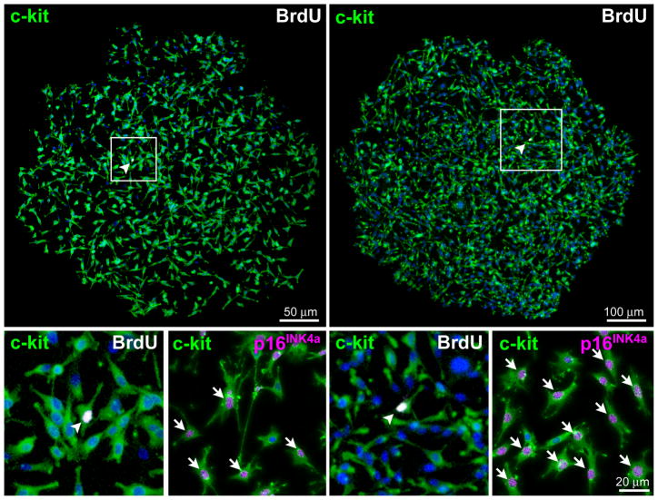
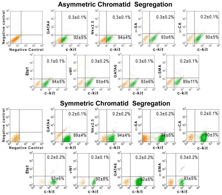
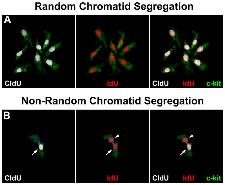
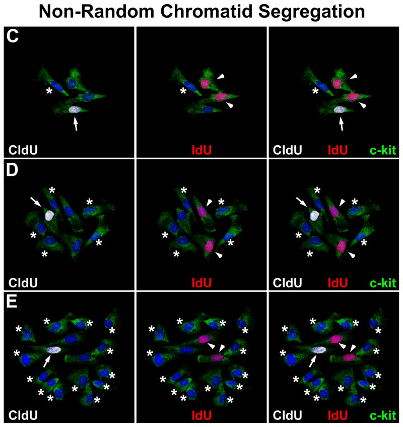
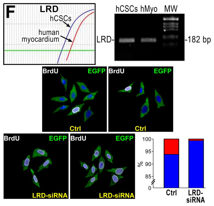
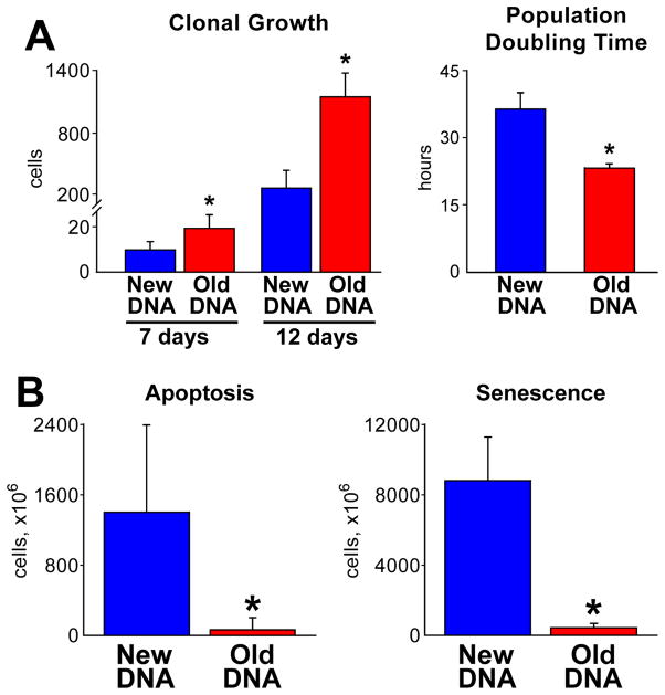
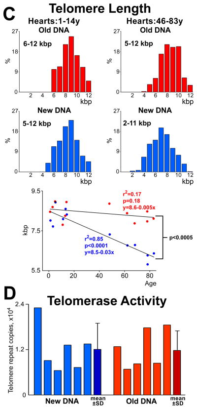
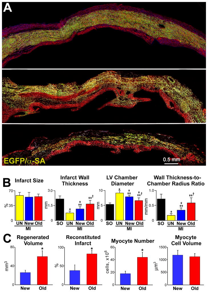
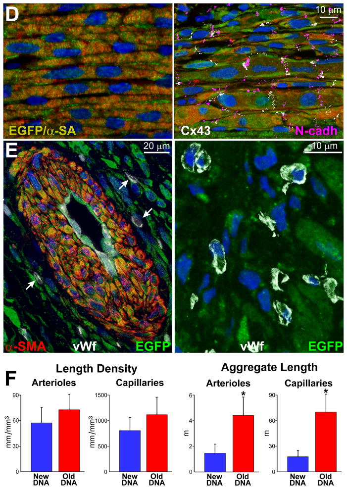
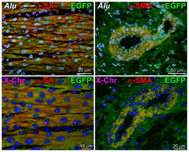
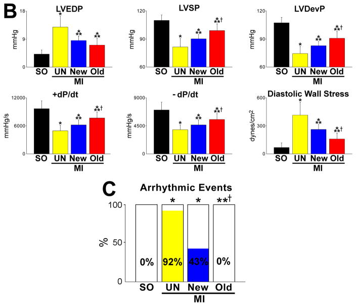
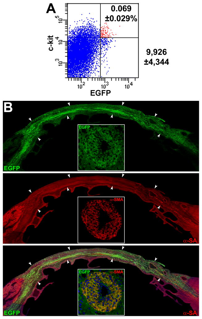
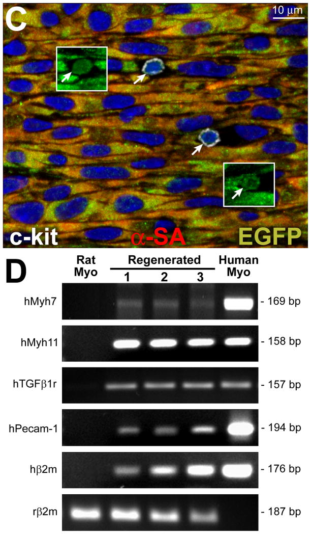
Methods and results: Division of human cardiac stem cells (hCSCs) by nonrandom and random segregation of chromatids was documented by clonal assay of bromodeoxyuridine-tagged hCSCs. Additionally, their growth properties were determined by a series of in vitro and in vivo studies. We report that a small class of hCSCs retain during replication the mother DNA and generate 2 daughter cells, which carry the old and new DNA, respectively. hCSCs with immortal DNA form a pool of nonsenescent cells with longer telomeres and higher proliferative capacity. The self-renewal and long-term repopulating ability of these cells was shown in serial-transplantation assays in the infarcted heart; these cells created a chimeric organ, composed of spared rat and regenerated human cardiomyocytes and coronary vessels, leading to a remarkable restoration of cardiac structure and function. The documentation that hCSCs divide by asymmetrical and symmetrical chromatid segregation supports the view that the human heart is a self-renewing organ regulated by a compartment of resident hCSCs.

Conclusions: The impressive recovery in ventricular hemodynamics and anatomy mediated by clonal hCSCs carrying the "mother" DNA underscores the clinical relevance of this stem cell class for the management of heart failure in humans.


Figures

Figure 1
Figure 1. hCSC growth and clonal assay
A, Length of the cell cycle in lineage-negative c-kit-positive hCSCs. Mean±SD. The sequence of the phases of the cell cycle, i.e., G2, S, and G1, reflects the order in which cells enter mitosis after the BrdU pulse. Cells in G2 are the first to reach mitosis; these cells, however, are BrdU-negative. Subsequently, cells that are in S phase will divide and the mitotic figures will be BrdU-positive, deflecting upwards the labeled mitosis curve. Finally, cells in G1 at the time of the BrdU pulse will reach mitosis; they will be BrdU negative and mitotic figures will be no longer labeled by the thymidine analog. B, Lineage-negative c-kit-positive hCSCs (green) are BrdU-positive (white) before plating for clonal analysis. C, Individual lineage-negative c-kit-positive hCSCs (green) were plated at limiting dilution (left panel), or seeded in single wells of Terasaki plates (right panel). BrdU, white. D, Clonal efficiency in each sample is shown together with the mean±SD.
Figure 2
Figure 2. Clonal growth of hCSCs
A, Clones derived from hCSCs carrying the old (red) and new (blue) DNA in each patient. Mean±SD is shown. B, Linear relationship between age and number of clones formed by hCSCs carrying the old (red) and new (blue) DNA. C, Two clones having each one BrdU-positive hCSC (arrowheads). The BrdU-positive hCSC is shown at higher magnification in the inset (lower left panel; arrowheads); BrdU-positive and BrdU-negative hCSCs are negative for p16INK4a. hCSCs exposed to doxorubicin were used as positive control for p16INK4a expression (lower right panel; magenta, arrows). D, Clonal hCSCs derived by asymmetric and symmetric chromatid segregation are predominantly undifferentiated and express at very low level epitopes of cardiomyocytes [GATA4, Nkx2.5, α-sarcomeric actin (α-SA), α-cardiac actinin (α-CA)], ECs [Ets1, von Willebrand factor (vWf)], and SMCs [GATA6, α-smooth muscle actin (α-SMA)]. E and F, Only one set (arrows) of anaphase/telophase chromosomes (PI, red) in hCSCs (c-kit, green) is labeled by BrdU (white) demonstrating non-random chromatid segregation; α-adaptin (blue) surrounds both sets of chromosomes (F), documenting symmetric stem cell division.
Figure 3
Figure 3. hCSC division
A, Small clone derived from hCSCs dividing by symmetric chromatid segregation; all hCSCs are positive for CldU (left, white), and IdU (center, red). Right, merge. BE, Clones derived from hCSCs dividing by asymmetric chromatid segregation (Online Figure IV illustrates schematically this mechanism of hCSC growth). B, Division of 1 parent CldU-positive hCSC synthesizes DNA during S-phase and, in the presence of IdU, incorporates the halogenated nucleotide, and generates 2 daughter stem cells: 1 positive for both CldU and IdU (left and center, arrows), and 1 positive for only IdU (center, arrowhead). Right, merge. C, By division of these 2 cells, 4 cells are formed; 1 positive for only CldU (left, arrow), 2 positive for only IdU (center, arrowheads), and 1 negative for CldU and IdU (left and center, asterisk). Right, merge. D, By division of these 4 cells, 8 cells are formed; 1 positive for only CldU (left, arrow), 2 positive for only IdU (center, arrowheads), and 5 negative for CldU and IdU (left and center, asterisks). Right, merge. E, By division of these 8 cells, 16 cells are formed; 1 positive for only CldU (left, arrow), 2 positive for only IdU (center, arrowheads), and 13 negative for CldU and IdU (left and center, asterisks). Right, merge. F, Representative tracings of transcript for LRD in hCSCs and human myocardium (hMyo). PCR products had the correct molecular size. EGFP (green) identifies hCSCs transduced with scrambled-siRNA (control, Ctrl) or LRD-siRNA. BrdU, white. Clones generated by hCSCs with intact LRD (Ctrl) and downregulated LRD (LRD-siRNA) are shown. Asymmetric chromatid segregation is present in a fraction of dividing control hCSCs (red bar) and is almost completely abrogated in dividing hCSCs transduced with LRD-siRNA. Blue bars, hCSCs dividing by symmetric segregation of chromatids.
Figure 4
Figure 4. Growth characteristics of hCSCs
A, Clonal growth and population doubling time of hCSCs dividing by asymmetric (old DNA, red) and symmetric (new DNA, blue) chromatid segregation, respectively. B, Rate of apoptosis and senescence of in hCSC classes; mean±SD. *P<0.05 vs. New DNA. C, Distribution of telomere length in hCSC subsets; in hearts 46–83 years (y) old, telomere length of hCSCs carrying the old DNA is shifted towards higher values. Telomere length decreases with age only in hCSCs carrying the new DNA. D, Telomerase activity in hCSC subsets measured by qPCR; individual values are shown together with mean±SD.
Figure 5
Figure 5. Growth characteristics and karyotype of hCSCs
A and B, PDT (A) and telomere length (B) of hCSCs dividing by asymmetric chromatid segregation (red) following exposure to UV light. Control cells (hCSCs labeled by BrdU but not exposed to UV light), blue. C, Euploid set of chromosomes in a metaphase spread of hCSCs after exposure to UV light.
Figure 6
Figure 6. Myocardial regeneration and hCSC classes
A, hCSCs carrying the old DNA reconstitute most of the infarct (upper and central: combination of EGFP and α-SA, yellowish. Labeling by EGFP, α-SA, and merge are shown in Online Figure XA through XC. hCSCs carrying the new DNA replace only in part the infarcted myocardium (lower). B, Infarct size is similar in all cases. hCSCs obtained by FACS-FRET or UV light produced similar results, which were combined. hCSCs with old DNA have a more positive effect on the thickness of the infarcted wall, chamber diameter, and wall thickness-to-chamber radius ratio than hCSCs carrying the new DNA. SO, sham-operated; MI, infarct; UN, untreated; New, treated with hCSCs with new DNA; Old, treated with hCSCs with old DNA. Values are mean±SD. *,**,†P<0.05 vs. SO, UN, and New, respectively. C, The amount of regenerated human myocardium is 2.4-fold larger with hCSCs with old DNA, reconstituting most of the infarcted wall; this was mediated by a 2.5-fold increase in the number of newly-formed myocytes of comparable size. *P<0.05 vs. New. D, Human myocytes are EGFP-positive and show sarcomere striation: EGFP and α-SA (yellowish). Human myocytes express the junctional proteins connexin 43 (Cx43, white) and N-cadherin (N-cadh, magenta). E, Human arteriole is EGFP-positive and is composed of several layers of SMCs (combination of EGFP and α-SMA, yellowish) and a thin layer of ECs (combination of EGFP and vWf, light green); EGFP, α-SMA, vWf, and merge are shown in Online Figure XD. Similarly, human capillaries are EGFP-positive and are composed of a thin layer of ECs (combination of EGFP and vWf, light green). F, Length density and aggregate length of arterioles and capillaries in the regenerated myocardium with hCSCs carrying the old and new DNA. *P<0.05 vs. New.
Figure 7
Figure 7. Human structures and ventricular function
A, Human myocytes and vessels show human DNA sequences (Alu probe, white dots in nuclei) and human X-chromosome (X-Chr, single magenta dots in nuclei). B, hCSCs with old DNA had a more positive effect on left ventricular (LV) end-diastolic pressure (LVEDP), LV systolic pressure (LVSP), LV developed pressure (LVDP), positive and negative dP/dt, and calculated diastolic wall stress than hCSCs carrying the new DNA. C, Number of arrhythmic events in SO, and untreated and treated infarcts. For abbreviation and statistics see Figure 6B.
Figure 8
Figure 8. Serial transplantation of clonal hCSCs with old DNA
A, c-kit-positive EGFP-positive hCSCs (red dots) isolated after regeneration of the infarcted heart are shown by scatter plot. B, Following serial transplantation, a large area of the infarcted myocardium is replaced by EGFP-positive (upper, green), α-SA-positive (central: red, arrowheads) cardiomyocytes (lower: merge, yellowish, arrowheads). A coronary vessel (inset) is positive for EGFP (upper, green), and α-SMA (central, red); merge (lower, yellowish). C, In a serially transplanted infarcted heart, EGFP-positive (insets) c-kit-positive (white) hCSCs are present (arrows). D, Expression of human genes by q-RT-PCR in cell treated infarcted hearts: cardiomyocyte (β-myosin heavy chain, hMyh7), SMC (SM-myosin-heavy-chain 11, hMyh 11; TGF-β1 receptor, hTGFβ1r), and EC (hPecam-1) genes are shown together with the housekeeping gene human β2-microglobulin (hβ2m) and rat β2-microglobulin (rβ2m). Rat and human myocardium (Myo) were used as negative and positive control, respectively. The PCR products had the expected molecular weight. For tracings and sequences of PCR products, see Online Figure XI.

Comment in

Similar articles

Cited by

References

    1. Murry CE, Lee RT. Development biology. Turnover after the fallout. Science. 2009;324:47–48. - PubMed
    1. Parmacek MS, Epstein JA. Cardiomyocyte renewal. N Engl J Med. 2009;361:86–88. - PMC - PubMed
    1. Minami E, Laflamme MA, Saffitz JE, Murry CE. Extracardiac progenitor cells repopulate most major cell types in the transplanted human heart. Circulation. 2005;112:2951–2958. - PubMed
    1. Bayes-Genis A, Roura S, Prat-Vidal C, Farré J, Soler-Botija C, de Luna AB, Cinca J. Chimerism and microchimerism of the human heart: evidence for cardiac regeneration. Nat Clin Pract Cardiovasc Med. 2007;4(Suppl 1):S40–S45. - PubMed
    1. Urbanek K, Cesselli D, Rota M, Nascimbene A, De Angelis A, Hosoda T, Bearzi C, Boni A, Bolli R, Kajstura J, Anversa P, Leri A. Stem cell niches in the adult mouse heart. Proc Natl Acad Sci USA. 2006;103:9226–9231. - PMC - PubMed

Publication types

MeSH terms