. 2012 Jul 31;3(4):e00166-12.
doi: 10.1128/mBio.00166-12. Print 2012.

Emergence of fatal avian influenza in New England harbor seals

Affiliations

Emergence of fatal avian influenza in New England harbor seals

S J Anthony et al. mBio. .

Abstract

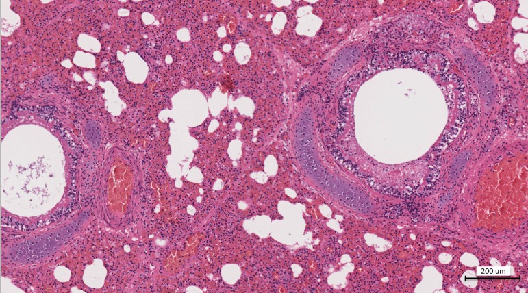
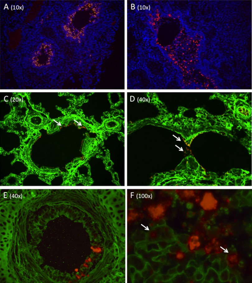
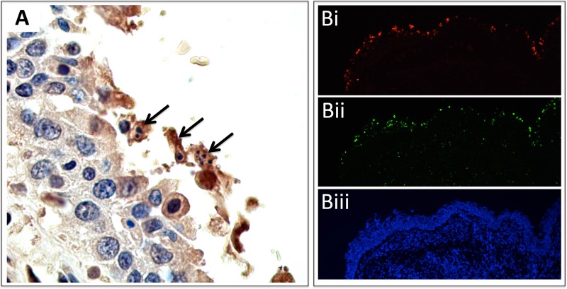
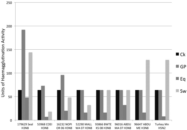
From September to December 2011, 162 New England harbor seals died in an outbreak of pneumonia. Sequence analysis of postmortem samples revealed the presence of an avian H3N8 influenza A virus, similar to a virus circulating in North American waterfowl since at least 2002 but with mutations that indicate recent adaption to mammalian hosts. These include a D701N mutation in the viral PB2 protein, previously reported in highly pathogenic H5N1 avian influenza viruses infecting people. Lectin staining and agglutination assays indicated the presence of the avian-preferred SAα-2,3 and mammalian SAα-2,6 receptors in seal respiratory tract, and the ability of the virus to agglutinate erythrocytes bearing either the SAα-2,3 or the SAα-2,6 receptor. The emergence of this A/harbor seal/Massachusetts/1/2011 virus may herald the appearance of an H3N8 influenza clade with potential for persistence and cross-species transmission.

Importance: The emergence of new strains of influenza virus is always of great public concern, especially when the infection of a new mammalian host has the potential to result in a widespread outbreak of disease. Here we report the emergence of an avian influenza virus (H3N8) in New England harbor seals which caused an outbreak of pneumonia and contributed to a U.S. federally recognized unusual mortality event (UME). This outbreak is particularly significant, not only because of the disease it caused in seals but also because the virus has naturally acquired mutations that are known to increase transmissibility and virulence in mammals. Monitoring the spillover and adaptation of avian viruses in mammalian species is critically important if we are to understand the factors that lead to both epizootic and zoonotic emergence.


Figures

FIG 1
FIG 1
Hematoxylin and eosin staining of the lung at a ×10 magnification. There is diffuse acute interstitial pneumonia and a mix of acute hemorrhagic alveolitis with necrotizing bronchitis. Multifocally, alveoli are either filled with hemorrhage and scant inflammatory cells or expanded with emphysema. There is irregularity to the bronchial mucosa due to necrosis, a mild to moderate edema, and mucous partially filling the bronchial lumen. There is mild to moderate expansion of the interlobular septa, with edema and hemorrhage.
FIG 2
FIG 2
(A and B) Fluorescent in situ hybridization (ISH) of H3N8 virus-infected seal cells with DAPI counterstaining. A probe targeting the viral hemagglutinin demonstrates diffuse infection of the bronchial mucosal epithelium. ISH was also performed using probes for the matrix gene. Staining was identical to that shown here for HA. (C and D) Lectin staining to demonstrate the distribution of SAα-2,3 and SAα-2,6 in seal pulmonary parenchyma. The SAα-2,6 (green) was detected using fluorescein-labeled Sambucus nigra agglutinin (SNA) lectin, while SAα-2,3 (red) was detected using Maackia amurensis II (MAL II) lectin. Both infected and uninfected control tissues were stained, and the results were consistent for both. High levels of SAα-2,6 are observed on bronchiole and alveolar epithelial cells and on endothelial cells. The images in panels C and D were selected because they show staining for both sialic acids; however, the expression of SAα-2,3 was rarely observed (arrows) and limited to bronchiole luminal (C) and occasional alveolar (D) epithelia. (E and F) Costaining of SAα-2,6 and H3N8 HA. SAα-2,6 (green) is expressed on the respiratory epithelium of an intrapulmonary bronchus (E). H3N8 virus-infected cells (red) are present. A serial section was also stained for SAα-2,3, and none was detected. A high-magnification image of infected mucosa clearly shows H3N8 virus infection of cells expressing SAα-2,6 (arrows). All composite images are presented separately (single stains) in Fig. S1 to S3.
FIG 3
FIG 3
(A) Immunohistochemistry (IHC) of seal bronchus. Polyclonal antibodies were raised against H3N8 virus-specific HA antigen. Brown staining (DAB reporter system) indicates the presence of viral antigen. There is irregularity of the mucosal surface, with sloughed epithelium. IHC demonstrates the presence of viral antigen, pyknosis, and apoptosis (arrows). No viral antigen or apoptosis was seen on negative-control tissue. (Bi) ISH staining of H3N8 virus in lung epithelium (HA probe). (Bii) TUNEL staining (green) in the same region (serial section) of the lung, showing the presence of apoptotic cells. Comparison of virus and TUNEL staining shows localization of apoptosis to virally infected cells. (Biii) DAPI (with dihydrochloride) staining for cell nuclei.
FIG 4
FIG 4
Phylogenetic trees of representative influenza H3N8 genome segments. Nucleotide sequence alignments for all genome segments were created using ClustalW, and trees were produced using neighbor-joining, maximum-likelihood, and Bayesian algorithms. Models of evolution were selected using ModelTest, and a tree was selected based on a consensus of the results of the three algorithms. Only the trees for HA and NA are shown; however, all eight segments showed strong association with sequences of avian origin. Trees are constructed with H3N8 viruses only, and published sequences were selected to represent variation in the year, host, and location of isolation. *, A/blue-winged teal/Ohio/926/2002; NY, New York; LA, Louisiana.
FIG 5
FIG 5
Structural-homology model showing the interaction of influenza HA with the SAα2,3 ligand. Seal H3 (gray), avian H5 (orange) (Protein Data Bank [PDB] accession number 1JSO), and swine H9 (pink) (PDB accession number 1JSH) were compared. The mutation G151E causes a conformational shift and interrupts H bonding between seal H3 S152 and SAα2,3, which suggests a reduction in SAα2,3 binding efficiency. A lost H bond in seal H3 is depicted in green.
FIG 6
FIG 6
Hemagglutination assays were performed on an isolate of the H3N8 seal virus (A/harbor seal/New Hampshire/179629/2011) to confirm sialic acid binding preferences. Viruses were tested for their ability to agglutinate erythrocytes that preferentially express SAα-2,3 (horse) or SAα-2,6 (guinea pig, pig). Average agglutination titers for seal H3N8 with horse erythrocytes (1:48) show that the virus can still bind to SAα-2,3, though weakly. Titers were appreciably higher with guinea pig (1:192) and pig (1:144) erythrocytes, demonstrating a preference for SAα-2,6. Given that horse erythrocytes express SAα-2,3, it is interesting that the avian H3N8 viruses did not agglutinate with horse RBCs (red blood cells) efficiently, even following repeated attempts. Ito et al. (25) showed that avian H3N8 viruses from Asia in the early 1980s could bind to horse RBCs, while Wiriyarat et al. (43) gave examples of avian viruses (albeit not H3 viruses) that did not bind to horse RBCs. It is not known whether the avian viruses included here simply have a preference for the N-acetyl (NeuAc) sialic acid species, which is not found on horse RBCs, while the seal virus uses N-glycolyl (NeuGc) SAα-2,3. Ck, chicken; GP, guinea pig; Eq, equine; Sw, swine; 179629 Seal H3N8, A/harbor seal/New Hampshire/179629/2011; 53968 COEI H3N8, A/common eider/Massachusetts/20507-001/2007 (H3N8) virus; 16232 NOPI OR 06 H3N8, A/northern pintail/Oregon/44249-547/2006 (H3N8) virus; 52290 MALL WA 07 H3N8, A/mallard/Washington/44338-052/2007 (H3N8) virus; 93866 BWTE KS 08 H3N8, A/blue-winged teal/Kansas/44440-003/2008 (H3N8) virus; 96016 ABDU MA 07 H3N8, A/American black duck/Maine/44411-174/2008 (H3N8) virus; 96647 ABDU ME H3N8, A/American black duck/Maine/44411-532/2008 (H3N8) virus; Turkey Mn H5N2, A/turkey/Minnesota/3689-1551/1981 (H5N2) virus.

Similar articles

Cited by

References

    1. Geraci JR, et al. 1982. Mass mortality of harbor seals: pneumonia associated with influenza A virus. Science 215:1129–1131 - PubMed
    1. Webster RG, et al. 1981. Characterization of an influenza A virus from seals. Virology 113:712–724 - PubMed
    1. Hinshaw VS, et al. 1984. Are seals frequently infected with avian influenza viruses? J. Virol. 51:863–865 - PMC - PubMed
    1. Callan RJ, Early G, Kida H, Hinshaw VS. 1995. The appearance of H3 influenza viruses in seals. J. Gen. Virol. 76(Part 1):199–203 - PubMed
    1. Vijaykrishna D, et al. 2010. Reassortment of pandemic H1N1/2009 influenza A virus in swine. Science 328:1529 - PMC - PubMed

Publication types

MeSH terms

Associated data