. 2012 Aug;140(2):189-205.
doi: 10.1085/jgp.201210806.

Regulation of voltage-gated potassium channels by PI(4,5)P2

Affiliations

Regulation of voltage-gated potassium channels by PI(4,5)P2

Martin Kruse et al. J Gen Physiol. 2012 Aug.

Abstract

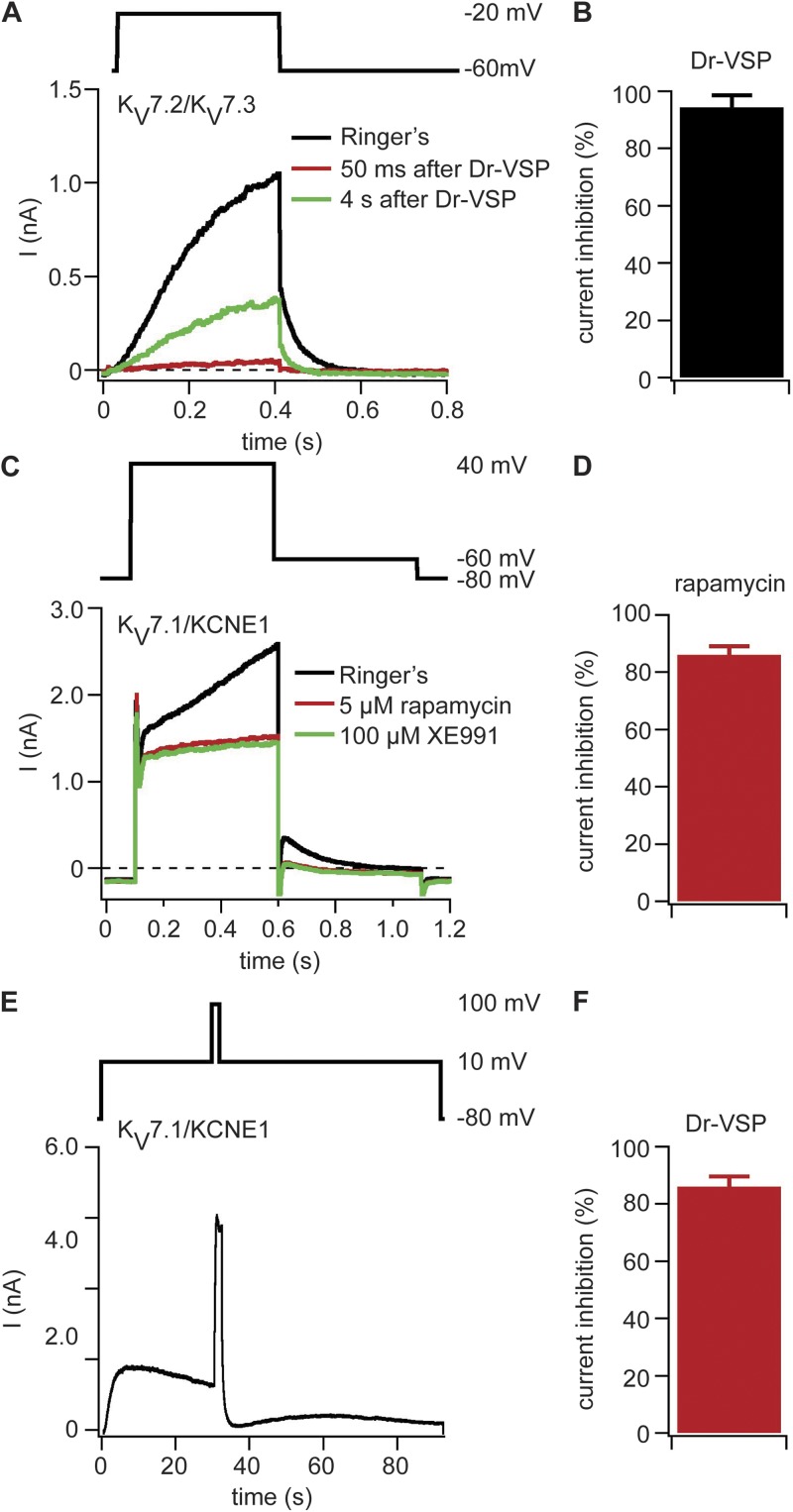
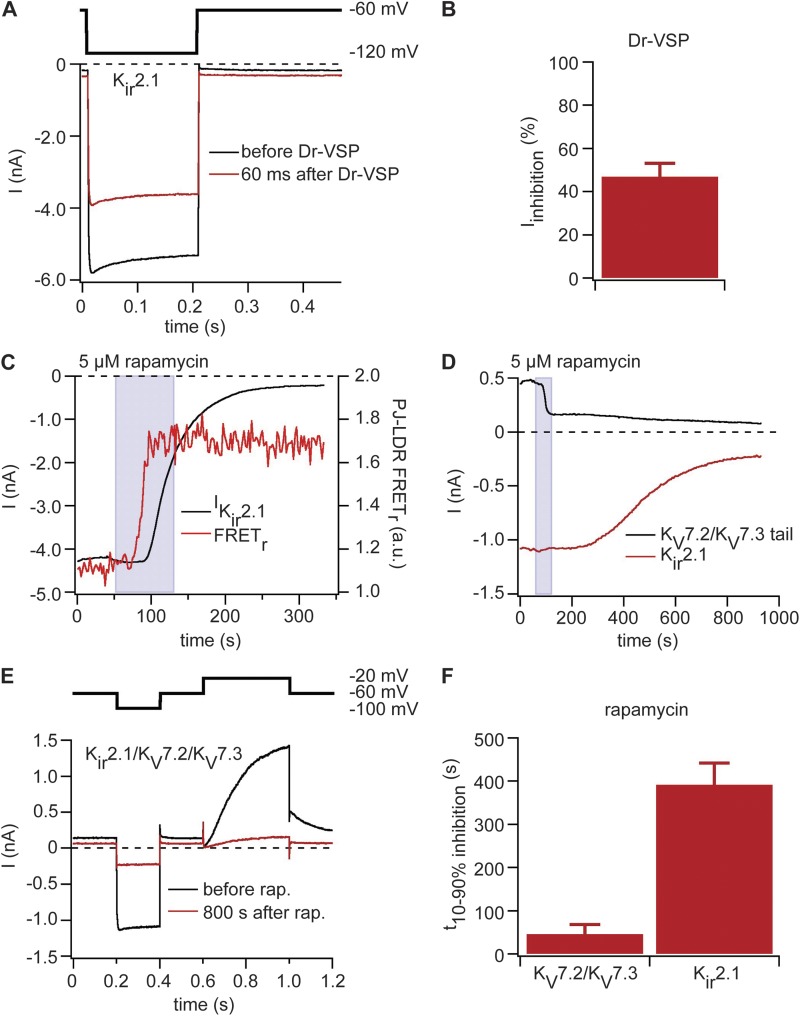
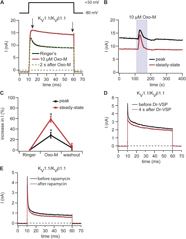
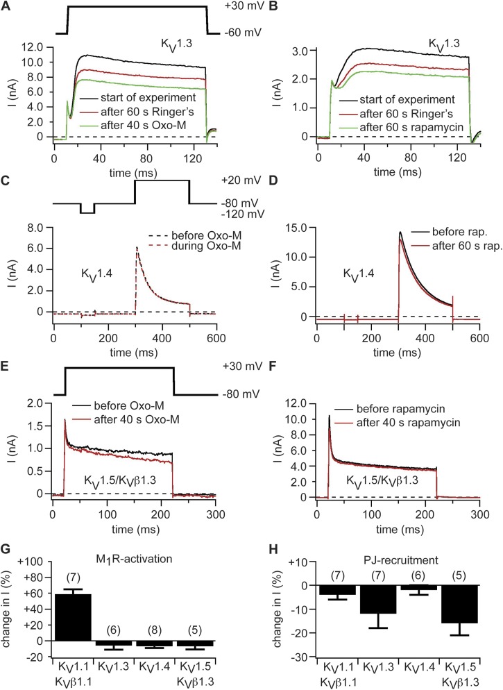
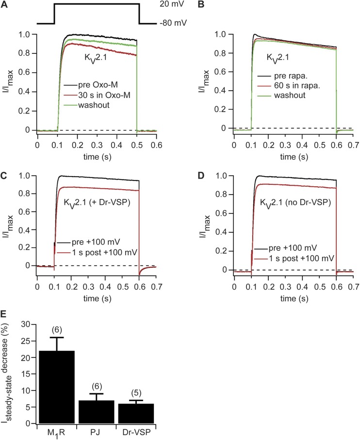
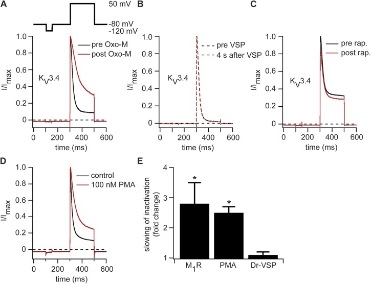
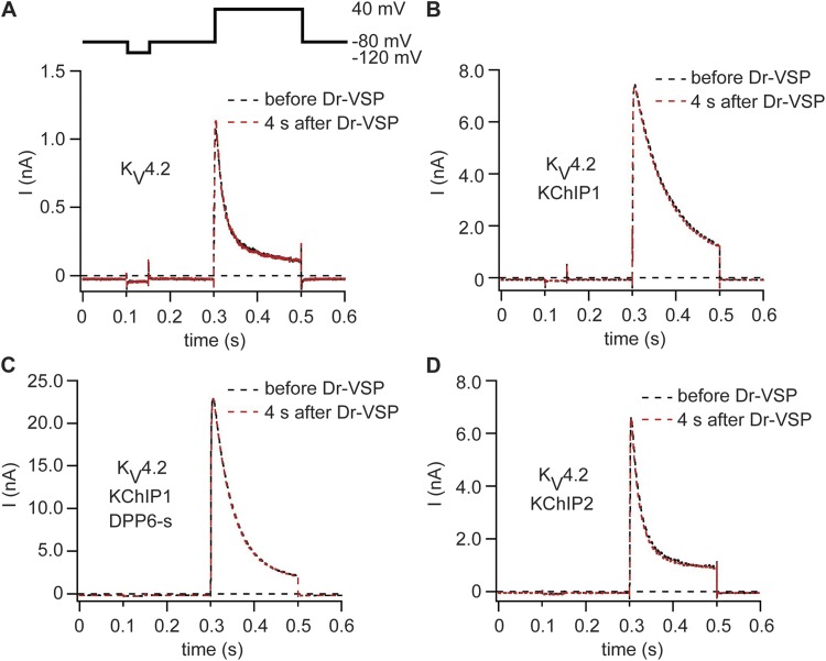
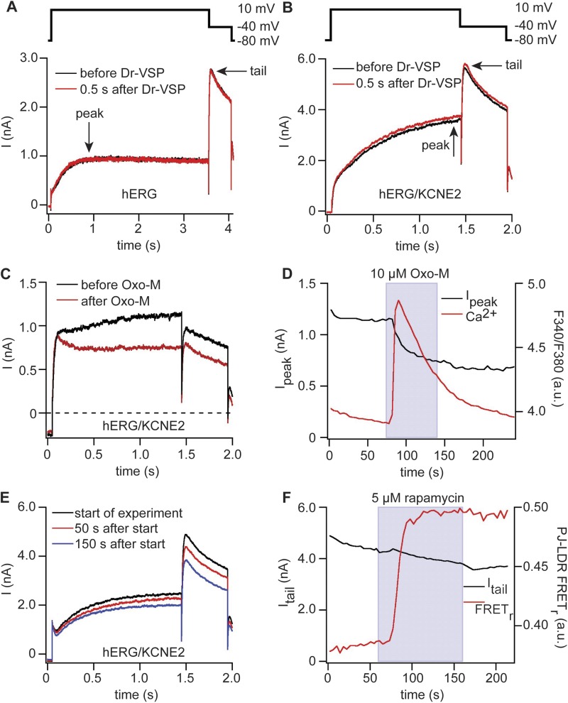
Phosphatidylinositol 4,5-bisphosphate (PI(4,5)P(2)) regulates activities of numerous ion channels including inwardly rectifying potassium (K(ir)) channels, KCNQ, TRP, and voltage-gated calcium channels. Several studies suggest that voltage-gated potassium (K(V)) channels might be regulated by PI(4,5)P(2). Wide expression of K(V) channels in different cells suggests that such regulation could have broad physiological consequences. To study regulation of K(V) channels by PI(4,5)P(2), we have coexpressed several of them in tsA-201 cells with a G protein-coupled receptor (M(1)R), a voltage-sensitive lipid 5-phosphatase (Dr-VSP), or an engineered fusion protein carrying both lipid 4-phosphatase and 5-phosphatase activity (pseudojanin). These tools deplete PI(4,5)P(2) with application of muscarinic agonists, depolarization, or rapamycin, respectively. PI(4,5)P(2) at the plasma membrane was monitored by Förster resonance energy transfer (FRET) from PH probes of PLCδ1 simultaneously with whole-cell recordings. Activation of Dr-VSP or recruitment of pseudojanin inhibited K(V)7.1, K(V)7.2/7.3, and K(ir)2.1 channel current by 90-95%. Activation of M(1)R inhibited K(V)7.2/7.3 current similarly. With these tools, we tested for potential PI(4,5)P(2) regulation of activity of K(V)1.1/K(V)β1.1, K(V)1.3, K(V)1.4, and K(V)1.5/K(V)β1.3, K(V)2.1, K(V)3.4, K(V)4.2, K(V)4.3 (with different KChIPs and DPP6-s), and hERG/KCNE2. Interestingly, we found a substantial removal of inactivation for K(V)1.1/K(V)β1.1 and K(V)3.4, resulting in up-regulation of current density upon activation of M(1)R but no changes in activity upon activating only VSP or pseudojanin. The other channels tested except possibly hERG showed no alteration in activity in any of the assays we used. In conclusion, a depletion of PI(4,5)P(2) at the plasma membrane by enzymes does not seem to influence activity of most tested K(V) channels, whereas it does strongly inhibit members of the K(V)7 and K(ir) families.


Figures

Figure 1.
Figure 1.
PI(4,5)P2 depletion at the plasma membrane inhibits KV7.x channels. (A) Currents with coexpression of KV7.2, KV7.3, and Dr-VSP in tsA-201 cells. The black current trace was recorded before Dr-VSP activation, the red trace was recorded 50 ms after Dr-VSP activation by a 2-s pulse to 100 mV, and the green trace was recorded 4 s after Dr-VSP activation. Test pulse protocol is shown above the traces. (B) Reduction of KV7.2/KV7.3-mediated tail currents by activation of Dr-VSP (n = 10). (C) Currents traces recorded from a cell expressing KV7.1, KCNE1, Ins-5-P-FKBP-CFP, and LDR-CFP. Traces are shown before application of 5 µM rapamycin (black), after 60 s of rapamycin application (red), and after application of 100 µM XE991 (green). (D) Rapamycin-induced inhibition of XE991-sensitive current (n = 5). (E) Current trace of a cell expressing KV7.1, KCNE1, and Dr-VSP. Currents were recorded at 10 mV, Dr-VSP was activated after 30 s with a 2-s long test pulse to 100 mV, and then the membrane potential was returned to 10 mV. At the end of the recording, XE991 was applied to determine the amount of KV7.1-mediated current (not depicted). (F) Inhibition of XE991-sensitive current by Dr-VSP activation (n = 6). (B, D, and F) Error bars represent ±SEM.
Figure 2.
Figure 2.
Dr-VSP and pseudojanin can inhibit PI(4,5)P2-sensitive Kir2.1 channels. (A) Currents in Kir2.1 channels coexpressed with Dr-VSP. Black indicates current trace before activation of Dr-VSP, and red indicates current trace 60 ms after activation of Dr-VSP by a 2-s pulse to 100 mV. Hyperpolarizing test pulse protocol is shown above the traces. (B) Inhibition of Kir2.1-mediated currents by activation of Dr-VSP (n = 5). (C) Currents with coexpression of Kir2.1, pseudojanin (PJ)-YFP, and LDR-CFP recorded with the protocol shown in A. Black trace shows steady-state current over time, and the red trace is FRET ratio between pseudojanin-YFP and LDR-CFP. (D) Same experiment as in C but with expression of KV7.2 and KV7.3 as well as Kir2.1. Black trace shows KV7.2/KV7.3-mediated tail currents, and red trace shows Kir2.1-mediated steady-state current over time. (E) Current traces and pulse protocol for the recording shown in D. Black trace shows currents at start of rapamycin (rap.) application (60 s), and red trace is at 900 s (end of recording). (F) Time needed to decrease either KV7.2/KV7.3 or Kir2.1-mediated currents from 90 to 10% original amplitude (n = 4). (B and F) Error bars represent ±SEM.
Figure 3.
Figure 3.
Modulation of KV1 family channels by depletion of PI(4,5)P2 at the plasma membrane. (A) Potassium outward currents in KV1.1/KVβ1.1 channel complexes coexpressed with M1R. Black indicates control current trace in Ringer’s solution, red indicates during application of 10 µM Oxo-M, and green indicates after washout of Oxo-M. Arrows indicate points at which peak and steady-state current amplitudes were measured. (B) Current amplitudes of peak and steady-state current of the cell shown in A over time. (C) Percent increase of peak and steady-state currents of KV1.1/KVβ1.1 channel complexes coexpressed with M1R upon stimulation with Oxo-M (n = 7). Error bars represent ±SEM. *, P < 0.05. (D) Current time course in a cell expressing KV1.1/KVβ1.1 channel complexes and Dr-VSP. Dr-VSP was activated by a 2-s pulse to 100 mV. Test pulse protocol as shown in A. (E) Same as D but with expression of pseudojanin-YFP and LDR-CFP instead of Dr-VSP and addition of 5 µM rapamycin.
Figure 4.
Figure 4.
KV1.3, KV1.4, and KV1.5 channels are insensitive to depletion of PI(4,5)P2 at the plasma membrane. (A) Current traces of KV1.3 channels coexpressed with M1R. Black indicates traces at start of experiment, red indicates after 60 s in Ringer’s, and green indicates traces after 40-s superfusion with 10 µM Oxo-M (100 s after start of experiment). (B) Currents in KV1.3 channels coexpressed with pseudojanin-YFP and LDR-CFP. Black indicates traces at start of experiment, red indicates after 60 s in Ringer’s, and green indicates traces after 60-s rapamycin application (120 s after start of experiment). Same pulse protocol as in A. (C) Currents in a cell expressing KV1.4 and M1R. Black indicates current trace in Ringer’s solution, and red indicates current trace during application of 10 µM Oxo-M. (D) KV1.4 channels coexpressed with pseudojanin-YFP and LDR-CFP. Black indicates current before application of rapamycin (rap.), and red indicates traces after 60 s of rapamycin application. (E and F) Currents in KV1.5 channels coexpressed with KVβ1.3 and M1R (E) or pseudojanin-YFP and LDR-CFP (F). Black indicates current before application of Oxo-M (E) or rapamycin (F), and red indicates current after 40-s application of Oxo-M (E) or rapamycin (F). (G and H) Percent changes in steady-state current amplitudes of KV1.x channels after activation of M1R (G) or recruitment of pseudojanin (PJ) to the plasma membrane (H). Numbers in parentheses indicate n numbers for individual experiments. Error bars represent ±SEM.
Figure 5.
Figure 5.
KV2.1 channels are not sensitive to depletion of PI(4,5)P2 by activation of pseudojanin or Dr-VSP. (A) Normalized current traces recorded before (black), during (red), and after (green) application of 10 µM Oxo-M in a cell transiently transfected with KV2.1 and M1R. (B) Current traces before (black), after 60 s of application (red), and after washout of 5 µM rapamycin (rapa.) to a cell expressing KV2.1, pseudojanin-YFP, and LDR-CFP. (C) Normalized KV2.1-mediated current traces before (black) and 1 s after (red) a 2-s depolarizing pulse to 100 mV in cells coexpressing Dr-VSP. (D) Same as in C but without coexpression of Dr-VSP. (E) Decrease in KV2.1-mediated steady-state currents at 20 mV after activation of M1R or Dr-VSP or recruitment of pseudojanin (PJ) to the plasma membrane. Numbers in parentheses indicate numbers of individual experiments. Error bars represent ±SEM.
Figure 6.
Figure 6.
KV3.4 channels are modulated by activation of M1R but not by activation of Dr-VSP or pseudojanin. (A) Normalized current traces recorded before (black) and during (red) application of 10 µM Oxo-M in a cell transiently transfected with KV3.4 and M1R. (B) Same as in A but with expression of Dr-VSP and an activating pulse instead of M1R. (C) Current traces before (black) and after (red) application of 5 µM rapamycin (rap.) to a cell expressing KV3.4, pseudojanin-YFP, and LDR-CFP. (D) Representative normalized current traces of cells expressing KV3.4 recorded under control conditions (black) or after 15-min treatment with 100 nM PMA in Ringer’s solution (red). (E) Fold slowing of inactivation after activation of M1R, application of 100 nM PMA, or activation of Dr-VSP (n = 4 [M1R] or 5 [PMA and Dr-VSP]). Error bars represent ±SEM. *, P < 0.05.
Figure 7.
Figure 7.
KV4.2 channels are insensitive to depletion of PI(4,5)P2 by Dr-VSP. (A) Current traces of a cell expressing KV4.2 and Dr-VSP. Black indicates before activation of Dr-VSP, and red indicates 4 s after activation of Dr-VSP. (B–D) Same protocol as in A but with coexpression of KChIP1 (B), KChIP1 and DPP6-s (C), or KChIP2 (D).
Figure 8.
Figure 8.
hERG channels are sensitive to M1R activation but not to activation of Dr-VSP or pseudojanin. (A) Currents in hERG channels coexpressed with Dr-VSP. Black indicates before activation of Dr-VSP, and red indicates directly after activation of Dr-VSP. Arrows indicate points at which peak and tail current amplitudes were measured. (B) Same as in A but with additional coexpression of KCNE2 and modified pulse protocol. (C) Currents in hERG channels coexpressed with KCNE2 and M1R. Black indicates before application of 10 µM Oxo-M, and red indicates after application of Oxo-M. Pulse protocol as in B. (D) Time course of current amplitudes at 10 mV and Fura-4F ratio of cell in C. (E) Currents in a cell expressing hERG, KCNE2, pseudojanin-YFP, and LDR-CFP. Currents are shown at the beginning of the recordings (black), directly before application of rapamycin (50 s after start of experiment; red), and at the end of rapamycin application (150 s after start of experiment; blue). Pulse protocol as in B. (F) Time course of current amplitudes of tail currents at −40 mV of the experiment in E (black) and FRET ratio between pseudojanin (PJ)-YFP and the anchor LDR-CFP (red).

Comment in

References

    1. Amarillo Y., De Santiago-Castillo J.A., Dougherty K., Maffie J., Kwon E., Covarrubias M., Rudy B. 2008. Ternary Kv4.2 channels recapitulate voltage-dependent inactivation kinetics of A-type K+ channels in cerebellar granule neurons. J. Physiol. 586:2093–2106 10.1113/jphysiol.2007.150540 - DOI - PMC - PubMed
    1. Beck E.J., Sorensen R.G., Slater S.J., Covarrubias M. 1998. Interactions between multiple phosphorylation sites in the inactivation particle of a K+ channel. Insights into the molecular mechanism of protein kinase C action. J. Gen. Physiol. 112:71–84 10.1085/jgp.112.1.71 - DOI - PMC - PubMed
    1. Bian J.S., McDonald T.V. 2007. Phosphatidylinositol 4,5-bisphosphate interactions with the HERG K(+) channel. Pflugers Arch. 455:105–113 10.1007/s00424-007-0292-5 - DOI - PubMed
    1. Bian J., Cui J., McDonald T.V. 2001. HERG K(+) channel activity is regulated by changes in phosphatidyl inositol 4,5-bisphosphate. Circ. Res. 89:1168–1176 10.1161/hh2401.101375 - DOI - PubMed
    1. Bian J.S., Kagan A., McDonald T.V. 2004. Molecular analysis of PIP2 regulation of HERG and IKr. Am. J. Physiol. Heart Circ. Physiol. 287:H2154–H2163 10.1152/ajpheart.00120.2004 - DOI - PubMed

Publication types

MeSH terms