. 2012 Oct;32(19):3978-89.
doi: 10.1128/MCB.00376-12. Epub 2012 Jul 30.

Sequential eukaryotic translation initiation factor 5 (eIF5) binding to the charged disordered segments of eIF4G and eIF2β stabilizes the 48S preinitiation complex and promotes its shift to the initiation mode

Affiliations

Sequential eukaryotic translation initiation factor 5 (eIF5) binding to the charged disordered segments of eIF4G and eIF2β stabilizes the 48S preinitiation complex and promotes its shift to the initiation mode

Chingakham Ranjit Singh et al. Mol Cell Biol. 2012 Oct.

Abstract

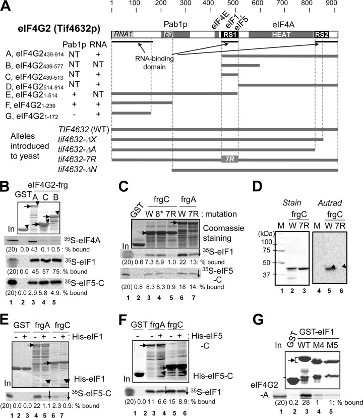
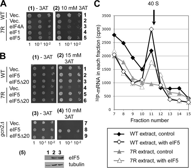
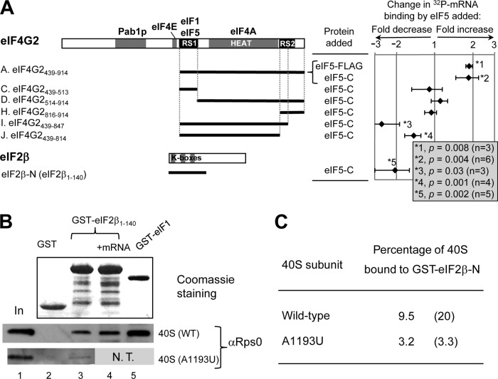
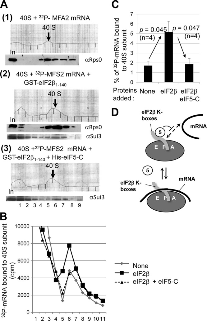
During translation initiation in Saccharomyces cerevisiae, an Arg- and Ser-rich segment (RS1 domain) of eukaryotic translation initiation factor 4G (eIF4G) and the Lys-rich segment (K-boxes) of eIF2β bind three common partners, eIF5, eIF1, and mRNA. Here, we report that both of these segments are involved in mRNA recruitment and AUG recognition by distinct mechanisms. First, the eIF4G-RS1 interaction with the eIF5 C-terminal domain (eIF5-CTD) directly links eIF4G to the preinitiation complex (PIC) and enhances mRNA binding. Second, eIF2β-K-boxes increase mRNA binding to the 40S subunit in vitro in a manner reversed by the eIF5-CTD. Third, mutations altering eIF4G-RS1, eIF2β-K-boxes, and eIF5-CTD restore the accuracy of start codon selection impaired by an eIF2β mutation in vivo, suggesting that the mutual interactions of the eIF segments within the PIC prime the ribosome for initiation in response to start codon selection. We propose that the rearrangement of interactions involving the eIF5-CTD promotes mRNA recruitment through mRNA binding by eIF4G and eIF2β and assists the start codon-induced release of eIF1, the major antagonist of establishing tRNA(i)(Met):mRNA binding to the P site.


Figures

Fig 1
Fig 1
Minimal binding domains of eIF4G2 (Tif4632p) for eIF1, eIF4A, eIF5, and mRNA. (A) Primary structure of yeast eIF4G2 mutations used in this study with defined binding sites for Pab1p, eIF4A, and eIF4E (gray boxes). Boldface underlining denotes RNA-binding segments. Dark boxes indicate the RS domains for RNA binding (43). Light gray boxes are homologous to RNA1, an RNA-binding domain in yeast eIF4G1 (9), and box 3, displaying high homology among fungal eIF4G (b3) (30). Tables to the left summarize the results of interaction assays with RNA and Pab1p. NT, not tested; ΔX, deletion of aa 847 to 914; ΔN, deletion of aa 1 to 237. (B, C, E, and F) GST-eIF4G2 fusion proteins (2 to 5 μg of full-length products, indicated by arrowheads in the top gel), listed across the top, were bound to 35S-labeled proteins synthesized in rabbit reticulocyte lysate in 200 μl binding buffer. GST fusion-containing complexes together with 20% input amounts of the reaction mixtures were analyzed by SDS-PAGE followed by Coomassie staining (top) and autoradiography (bottom). Horizontal arrows indicate the full-length products. The downward arrow indicates a reproduced decrease in the interaction compared to the control (n = 3). (B) Binary interactions of GST or the indicated GST-eIF4G2 segments with 35S-eIF4A, eIF1, and eIF5-C. (C) The effect of RS1 mutation tif4632-8* (8*) or tif4632-7R (7R) (as defined in Table S3 in the supplemental material) on eIF4G2 binding to eIF1 and eIF5. W, wild type. (D) The effect of tif4632-7R on GST-eIF4G-C binding to 32P-β-globin mRNA, as examined by Northwestern blotting (8). Left, Coomassie staining of proteins used. Right, autoradiography of proteins bound to 32P-β-globin mRNA. Arrowheads, locations of full-length products. W, wild type. (E and F) Indicated GST-eIF4G2 proteins were bound to 35S-eIF5-C in the presence of 20 μg His-eIF1 (E) or 35S-eIF1 in the presence of 20 μg His-eIF5-C (F). Arrowheads indicate locations of bands representing His-eIF1 or His-eIF5-C bound. (G) GST-eIF1 and its M4 and M5 mutant derivatives were allowed to bind 35S-eIF4G2-A, and the 35S proteins bound were analyzed together with 20% input amounts (lane 1).
Fig 2
Fig 2
Yeast phenotypes of mutants with altered RNA-binding sites of eIF4G2. (A) The expression of eIF4G2 mutants used in this study (YAS1955 derivatives) (Fig. 1A; also see Table S3 in the supplemental material). Twenty and 40 μg total protein from whole-cell extracts of the indicated strains were used for immunoblotting with anti-HA (to detect HA-eIF4G2) and antitubulin antibodies (loading control). (B) Temperature- and 3AT-sensitive growth of tif4632-7R was assayed by spotting 5 μl of 0.15 A600 U culture (columns 1) and 10-fold serial dilutions (columns 10−1 and 10−2) onto SC-His plates without (−) and with (+) 10 mM 3AT and incubated at the indicated temperatures for 3 days. (C) eIF4G2 mutations impair mRNA recruitment to the 40S subunit in vitro. Cell-free translation mixture with 32P-labeled poly(A)-tailed MFA2 mRNA was fractionated by sucrose gradient velocity sedimentation. 32P counts in relevant fractions are shown with an arrow indicating the fraction containing free 40S subunit. (D) Effect of tif4632-7R on GCN4-lacZ expression from p180. The graph summarizes β-galactosidase activity of YAS1955 (WT) and KAY901 (tif4632-7R [7R]) transformants carrying p180 in the presence (+) or absence (−) of 10 mM 3AT. The P value for differences between the values in columns 1 and 3 is presented.
Fig 3
Fig 3
Interaction of eIF4G2-RS1 with eIF5 mediates mRNA recruitment to the 43S complex. (A and B) Transformants bearing the indicated mutations (WT, YAS1955; 7R, KAY874; and gcn2Δ, KAY24) were diluted and spotted onto SC-His plates without (−3AT) or with (+3AT) the indicated concentrations of 3AT, and the plates were incubated for 3 days (−3AT) and 5 days (+3AT), respectively. In row 5 of panel B are immunoblots of 5 μg whole-cell extracts from KAY24 transformants (rows 7 and 9) with antibodies against the proteins labeled to the right. (C) eIF4G interaction with eIF5 promotes mRNA recruitment to the 40S subunit in vitro. Cell-free translation extracts prepared from strains with the indicated mutations (lower box) were incubated with 32P-labeled poly(A)-tailed MFA2 mRNA with or without FLAG-eIF5 and fractionated by sucrose gradient velocity sedimentation. The assay was analyzed and presented as described for Fig. 2E, except the reaction volume was 200 μl and 80 μg of eIF5 was added.
Fig 4
Fig 4
eIF5 regulates mRNA binding by eIF4G2 and eIF2β in vitro. (A) Primary structure of eIF4G2 with defined binding sites for the indicated initiation factors. Thick lines below the structure represent the portion of eIF4G2 polypeptide found in each construct (see Fig. S3 in the supplemental material for experimental design). The chart on the right summarizes the fold change in mRNA binding caused by eIF5 in a logarithmic scale, with P values listed in the box to the right. A positive value indicates fold increase, while a negative value indicates fold decrease. Bars indicate standard deviations (SD). GST-eIF2β-Ν was also tested, as shown at the bottom. (B) Interaction of eIF2β-NTT with the 40S subunit. GST (5 μg), GST-eIF2β-N (10 μg), and GST-eIF1 (5 μg) were bound to the 40S subunit purified from the wild-type (WT) or A1193U strain with (+mRNA) or without (blank) nonradiolabeled β-globin mRNA (10 μg). The bound 40S subunit was detected by anti-Rps0 antibodies (bottom gels). Top gel, Coomassie staining of proteins used in this study. In, 25% input amount of the 40S subunit. N.T., not tested; however, the same preparation of the mutant 40S subunit used here did not bind to GST-eIF1 in a different experiment (28). (C) Summary of percentages of the 40S subunit bound to GST-eIF2β-N calculated from lanes 1 and 3 in panel B. Parentheses indicate values from an independent experiment.
Fig 5
Fig 5
eIF5-CTD inhibits eIF2β-mediated 32P-MFA2 mRNA binding to the 40S subunit in vitro. (A) Purified wild-type 40S subunit was incubated in the reaction buffer at 4°C for 90 min with 32P-MFA2 mRNA in the absence (section 1) or presence of GST-eIF2β-N (section 2) or in the presence of GST-eIF2β-N and eIF5-C (section 3). The binding reaction was terminated by cross-linking with formaldehyde (5 min on ice), followed by glycine treatment and then fractionation by sucrose gradient velocity sedimentation; the A254 profile of gradient fractions is shown. The relevant 40S subunit-containing fractions are shown with a thick arrow. Proteins from one-half of the gradient samples were precipitated by ethanol, followed by immunoblot analysis with antibodies raised against proteins listed to the right. In, 10% input amount. The resulting immunoblot patterns were shown under the corresponding A254 profile. (B) Scintillation count of the 32P-MFA2 mRNA present in the other half of the gradient samples from the experiments shown in panel A. Gray line, section 1; black solid line, section 2; black dotted line, section 3. (C) Amount of 32P-MFA2 mRNA found in 40S subunit fractions in the absence or presence of GST-eIF2β-N (eIF2β) or GST-eIF2β-N and eIF5-CTD (eIF2β eIF5-C). Bars indicate standard errors. (D) Model showing that eIF5-C (empty oval) competes with mRNA (thick line) binding without impeding eIF2β K-box (gray tube) binding to the 40S subunit (gray oval with E P A denoting the decoding site).
Fig 6
Fig 6
Mutations in eIF4G2-RS1 and eIF5-CTD suppress relaxed start codon selection caused by eIF2β mutation (SUI3-2). (A) Five microliters of 0.15 and 0.015 A600 U cultures of transformants of KAY220 (WT) and KAY862 (7R) carrying SUI3 or SUI3-2 URA3 plasmids (see Table S1 in the supplemental material) were spotted onto SC-Ura (+His) and SC-Ura-His (−His) plates and incubated at 30°C for 3 and 7 days, respectively. (B) Translation initiation from UUG codons in tif4632-7R. Double transformants of YAS1955 (WT) and KAY901 (7R) with a HIS4-lacZ reporter plasmid, p367 with its natural start codon, HIS4AUG-lacZ, p400 with HIS4UUG-lacZ, and with SUI3 or SUI3-2 ADE2 plasmids (see Table S1) were grown in SC-Ura-Ade medium and assayed for β-galactosidase activity. Shown are the averages and standard errors from 6 independent measurements. (C) Bars indicate the percentage of the HIS4UUG-lacZ values relative to HIS4AUG-lacZ values shown in panel B, calculated as UUG suppression activity. (D) Five microliters of 0.15 A600 U culture and its 10-fold serial dilutions of transformants of KAY976 (WT) and tif5 derivatives were assayed as described for panel A, except +His and −His plates were incubated for the times indicated above the images.
Fig 7
Fig 7
Hypothetical model of eIF assembly rearrangement within the PIC. (a) Summary of interaction between eIF1 (1), eIF2β-NTT (2β-NTT), eIF3c-NTT (3c-NTT), eIF5-CTD (5-CTD), and eIF4G-CTD (4G-CTD), as well as that between eIF1 (with K60, the ribosome contact site) and the 40S subunit (gray oval with E P A denoting the decoding site). Boxes indicate unstructured charged segments, K-boxes in eIF2β, RS1 in eIF4G, and glutamate (E)- and lysine (K)-rich segments (boxes 2, 6, and 12) in eIF3c. Circles indicate the folded domains of these eIFs, with charged amino acids (E or K) in designated surface areas. A dotted line or arrow indicates proposed interaction between the indicated amino acids or surface area (a question mark indicates that the interaction surface has not been determined). Orange line, interaction important for mRNA recruitment to or stabilization within the PIC identified in this study. Purple line, mutational disruption of this interaction causes the Sui phenotype (10, 14, 23, 33, 42). Blue line, mutational disruption of this interaction causes the Ssu phenotype (, , , and this study). (b and c) The network of interactions stabilizing the open and closed states of the PIC, respectively. The interactions whose mutations cause the Sui phenotype are proposed to assist anchoring eIF1 to the 40S subunit shown in panel b. The interactions whose mutations cause the Ssu phenotype (except for eIF5-eIF3c interactions [see the text]) are proposed to promote eIF1 release in panel c.

References

    1. Acker MG, Kolitz SE, Mitchell SF, Nanda JS, Lorsch JR. 2007. Reconstitution of yeast translation initiation. Methods Enzymol. 430: 111– 145 - PubMed
    1. Algire MA, Maag D, Lorsch JR. 2005. Pi release from eIF2, not GTP hydrolysis, is the step controlled by start-site selection during eukaryotic translation initiation. Mol. Cell 20: 251– 262 - PubMed
    1. Asano K, Clayton J, Shalev A, Hinnebusch AG. 2000. A multifactor complex of eukaryotic initiation factors eIF1, eIF2, eIF3, eIF5, and initiator tRNAMet is an important translation initiation intermediate in vivo. Genes Dev. 14: 2534– 2546 - PMC - PubMed
    1. Asano K, Krishnamoorthy T, Phan L, Pavitt GD, Hinnebusch AG. 1999. Conserved bipartite motifs in yeast eIF5 and eIF2Bε, GTPase-activating and GDP-GTP exchange factors in translation initiation, mediate binding to their common substrate eIF2. EMBO J. 18: 1673– 1688 - PMC - PubMed
    1. Asano K, et al. 2002. Analysis and reconstitution of translation initiation in vitro. Methods Enzymol. 351: 221– 247 - PubMed

Publication types

MeSH terms

Substances