. 2012 Sep 1;189(5):2478-87.
doi: 10.4049/jimmunol.1103786. Epub 2012 Jul 30.

Human H-ficolin inhibits replication of seasonal and pandemic influenza A viruses

Affiliations

Human H-ficolin inhibits replication of seasonal and pandemic influenza A viruses

Anamika Verma et al. J Immunol. .

Abstract

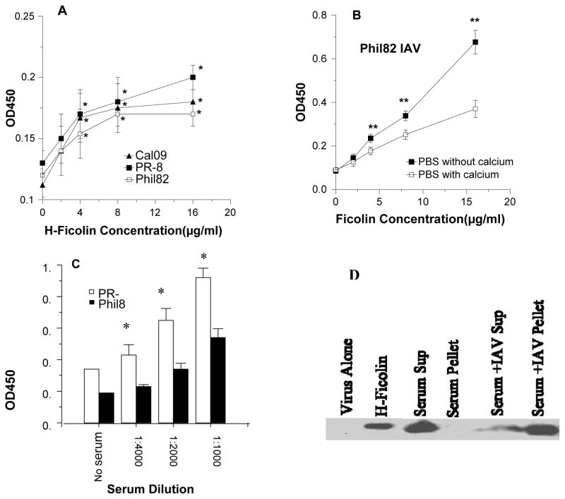
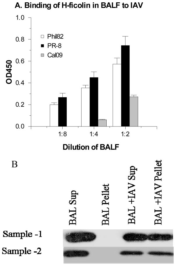
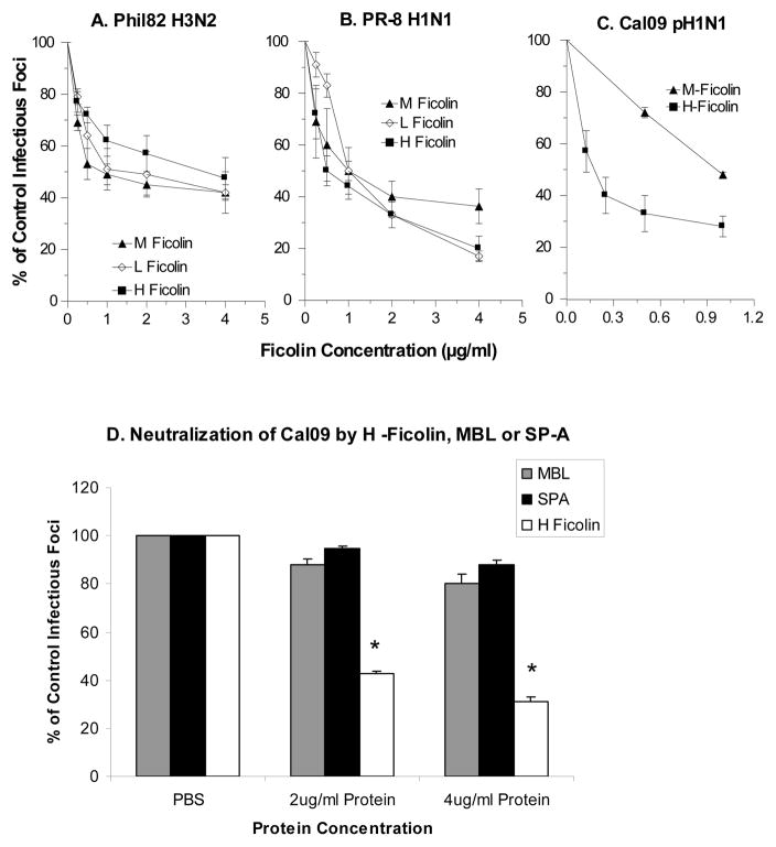
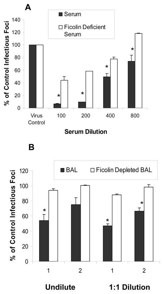
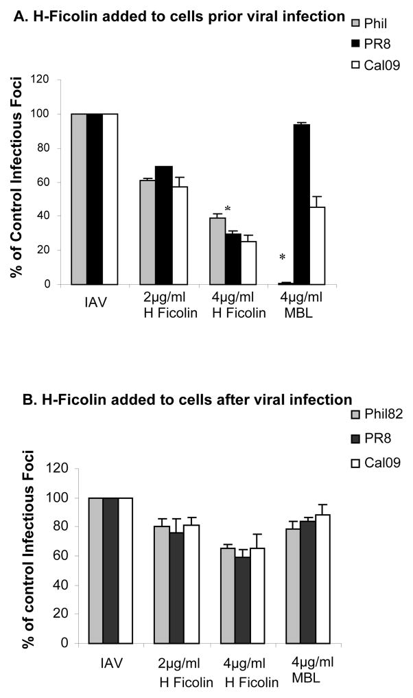
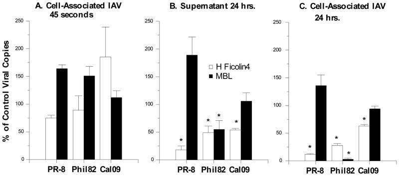
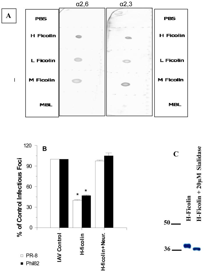
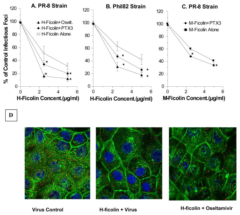
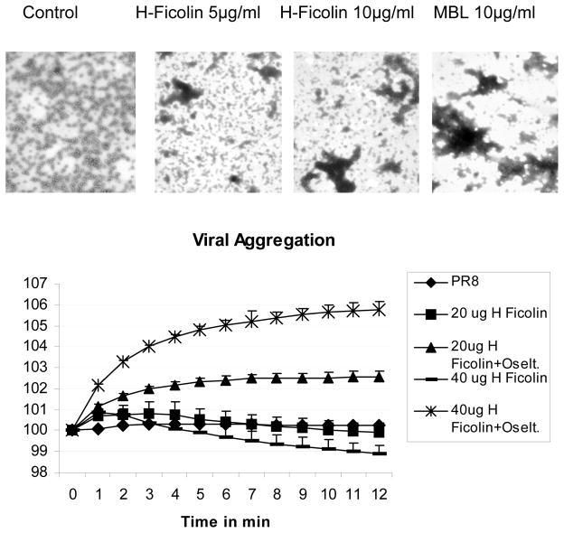
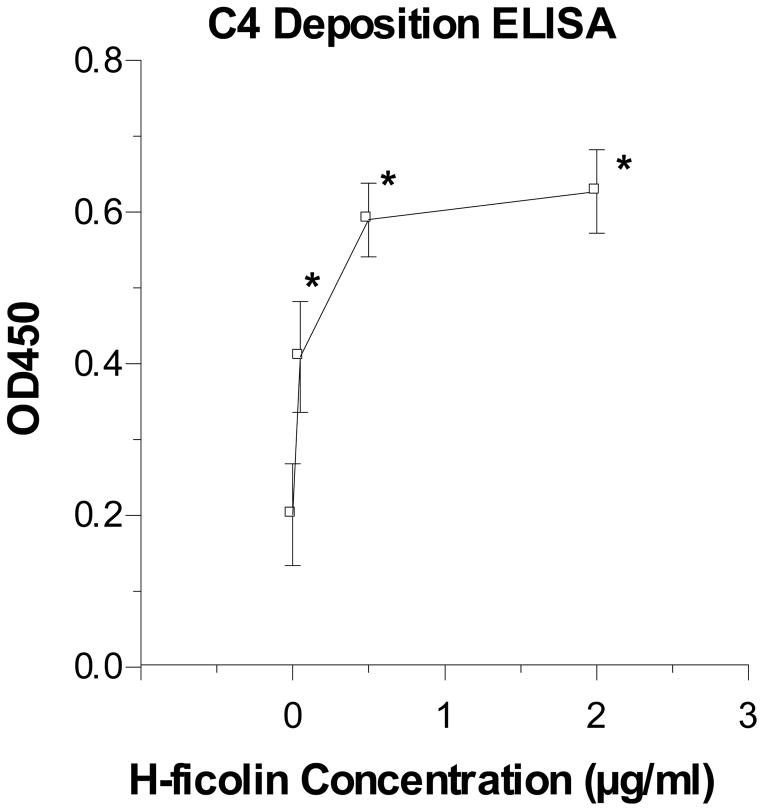
The collectins have been shown to have a role in host defense against influenza A virus (IAV) and other significant viral pathogens (e.g., HIV). The ficolins are a related group of innate immune proteins that are present at relatively high concentrations in serum, but also in respiratory secretions; however, there has been little study of the role of ficolins in viral infection. In this study, we demonstrate that purified recombinant human H-ficolin and H-ficolin in human serum and bronchoalveolar lavage fluid bind to IAV and inhibit viral infectivity and hemagglutination activity in vitro. Removal of ficolins from human serum or bronchoalveolar lavage fluid reduces their antiviral activity. Inhibition of IAV did not involve the calcium-dependent lectin activity of H-ficolin. We demonstrate that H-ficolin is sialylated and that removal of sialic acid abrogates IAV inhibition, while addition of the neuraminidase inhibitor oseltamivir potentiates neutralization, hemagglutinin inhibition, and viral aggregation caused by H-ficolin. Pandemic and mouse-adapted strains of IAV are generally not inhibited by the collectins surfactant protein D or mannose binding lectin because of a paucity of glycan attachments on the hemagglutinin of these strains. In contrast, H-ficolin inhibited both the mouse-adapted PR-8 H1N1 strain and a pandemic H1N1 strain from 2009. H-ficolin also fixed complement to a surface coated with IAV. These findings suggest that H-ficolin contributes to host defense against IAV.


Figures

Figure 1
Figure 1. Binding of recombinant and human serum H-ficolin to IAV
In panel A ELISA plates were coated with three IAV strains indicated (California 2009 H1N1, PR-8 1934 H1N1, and Philippines 1982 H3N2) and then incubated with increasing concentrations of recombinant H-ficolin. Background binding to BSA coated plates was subtracted from the values shown. There was significant binding (p<0.05; indicated by *) of H-ficolin to all viruses at all the doses shown. Panel B shows binding of H-ficolin to the PR-8 strain of IAV in presence or absence of calcium and magnesium. Binding was significantly greater in the absence of calcium and magnesium. Panel C shows results of incubation of various dilutions of human serum with similar ELISA plates, followed by antibody detection of bound H-ficolin. Here again there was significant binding of H-ficolin at all dilutions shown, although binding to PR-8 was significantly greater than binding to Phil82. Results are mean±SEM of 4 experiments. Panel D shows a Western blot of H-ficolin in serum. In this experiment the serum was incubated with the PR-8 strain of IAV or in control buffer, followed by centrifugation to pellet the virus. Supernatant and pellet samples (resuspended in the same amount of buffer) were analyzed by Western blot. This experiment is representative of 3 similar experiments.
Figure 2
Figure 2. Binding of H-ficolin in human BAL fluid to IAV
Binding was demonstrated using two methods. In panel A, BAL fluid was incubated with IAV coated plates and binding of H-ficolin in BAL fluid was detected by ELISA. Binding was significant (p<0.05 for all tested dilutions of BAL fluid). Results are mean±SEM of 4 experiments. In panel B, virus (PR-8 strain) was incubated with BAL fluid for 45 min followed by centrifugation to pellet virus particles. The presence of H-ficolin in the supernatant and pellet was demonstrated by Western blot. Results are representative of 3 experiments.
Figure 3
Figure 3. Neutralization of IAV strains by ficolins
Viral neutralization was tested using a fluorescent focus assay and MDCK cells as described. The number of fluorescent (infected) cells was counted after 7 hours of infection. Diluted viral strains were incubated with control buffer (PBS with 2mM calcium and magnesium) or different concentrations of ficolins, MBL, or SP-A, followed by addition of these samples to cells. The multiplicity of infection (MOI) for these experiments was 1. In panels A and B, M-, L-, and H-ficolin were all tested for their ability to inhibit the Phil82 (panel A) or PR-8 (panel B) strains of IAV. In panel C, H-ficolin was tested for its activity against Cal09. All ficolins caused significant inhibition of all IAV strains at the concentrations tested (p<0.05 vs control). Panel D shows an additional set of experiments in which the activity of H-ficolin was compared to that of human SP-A (native protein from BAL) or recombinant human MBL. H-ficolin caused significantly greater inhibition of Cal09 than either SP-A or MBL at the concentrations tested (* indicates significant difference by ANOVA). Results are mean±SEM of 4 experiments.
Figure 4
Figure 4. Effect of ficolin depletion on viral neutralizing activity of human serum or BAL fluid
Normal donor human serum or BAL fluid was incubated with N-Acetyl-D-Galactosamine-Agarose as described to remove ficolins and the serum or BALF pre- or post-depletion were compared for viral neutralizing activity using the infectious focus assay and PR-8. The serum or BALF was pre-incubated with the virus for 45min followed by titration on MDCK cells. Panel A shows results obtained with serum from one donor at the indicated dilutions. Panel B shows results obtained with BALF from two separate donors (labeled 1 and 2). The BALF was used either undiluted or at 1:1 dilution in PBS as indicated. The results are mean±SEM of 3 experiments with each dilution of serum or BALF. * indicates were depleted BALF or serum had significantly less neutralizing activity than the untreated samples
Figure 5
Figure 5. Effect of adding H-ficolin to MDCK cells before or after virus infection on neutralizing activity
The neutralization assay was performed as in figure 3 except that in panel A the MDCK cells were pre-incubated with the ficolin prior to infection with IAV, and in panel B ficolin was added to MDCK cells after viral infection. (In figure 3 virus was pre-incubated with ficolins prior to infection of MDCK cells). Pre-incubation of MDCK cells with H-ficolin caused comparable inhibition of viral infection with the three indicated viral strains as in figure 3. Results with human MBL are shown for comparison. H-ficolin caused significantly greater inhibition of the PR-8 strain than MBL, while MBL caused significantly greater inhibition of the Phil82 strain than H-ficolin at 4μg/ml (* indicates these differences as assessed by ANOVA). Results in panel B show that addition of H-ficolin or MBL after viral infection for 45 min causes much less inhibition than found in panel A or in figure 3. Note, however, that H-ficolin still caused significant inhibition compared to control using this method (p<0.05 for all viruses at 4μg/ml). Results are mean±SEM of 4 experiments.
Figure 6
Figure 6. Effect of recombinant H-ficolin or MBL on viral uptake and replication in A549 cells as assessed by RT-PCR
The indicated viral strains were pre-incubated with H-ficolin or MBL followed by infection of A549 cells for 45 min. The MOI used in these experiments was 1 as in the infectious focus assays. Cell associated virus or virus in culture supernatant were then measured using RT-PCR to detect the viral RNA encoding M protein. To assess viral uptake cell associated virus was measured just after completion of viral infection (45 min) in panel A. Neither H-ficolin nor MBL reduced the amount of cell-associated virus at this time point. In contrast, H-ficolin significantly reduced the amount of virus detectable in the culture supernatant (panel B) or cells (panel C) for all viruses tested (* indicates p<0.05 vs control). MBL only caused significant inhibition of the Phil82 strain in panels B and C. Results are mean±SEM of 4 experiments.
Figure 7
Figure 7. Presence of α(2,3) or α(2,6)-linked sialic acids on recombinant ficolins and effect of neuraminidase treatment of H-ficolin on antiviral activity
In panel A, the presence of α(2,3) or α(2,6)-linked sialic acids on recombinant H-, L-, and M-ficolins was assessed by lectin blotting using SNA and MAA as sialic acid detecting molecules as described in methods. For comparison, MBL (which has no N-linked oligosaccharide attachments) was tested as well. In panel B, untreated H-ficolin, neuraminidase treated H-ficolin, or neuraminidase alone was pre-incubated with PR-8 or Phil82 IAV followed by infection of MDCK cells with the virus samples and infectious focus assay. Only untreated H-ficolin significantly inhibited viral infectivity (* indicates p<0.01 vs control). Panel C shows SDS-PAGE of untreated and neuraminidase treated H-ficolin. Results in panel B are mean±SEM of 4 experiments.
Figure 8
Figure 8. Effect of oseltamivir or PTX-3 on viral neutralizing activity of H-ficolin or on effects of H-ficolin on viral attachment or uptake by A549 cells as assessed with confocal microscopy
Panel A–C show results of infectious focus assays using H- or M-ficolin alone or combined with either oseltamivir (5μg/ml) or PTX-3 (5μg/ml). Results in panel A–C are mean±SEM of 4 experiments. Addition of both oseltamivir and PTX3 significantly increased neutralizing activity compared to H-ficolin alone at either the 2.5 or 5μg/ml concentration of H-ficolin. This was true both for the Phil82 (panel A) or PR-8 (panel B) strains of IAV. PTX3 or oseltamivir alone did not reduce viral infectivity in this assay. The results for PTX3 are shown at the zero concentration of ficolins in the curves labeled PTX3. PTX3 significantly increased the neutralizing activity of M-ficolin as shown in panel C. Panel D shows confocal microscopic pictures of virus (red) after 45 min incubation with A549 cells (cell membrane green and nucleus blue). The MOI for the confocal experiments was 200 (i.e., higher than in infectious focus and Q-PCR assays). Results are representative of three experiments.
Figure 9
Figure 9. Viral aggregation induced by H-ficolin or MBL
Panel A shows representative (of 4 experiments) electron microscopic images of PR-8 IAV alone (control) vs. IAV pre-treated with the indicated concentrations of H ficolin or MBL. In these experiments, 10μg/ml of H-ficolin caused a similar degree of viral aggregation as MBL. In panel B the ability of H-ficolin to cause viral aggregation was also tested by light absorbance assay. In this assay increased light transmission results from viral aggregation. This assay is less sensitive for detecting viral aggregation than EM; however, 20μg/ml and 40μg/ml of H-ficolin did cause significant increase in light transmission two minutes after addition to the viral suspension. Addition of oseltamivir (10μg/ml) during incubation of IAV with H-ficolin resulted in much more pronounced and sustained viral aggregation. No aggregation was seen with oseltamivir alone or with the PR-8 virus alone (black diamonds in panel B). Results in panel B are mean±SEM of 4 experiments.
Figure 10
Figure 10. H-ficolin/MASP-2 complexes fix complement in presence of IAV
ELISA plates were coated with IAV as in figure 1. H-ficolin/MASP-2 complexes were purified from human serum and incubated with the surface bound IAV as described in “Materials and Methods”. Complement fixation was detected using antibody to complement component C4. H-ficolin caused dose related deposition of C4 onto the viral surface (n=5; * indicates p<0.05 vs. IAV-coated plates incubated with complement in the absence of H-ficolin. As a positive control 2μg/ml of the H-ficolin/MASP-2 complex was incubated with wells coated with acetylated BSA. This resulted in fixation of complement as reported (mean±SEM OD450 for C4 was 1.2±0.1).

Similar articles

Cited by

References

    1. Thiel S. Complement activating soluble pattern recognition molecules with collagen-like regions, mannan-binding lectin, ficolins and associated proteins. Mol Immunol. 2007;44:3875–3888. - PubMed
    1. Garred P, Borregaard N. The ficolins. J Innate Immun. 2009;2:1–2. - PubMed
    1. Akaiwa M, Yae Y, Sugimoto R, Suzuki SO, Iwaki T, Izuhara K, Hamasaki N. Hakata antigen, a new member of the ficolin/opsonin p35 family, is a novel human lectin secreted into bronchus/alveolus and bile. J Histochem Cytochem. 1999;47:777–786. - PubMed
    1. Zacho RM, Jensen L, Terp R, Jensenius JC, Thiel S. Studies of the pattern recognition molecule H-ficolin: specificity and purification. J Biol Chem. 2012;287:8071–8081. - PMC - PubMed
    1. Sallenbach S, Thiel S, Aebi C, Otth M, Bigler S, Jensenius JC, Schlapbach LJ, Ammann RA. Serum concentrations of lectin-pathway components in healthy neonates, children and adults: mannan-binding lectin (MBL), M-, L-, and H-ficolin, and MBL-associated serine protease-2 (MASP-2) Pediatr Allergy Immunol. 2011;22:424–430. - PubMed

Publication types

MeSH terms