. 2012 Sep 1;189(5):2290-9.
doi: 10.4049/jimmunol.1103495. Epub 2012 Jul 30.

An NKp30-based chimeric antigen receptor promotes T cell effector functions and antitumor efficacy in vivo

Affiliations

An NKp30-based chimeric antigen receptor promotes T cell effector functions and antitumor efficacy in vivo

Tong Zhang et al. J Immunol. .

Abstract

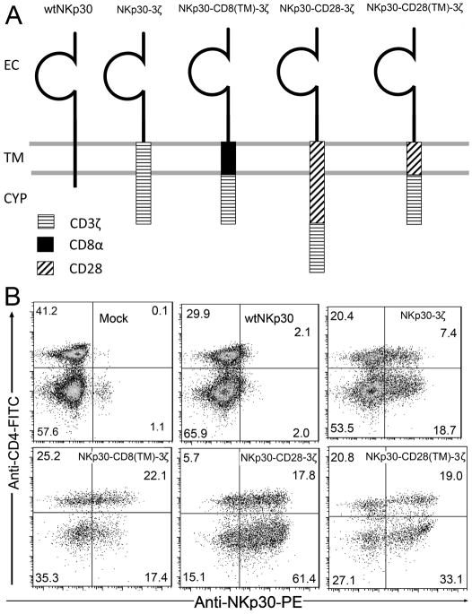
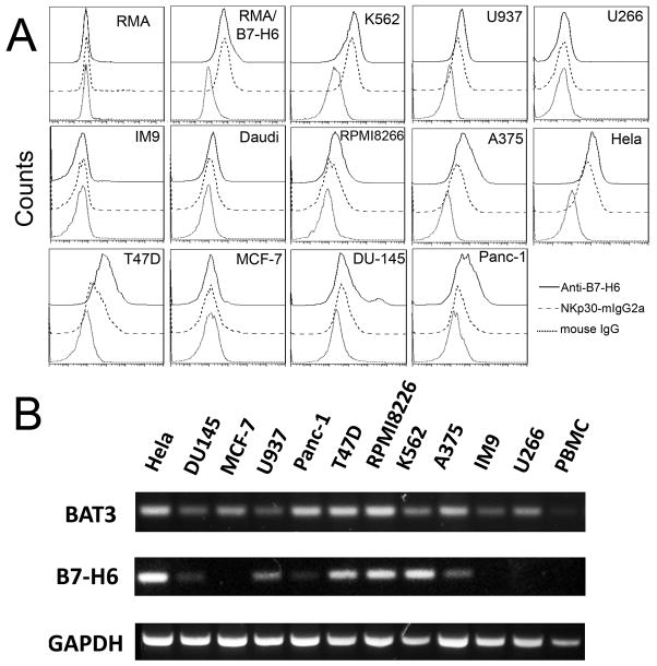
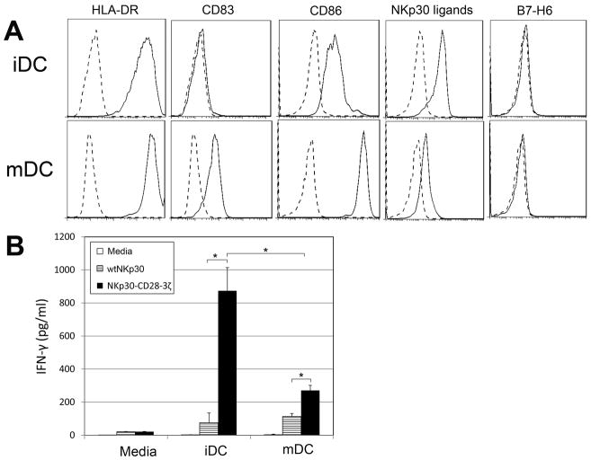
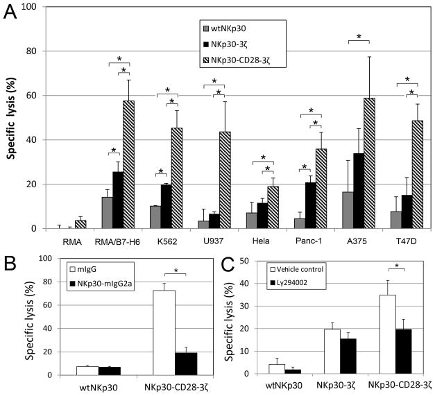
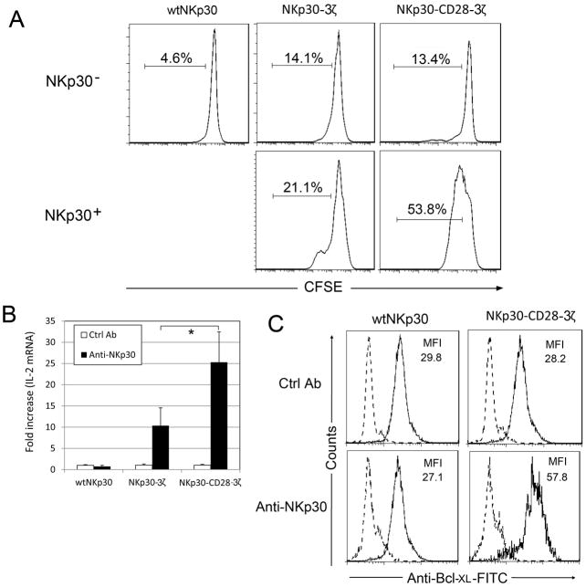
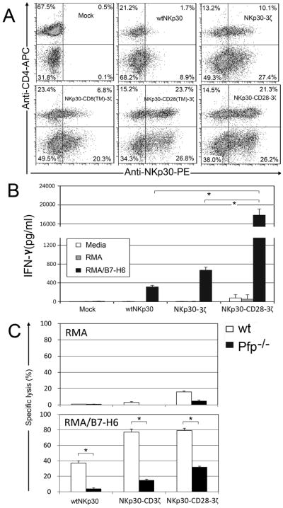
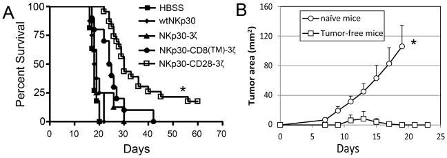
NKp30 is a natural cytotoxicity receptor that is expressed on NK cells and recognizes B7-H6, which is expressed on several types of tumors but few normal cells. To target effector T cells against B7-H6+ tumors, we developed several chimeric AgRs (CARs) based on NKp30, which contain the CD28- and/or CD3ζ-signaling domains with the transmembrane domains from CD3ζ, CD28, or CD8α. The data show that chimeric NKp30-expressing T cells responded to B7-H6+ tumor cells. The NKp30 CAR-expressing T cells produced IFN-γ and killed B7-H6 ligand-expressing tumor cells; this response was dependent upon ligand expression on target cells but not on MHC expression. PBMC-derived dendritic cells also express NKp30 ligands, including immature dendritic cells, and they can stimulate NKp30 CAR-bearing T cells to produce IFN-γ, but to a lesser extent. The addition of a CD28-signaling domain significantly enhanced the activity of the NKp30 CAR in a PI3K-dependent manner. Adoptive transfer of T cells expressing a chimeric NKp30 receptor containing a CD28-signaling domain inhibited the growth of a B7-H6-expressing murine lymphoma (RMA/B7-H6) in vivo. Moreover, mice that remained tumor-free were resistant to a subsequent challenge with the wild-type RMA tumor cells, suggesting the generation of immunity against other tumor Ags. Overall, this study demonstrates the specificity and therapeutic potential of adoptive immunotherapy with NKp30 CAR-expressing T cells against B7-H6+ tumor cells in vivo.


Figures

Figure 1
Figure 1. Structure and expression of NKp30 CARs
Schematic diagram of wild type (wt) and chimeric NKp30 proteins are shown in (A). The extracellular (EC), transmembrane (TM) and cytoplasmic regions (CYP) are indicated. In the NKp30-3ζ, the CD3ζ chain (TM plus CYP domains) was fused to the EC domain of the NKp30 molecule. The TM domains of CD8α and CD28 were inserted between NKp30 EC and CD3ζ CYP domains to make NKp30-CD8(TM)-3ζ and NKp30-CD28(TM)-3ζ, respectively. NKp30-CD28-3ζ receptor contains the human CD28 TM and CYP domains in between the NKp30 EC and CD3ζ CYP domains. (B) NKp30 expression on human T cells 7 days after transduction with NKp30 CARs. NKp30 expression was measured using the PE-conjugated anti-NKp30 mAbs in combination with anti-CD4-FITC mAbs. More than 99% of cells were CD3+ T cells (data not shown). CD4- T cells are CD8+ T cells. The data are representative of three experiments.
Figure 2
Figure 2. Human tumor cell lines express varying amounts of NKp30 ligands on the cell surface
(A) NKp30 ligand expression on the surface of human tumor cell lines was measured by flow cytometry using anti-B7H6 mAbs (solid line) or a soluble NKp30 receptor fused to a mouse IgG2a Fc region (NKp30-mIgG2a, dashed line) followed by staining with APC-conjugated goat anti-mouse IgG. Isotype controls are shown as a dotted line. (B) Detection of NKp30 ligands B7-H6 and BAT3 by RT-PCR. mRNA expression of human NKp30 ligands B7-H6 and BAT3 in human cell lines and human PBMCs were determined by RT-PCR. GAPDH was used as an internal control.
Figure 3
Figure 3. NKp30 CAR-modified T cells respond to NKp30 ligand positive cells by producing IFN-γ
Five to seven days after retroviral transduction, NKp30 CAR-modified T cells (105 cells) were co-cultured with either irradiated or mitomycin-C treated tumor cells for 24 h. (A) Suspension tumor cells (105 cells) and (B) adherent tumor cells (2.5×10⁴ cells) were used. RMA and MCF-7 cells were used as negative controls. IFN-γ amounts in the supernatants were analyzed with ELISA. Results are shown in mean + SD. *: p<0.05
Figure 4
Figure 4. Human DCs bind to NKp30 and can stimulate autologous NKp30-CD28-3ζ-modified T cells to produce IFN-γ
(A) The cell surface phenotype and binding to NKp30 of PBMC-derived human DCs (both iDCs and mDCs) was determined by flow cytometry. Specific mAb or NKp30-Ig as indicated (solid line) or an isotype control Ab staining (dashed line) are shown. (B) Five to seven days after retroviral transduction, NKp30 CAR-modified T cells (105 cells) were co-cultured with either iDCs or mDCs cells at a ratio of 5:1 (T: DC) for 24 h. IFN-γ amounts in the supernatants were determined by ELISA. Results shown (mean + SD) are representative of two experiments shown. *: p<0.05
Figure 5
Figure 5. NKp30 CAR-modified T cells lyse NKp30 ligand-positive tumor cells in vitro
(A) Effector T cells derived from human PBMCs were modified with either wtNKp30(grey), NKp30-3ζ (black) or NKp30-CD28-3ζ(thatch) were cocultured with tumor cells at a ratio of 5:1 in 5-hr LDH release assays. The data are presented as mean + SD of triplicates from 2 independent experiments. (B) Effector T cells modified with wtNKp30 or NKp30-CD28-3ζ were cocultured with target cells K562 in the presence of 10μg/ml NKp30-mIgG2a (■) or mouse IgG (□) at a ratio of 5:1, and percent specific lysis determined after a 5-hr LDH release assay. The data are presented as mean + SD and are representative of 2 independent experiments. (C) PI3-kinase is involved in NKp30-CD28-3ζ receptor-mediated cytotoxicity. NKp30-modified effector T cells were preincubated with a PI3-kinase inhibitor LY294002 (10 μM) at 37°C for 1 h before co-culture with K562 target cells at a E:T ratio of 5:1 in 5-hr LDH release assays. Vehicle controls are 0.1% DMSO. The data shown are presented as mean + SD of triplicates and are representative of two independent experiments. *: p<0.05
Figure 6
Figure 6. Engagement of NKp30-CD28-3ζ receptor led to increased T cell proliferation and up-regulation of IL-2 and Bcl-XL
(A) NKp30 receptor (either wtNKp30 or chNKp30)-modified human T cells were labeled with CFSE as described in Methods and Materials and were cocultured with Hela cells (105 cells, NKp30 ligand-positive) in the presence of a low amount of IL-2 (25 U/ml) for 3 days. Analysis of T cell proliferation (i.e. CFSE dilution) was performed on both NKp30+ (FL4) and NKp30 cells within the same mixed T cell population by flow cytometry. (B) NKp30-modified T cells (2.5×10⁵ cells) were cultured in anti-NKp30 mAb (4μg)-coated 24-well plates for 24 h. Mouse IgG was used as a negative control. IL-2 gene expression was determined by real-time PCR as described in Methods and Materials. Results are shown as fold increase, in which the IL-2 gene expression in the control mAb-treated T cells was normalized to 1. Data are presented as mean + SD from two independent experiments. *: p<0.05 (C) Twenty-four hours after cross-linking with immobilized anti-NKp30 mAbs, as described above, T cells were collected. Bcl-XL expression was determined by flow cytometry after intracellular staining with anti-Bcl-XL-FITC (solid line) or isotype control mAbs (broken line).
Figure 7
Figure 7. Human NKp30 CARs can be expressed and function on murine T cells
(A) Human NKp30 expression on mouse T cells 7 days after transduction. NKp30 expression was detected using the PE-conjugated anti-NKp30 mAb in combination with anti-mouse CD4-FITC mAb. CD4-T cells are CD8+ T cells. The data are representative of three experiments. (B) Seven days after retroviral transduction, NKp30-modified T cells (105 cells) were co-cultured with irradiated RMA/B7-H6 cells (105 cells) for 24 h. Mouse lymphoma cell line RMA used as a negative control. IFN-γ amounts in the supernatants were analyzed by ELISA. Results are shown in mean + SD. (C) Effector T cells derived from WT B6 (open bar) and perforin deficient (Pfp−/−, filled) mice that were modified with either wtNKp30, NKp30-3ζ or NKp30-CD28-3ζ were cocultured with RMA or RMA/B7-H6 cells, respectively, at a ratio of 1:1 in 5-hr LDH release assays. The data are presented as mean + SD of triplicates from a representative experiment of two independent experiments. *: p<0.05
Figure 8
Figure 8. Adoptive transfer of NKp30-CD28-3ζ-modified T cells promotes the survival of RMA/B7-H6 tumor-bearing mice and induces memory responses
(A) B6 mice were inoculated with RMA/B7-H6 (105 cells, i.v., day 0). Five days after tumor inoculation, mice were treated with either wtNKp30-(◆, n=8), NKp30-3ζ-( ▲, n=8), NKp30-CD8(TM)-3ζ-( ●, n=10) or NKp30-CD28-3ζ (□, n=19)-modified T cells (5×10⁶ cells). T-cell transfer was repeated on days 7 and 9. HBSS (■, n=11) treatment was used as negative control. Data pooled from two independent experiments are presented in Kaplan-Meier survival curves. *: p<0.01. (B) Tumor free mice (□) in the NKp30-CD28-3ζ-treated RMA lymphoma model (shown in panel A) and age-matched naïve mice (○) were re-challenged with wild type RMA cells (104 cells, s.c.) into the left flank. The tumor areas are represented as Mean + SEM. *: p<0.01.

References

    1. Rossig C, Brenner MK. Genetic modification of T lymphocytes for adoptive immunotherapy. Mol Ther. 2004;10:5–18. - PubMed
    1. Ho WY, Blattman JN, Dossett ML, Yee C, Greenberg PD. Adoptive immunotherapy: engineering T cell responses as biologic weapons for tumor mass destruction. Cancer Cell. 2003;3:431–437. - PubMed
    1. Porter DL, Levine BL, Kalos M, Bagg A, June CH. Chimeric antigen receptor-modified T cells in chronic lymphoid leukemia. N Engl J Med. 2011;365:725–733. - PMC - PubMed
    1. Kalos M, Levine BL, Porter DL, Katz S, Grupp SA, Bagg A, June CH. T cells with chimeric antigen receptors have potent antitumor effects and can establish memory in patients with advanced Leukemia. Sci Transl Med. 2011;3:95ra73. - PMC - PubMed
    1. Park TS, Rosenberg SA, Morgan RA. Treating cancer with genetically engineered T cells. Trends Biotechnol. 2011;29:550–557. - PMC - PubMed

Publication types

MeSH terms