. 2012 Oct;80(10):3533-44.
doi: 10.1128/IAI.00550-12. Epub 2012 Jul 30.

Vaccine-induced th17 cells are maintained long-term postvaccination as a distinct and phenotypically stable memory subset

Affiliations

Vaccine-induced th17 cells are maintained long-term postvaccination as a distinct and phenotypically stable memory subset

Thomas Lindenstrøm et al. Infect Immun. 2012 Oct.

Abstract

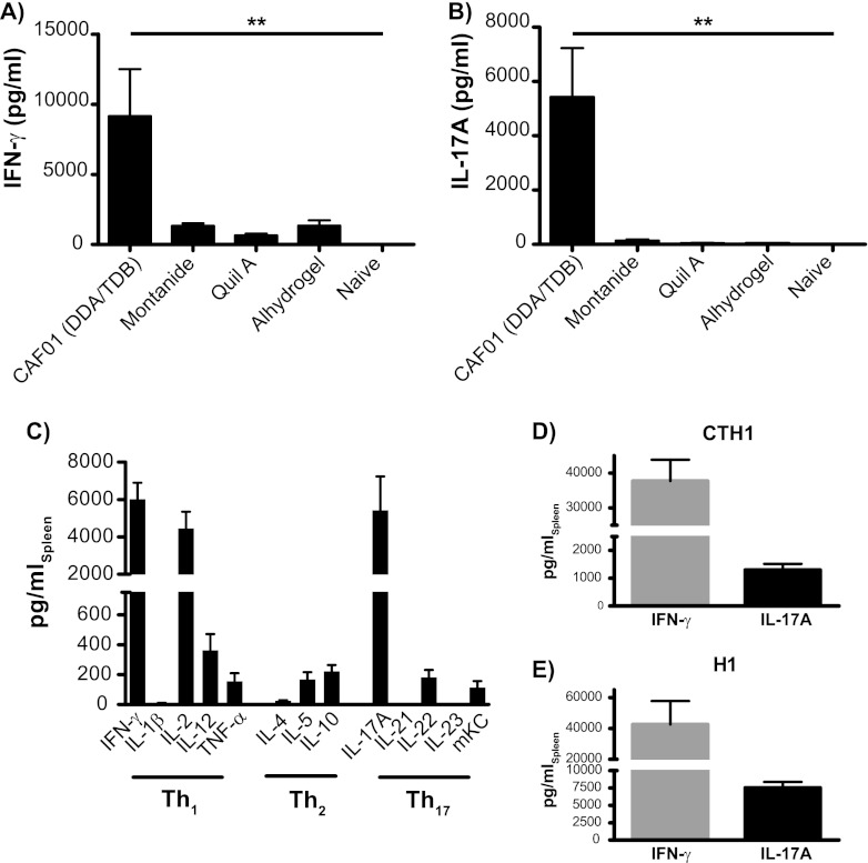
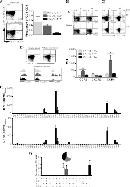
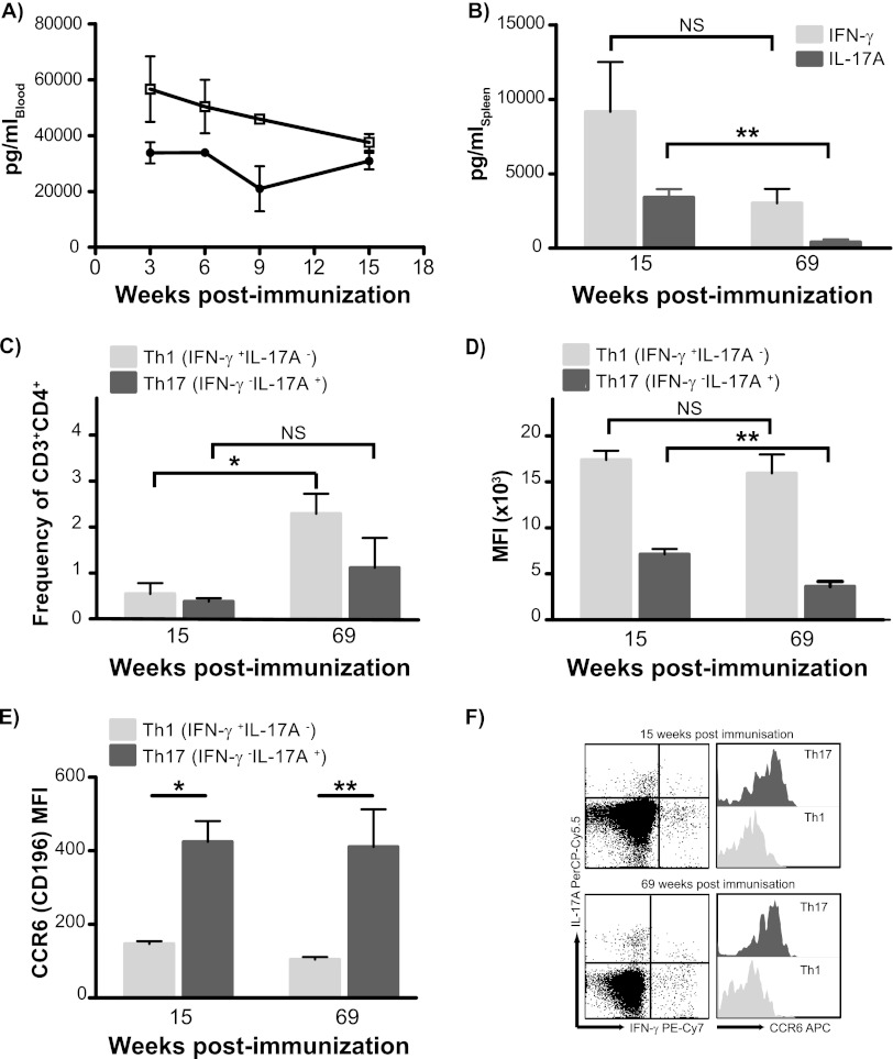
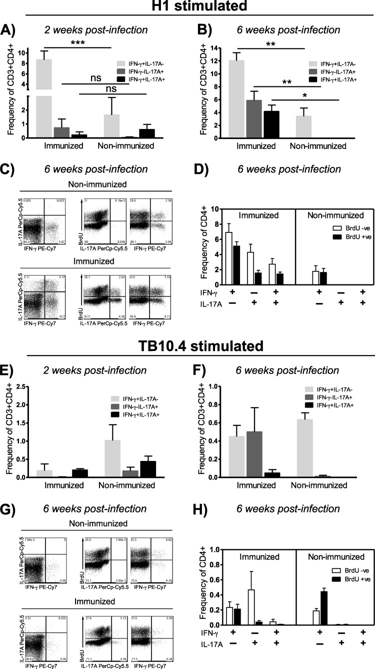
Th17 cells are increasingly being recognized as an important T helper subset for immune-mediated protection, especially against pathogens at mucosal ports of entry. In several cases, it would thus be highly relevant to induce Th17 memory by vaccination. Th17 cells are reported to exhibit high plasticity and may not stably maintain their differentiation program once induced, questioning the possibility of inducing durable Th17 memory. Accordingly, there is no consensus as to whether Th17 memory can be established unless influenced by continuous Th17 polarizing conditions. We have previously reported (T. Lindenstrøm, et al., J. Immunol. 182:8047-8055, 2009) that the cationic liposome adjuvant CAF01 can prime both Th1 and Th17 responses and promote robust, long-lived Th1 memory. Here, we demonstrate that subunit vaccination in mice with CAF01 leads to establishment of bona fide Th17 memory cells. Accordingly, Th17 memory cells exhibited lineage stability by retaining both phenotypic and functional properties for nearly 2 years. Antigen-specific, long-term Th17 memory cells were found to be mobilized from lung-draining lymph nodes to the lung following an aerosol challenge by Mycobacterium tuberculosis nearly 2 years after their induction and proliferated at levels comparable to those of Th1 memory cells. During the infection, the vaccine-induced Th17 memory cells expanded in the lungs and adapted Th1 characteristics, implying that they represent a metastable population which exhibits plasticity when exposed to prolonged Th1 polarizing, inflammatory conditions such as those found in the M. tuberculosis-infected lung. In the absence of overt inflammation, however, stable bona fide Th17 memory can indeed be induced by parenteral immunization.


Figures

Fig 1
Fig 1
Immunization with CAF01 leads to responses dominated by expression of Th1 and Th17 cytokines. Groups of four C57BL/6 mice were immunized s.c. three times with 2 μg H28 plus the indicated adjuvant. Levels of Ag-specific IFN-γ (A) and IL-17A (B) were measured by ELISA in splenocyte cultures 13 weeks after immunization. Data are means ± standard errors (SE). **, P < 0.01 by one-way ANOVA using Tukey's posttest. The experiment was performed twice with comparable results. (C) Levels of antigen-specific cytokine release were measured 13 weeks after immunization by either MSD multiplex analysis or conventional ELISA (IFN-γ, IL-17A, IL-21, IL-22, and IL-23 [p19/40]); data are means ± SE. (D) Groups of four C57BL/6 mice were immunized s.c. three times with 10 μg CTH1 plus CAF01. Levels of antigen-specific IFN-γ and IL-17A were measured 2 weeks after immunization by conventional ELISA. Data are means ± SE. (E) Groups of four C57BL/6 mice were immunized s.c. three times with 2 μg H1 plus CAF01. Three weeks postimmunization, levels of antigen-specific IFN-γ and IL-17A were measured as described above. Data are means ± SE and are representative of three repetitive experiments.
Fig 2
Fig 2
CAF01 induces distinct Th1 and Th17 subsets with functional differences. Groups of four C57BL/6 mice received three immunizations with 2 μg H28 plus CAF01. (A) Fifteen weeks after immunization, the frequency of Th1 (IFN-γ+ IL-17A; light gray bars), Th17 (IFN-γ IL-17A+; dark gray bars), and Th1/Th17 (IFN-γ+ IL-17A+; black bars) within the CD3+ CD4+ population was determined by intracellular flow cytometry (ICS). The left panels show representative plots of IL-17A versus IFN-γ expression in CD4 lymphocytes (singlets > lymphocytes > CD3+ > CD4+) from unstimulated and H28 stimulated splenocytes. The experiment was independently performed three times with similar results. (B) Distinct Th1 and Th17 cell populations predominate postvaccination, regardless of ex vivo stimulation and the intracellular staining protocol used. Mice received 2 to 3 s.c. immunizations of 2 μg H1 plus CAF01. PBMCs pooled from 8 C57BL/6 mice after 22 weeks were stimulated with H1 (top) or were left unstimulated for either 1 h (left) or 4 h (right) before 6 h of incubation in the presence of brefeldin A (BFA). (C) Splenocytes pooled from 4 B6 × BALB/c F1 mice 3 weeks postimmunization were stimulated with H1 for either 1 h (left) or 4 h (right) before 5 or 12 h of incubation in the presence of BFA. Representative plots of gated CD4 T cells are shown. (D) Expression levels of CCR6, CXCR3, and CCR4 were determined in H28-specific Th1 (IFN-γ+ IL-17A; light gray), Th17 (IFN-γ IL-17A+; dark gray), and Th1/Th17 (IFN-γ+ IL-17A+; black) cells within the CD3+ CD4+ lymphocyte gate by their geometric mean fluorescent intensity. The left panels show representative plots of IL-17A versus IFN-γ expression in CD4 lymphocytes (singlets > lymphocytes > CD3+ > CD4+) from unstimulated and H28-stimulated splenocytes and histogram overlays of chemokine receptor expression of H28-specific Th1, Th17, and Th1/Th17 subsets. Geometric mean MFI values of each marker were compared by one-way ANOVA with Tukey's posttest (right). *, P < 0.05; **, P < 0.01. The graph is representative of three independent experiments. (E) Splenocytes from 3 individual mice were restimulated with overlapping peptides spanning the entire Ag85B protein. Levels of secreted IFN-γ and IL-17A were subsequently measured by ELISA. Data are means ± SE. Peptides recognized by Th1 and Th17 included P30/P31 (SSFYSDWYSPACGKA/DWYSPACGKAGCQTY), P36/P37 PQWLSANRAVKPTGS/ANRAVKPTGSAAIGL), P63 QDAYNAAGGHNAVFN), and P67 (THSWEYWGAQLNAMK). (F) Cytokine coexpression profiles in vaccinated mice 15 weeks postimmunization. By Boolean gating, cytokine-producing cells (IL-17A, TNF-α, IL-21, and IL-22) within the CD3+ CD4+ population were divided into 15 distinct subpopulations based on their production of these cytokines in any combination, and their frequency was determined. The relative contribution of each of these subpopulations to the responding T cell population is shown by the inserted pie chart. The experiment was performed three times with comparable results.
Fig 3
Fig 3
CAF01-induced Th17 cells are established as a distinct and stable memory subset. (A) C57BL/6 mice received 3 s.c. immunizations of 2 μg H28 plus CAF01 spaced 2 weeks apart. Blood was obtained at weeks 3, 6, 9, and 15 after immunization by submandibular bleeding. PBMCs pooled from 8 mice were stimulated with H28 at a concentration of 0.5 μg/ml, and cell culture supernatants were harvested after 72 h of incubation. The levels of secreted antigen-specific IFN-γ and IL-17A were determined by capture ELISA in triplicate readings; data are means ± standard deviations. Open squares, IFN-γ; black circles, IL-17A. Groups of four mice received three immunizations with 2 μg H28 plus CAF01, and Th1 versus Th17 memory responses were measured at short (15 weeks) and long (69 weeks) intervals after vaccination. (B) Release of IL-17A and IFN-γ from spleen cells isolated after H28 immunization was determined by ELISA; data are means ± SE. P values were calculated using Student's t test (*, P < 0.05; **, P < 0.01). (C) The frequency of Th1 (IFN-γ+ IL-17A; light gray bars) and Th17 (IFN-γ IL-17A+; dark gray bars) at short (15 weeks) and long (69 weeks) intervals after vaccination was determined by ICS. The percentage of each subpopulation within the CD3+ CD4+ gated lymphocyte population was determined. Significant differences were calculated by two-way ANOVA with Bonferroni's posttest (*, P < 0.05). (D) Expression levels of IL-17A in H28-specific Th17 (IFN-γ IL-17A+; dark gray bars) and of IFN-γ in Th1 (IFN-γ+ IL-17A; light gray bars) cells within the CD3+ CD4+ lymphocyte gate were determined in four individual mice by their geometric mean fluorescent intensity at both short (15 weeks) and long (69 weeks) intervals. Differences in mean MFI values over time within each population were compared by Student's t test. **, P < 0.01. (E) Expression levels of CCR6 were determined in H28-specific Th1 (IFN-γ+ IL-17A; light gray bars) and Th17 (IFN-γ IL-17A+; dark gray bars) cells within the CD3+ CD4+ lymphocyte gate by their geometric mean fluorescent intensity. Significant differences in MFI values were calculated by two-way ANOVA with Bonferroni's posttest. *, P < 0.05; **, P < 0.01. (F) Representative plots of distinctive antigen-specific Th1 (IFN-γ+ IL-17A) and Th17 (IFN-γ IL-17A+) cells and histogram overlays of their CCR6 expression levels at 15 and 69 weeks postimmunization. This experiment was repeated once with comparable results, though with H1 (Ag85B-ESAT-6) as the antigen.
Fig 4
Fig 4
Long-term Th17 memory cells are rapidly mobilized from lung-draining lymph nodes following challenge with Mycobacterium tuberculosis. (A and B) C57BL/6 mice were immunized three times with 2 μg H1 plus CAF01 and 89 weeks later were aerosol challenged with M. tuberculosis Erdman. Mice were euthanized 2 weeks into the infection, and H1-specific Th1 and Th17 responses were determined by ICS in both immunized (3 mice) and aged-matched, nonimmunized animals (4 mice). (A) The frequency of Th1 (IFN-γ+ IL-17A; light gray bars), Th17 (IFN-γ IL-17A+; dark gray bars), and Th1/Th17 (IFN-γ+ IL-17A+; black bars) cells mobilized from the tracheobronchial lymph nodes was determined by ICS in both long-term memory and aged-matched, nonimmunized mice. (B) Expression levels of CCR6 were determined by geometric CCR6 mean fluorescent intensity in infection-mobilized, H1-specific Th1 (IFN-γ+ IL-17A; light gray bars), Th17 (IFN-γ IL-17A+; dark gray bars), and Th1/Th17 (IFN-γ+ IL-17A+; black bars) cells within the CD3+ CD4+ lymphocyte gate from the tracheobronchial lymph nodes of memory immune mice. Differences were found to be nonsignificant (ns; one-way ANOVA). (C to E) C57BL/6 mice were immunized three times with 2 μg H1 plus CAF01 and ∼2 years later were aerosol challenged with M. tuberculosis Erdman. Mice were euthanized 6 weeks into the infection, and H1-specific Th1 and Th17 responses and their proliferative capacity were determined by ICS in pooled tracheobronchial lymph nodes from both immunized and aged-matched, nonimmunized animals. (C) IFN-γ versus IL-17A expression in pooled lung-draining lymph node CD4+ T cells after stimulation with H1, medium, and phorbol myristate acetate-ionomycin. Numbers denote the frequency of CD4+ T cells. (D) Proportion of BrdU-incorporating H1-specific Th17 (left plots) versus Th1 (right plots) CD4 T cells from lung-draining lymph nodes 6 weeks into M. tuberculosis infection in nonimmunized (upper part) and immunized mice (lower part). Numbers denote the frequency of CD4+ T cells. (E) Histogram overlay showing the degree of BrdU incorporation (left) and CCR6 expression levels (right) in H1-specific Th1 (IFN-γ+ IL-17A) and Th17 (IFN-γ IL-17A+) CD4 T cells from tracheobronchial lymph nodes isolated from memory immune mice 6 weeks into infection.
Fig 5
Fig 5
Challenge with Mycobacterium tuberculosis mediates proliferation of long-term Th17 memory cells and recruitment to the lung, where they adopt Th1 cell characteristics as infection progresses into chronic stages. (A) C57BL/6 mice were immunized three times with 2 μg H1 plus CAF01, and 89 weeks later aerosol was challenged with M. tuberculosis Erdman. Mice were euthanized 2 weeks into the infection, and the frequency of H1-specific Th1 (IFN-γ+ IL-17A; light gray bars), Th17 (IFN-γ IL-17A+; dark gray bars), and Th1/Th17 (IFN-γ+ IL-17A+; black bars) cells in both immunized (3 mice) and aged-matched, nonimmunized animals (4 mice) were determined by ICS. Significant differences were calculated by two-way ANOVA with Bonferroni's posttest (***, P < 0.001). (B to H) C57BL/6 mice were immunized three times with 2 μg H1 plus CAF01 and ∼2 years later were aerosol challenged with M. tuberculosis Erdman. Mice were euthanized 6 weeks into the infection, and antigen-specific Th1 and Th17 responses and their proliferative capacity were determined by ICS in individual lungs from both immunized and aged-matched, nonimmunized animals (3 mice/group). (B) Frequency of H1-specific CD3+ CD4+ Th1 (IFN-γ+ IL-17A; light gray bars), Th17 (IFN-γ IL-17A+; dark gray bars), and Th1/Th17 (IFN-γ+ IL-17A+; black bars) cells from the lung 6 weeks into infection in both long-term memory and aged-matched, nonimmunized mice. Significant differences were calculated by two-way ANOVA with Bonferroni's posttest (***, P < 0.001; **, P < 0.01; *, P < 0.05). (C) Representative plots showing IL-17A versus IFN-γ expression (left) and the proportion of BrdU incorporating H1-specific Th17 (middle) and Th1 (right) cells 6 weeks into infection. (D) Mean frequencies of H1-specific Th1 (IFN-γ+ IL-17A), Th17 (IFN-γ IL-17A+), and Th1/Th17 (IFN-γ+ IL-17A+) subsets being either BrdU negative or BrdU positive 6 weeks into challenge. (E) Frequency of TB10.4-specific Th1 (IFN-γ+ IL-17A; light gray bars), Th17 (IFN-γ IL-17A+; dark gray bars), and Th1/Th17 (IFN-γ+ IL-17A+; black bars) cells 2 weeks after infection (ns, not significant by two-way ANOVA). (F) Frequency of TB10.4-specific Th1 (IFN-γ+ IL-17A; light gray bars), Th17 (IFN-γ IL-17A+, dark gray bars), and Th1/Th17 (IFN-γ+ IL-17A+; black bars) cells 6 weeks into infection. (G) Representative plots showing IL-17A versus IFN-γ expression (left) and the proportion of BrdU incorporating TB10.4-specific Th17 (middle) and Th1 (right) cells 6 weeks into infection. (H) Mean frequencies of TB10.4-specific Th1 (IFN-γ+ IL-17A), Th17 (IFN-γ IL-17A+), and Th1/Th17 (IFN-γ+ IL-17A+) subsets being either BrdU negative or BrdU positive 6 weeks into challenge.

References

    1. Aagaard C, et al. 2011. A multistage tuberculosis vaccine that confers efficient protection before and after exposure. Nat. Med. 17:189–194 - PubMed
    1. Acosta-Rodriguez EV, et al. 2007. Surface phenotype and antigenic specificity of human interleukin 17-producing T helper memory cells. Nat. Immunol. 8:639–646 - PubMed
    1. Agger EM, et al. 2008. Cationic liposomes formulated with synthetic mycobacterial cordfactor (CAF01): a versatile adjuvant for vaccines with different immunological requirements. PLoS One 3:e3116 doi:10.1371/journal.pone.0003116 - DOI - PMC - PubMed
    1. Aujla SJ, et al. 2008. IL-22 mediates mucosal host defense against Gram-negative bacterial pneumonia. Nat. Med. 14:275–281 - PMC - PubMed
    1. Chen K, et al. 2011. Th17 cells mediate clade-specific, serotype-independent mucosal immunity. Immunity 35:997–1009 - PMC - PubMed

Publication types

LinkOut - more resources