

Intraductal neoplasm of the intrahepatic bile duct: clinicopathological study of 24 cases
- PMID: 22851859
- PMCID: PMC3406419
- DOI: 10.3748/wjg.v18.i28.3673
Intraductal neoplasm of the intrahepatic bile duct: clinicopathological study of 24 cases
Abstract
Aim: To investigate the clinicopathological features of intraductal neoplasm of the intrahepatic bile duct (INihB).
Methods: Clinicopathological features of 24 cases of INihB, which were previously diagnosed as biliary papillomatosis or intraductal growth of intrahepatic biliary neoplasm, were reviewed. Mucin immunohistochemistry was performed for mucin (MUC)1, MUC2, MUC5AC and MUC6. Ki-67, P53 and β-catenin immunoreactivity were also examined. We categorized each tumor as adenoma (low grade), borderline (intermediate grade), and malignant (carcinoma in situ, high grade including tumors with microinvasion).
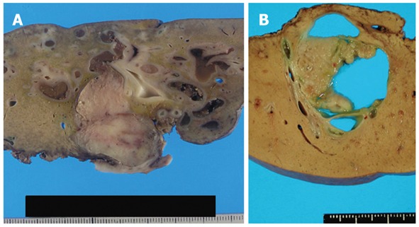
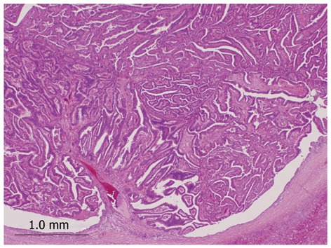
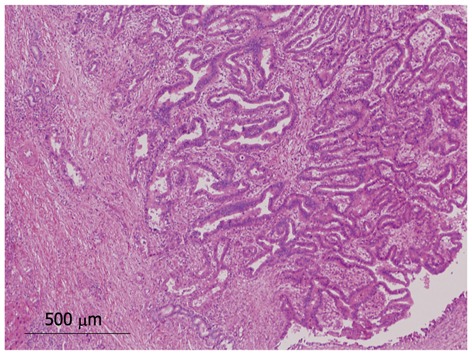
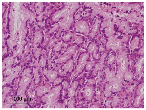
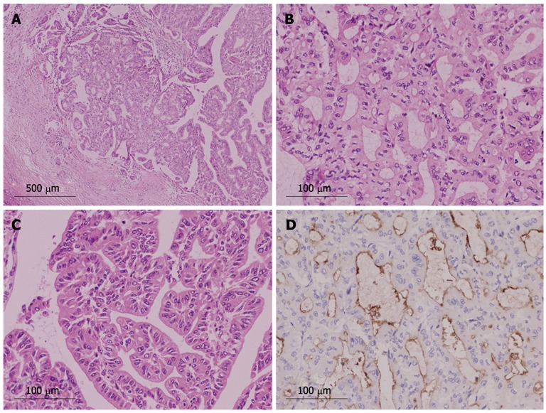
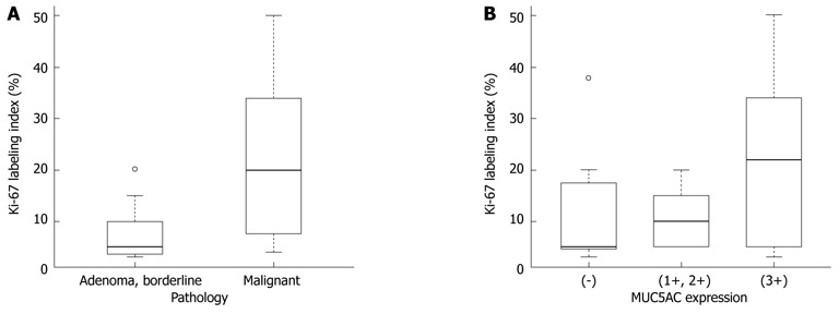
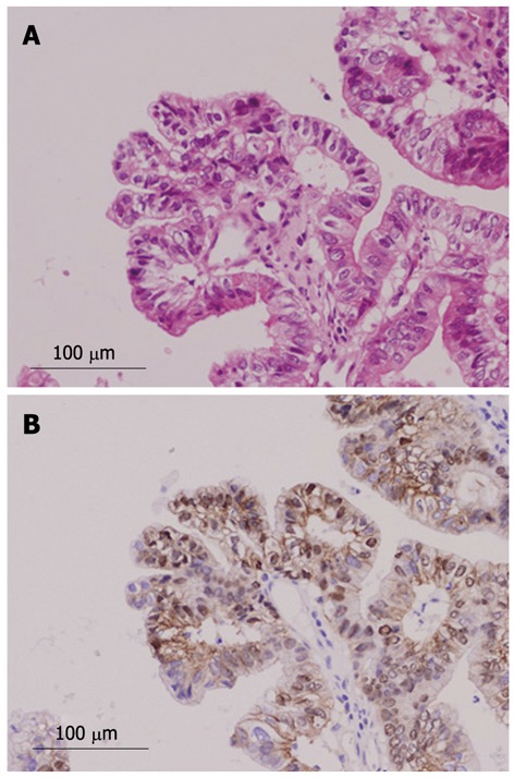
Results: Among 24 cases of INihB, we identified 24 tumors. Twenty of 24 tumors (83%) were composed of a papillary structure; the same feature observed in intraductal papillary neoplasm of the bile duct (IPNB). In contrast, the remaining four tumors (17%) showed both tubular and papillary structures. In three of the four tumors (75%), macroscopic mucin secretion was limited but microscopic intracellular mucin was evident. Histologically, 16 tumors (67%) were malignant, three (12%) were borderline, and five (21%) were adenoma. Microinvasion was found in four cases (17%). Immunohistochemical analysis revealed that MUC1 was not expressed in the borderline/adenoma group but was expressed only in malignant lesions (P = 0.0095). Ki-67 labeling index (LI) was significantly higher in the malignant group than in the borderline/adenoma group (22.2 ± 15.5 vs 7.5 ± 6.3, P < 0.01). In the 16 malignant cases, expression of MUC5AC showed borderline significant association with high Ki-67 LI (P = 0.0622). Nuclear expression of β-catenin was observed in two (8%) of the 24 tumors, and these two tumors also showed MUC1 expression. P53 was negative in all tumors.
Conclusion: Some cases of INihB have a tubular structure, and are subcategorized as IPNB with tubular structure. MUC1 expression in INihB correlates positively with degree of malignancy.
Keywords: Intraductal biliary neoplasm; Intraductal papillary neoplasm of the bile duct; Intraductal tubular neoplasm of the bile duct; Intraductal tubulopapillary neoplasm of the bile duct; Mucin expression.
Figures
References
-
- Ohtsubo K, Ohta H, Sakai J, Mouri H, Nakamura S, Ikeda T, Kifune K, Yoshikawa J, Harada K, Nakanuma Y, et al. Mucin-producing biliary papillomatosis associated with gastrobiliary fistula. J Gastroenterol. 1999;34:141–144. - PubMed
-
- Lim JH, Kim YI, Park CK. Intraductal mucosal-spreading mucin-producing peripheral cholangiocarcinoma of the liver. Abdom Imaging. 2000;25:89–92. - PubMed
-
- Sakamoto E, Hayakawa N, Kamiya J, Kondo S, Nagino M, Kanai M, Miyachi M, Uesaka K, Nimura Y. Treatment strategy for mucin-producing intrahepatic cholangiocarcinoma: value of percutaneous transhepatic biliary drainage and cholangioscopy. World J Surg. 1999;23:1038–1043; discussion 1043-1044. - PubMed
-
- Kokubo T, Itai Y, Ohtomo K, Itoh K, Kawauchi N, Minami M. Mucin-hypersecreting intrahepatic biliary neoplasms. Radiology. 1988;168:609–614. - PubMed
-
- Kim HJ, Kim MH, Lee SK, Yoo KS, Park ET, Lim BC, Park HJ, Myung SJ, Seo DW, Min YI. Mucin-hypersecreting bile duct tumor characterized by a striking homology with an intraductal papillary mucinous tumor (IPMT) of the pancreas. Endoscopy. 2000;32:389–393. - PubMed
MeSH terms
Substances
LinkOut - more resources
Full Text Sources
Medical
Research Materials
Miscellaneous
