

Dysplasia of the hip in adolescent patients successfully treated for developmental dysplasia of the hip
- PMID: 22852032
- PMCID: PMC3234894
- DOI: 10.1007/s11832-011-0356-0
Dysplasia of the hip in adolescent patients successfully treated for developmental dysplasia of the hip
Abstract
Background: The purpose of this study was to analyze whether hips treated for developmental dysplasia of the hip (DDH) during infancy, which were clinically and radiologically fully normalized by walking age, may become dysplastic again during later growth.
Materials and methods: A total of 150 patients were randomly selected out of a collective of 386 patients treated for DDH at the Department of Orthopaedics at the University of Zurich between 1993 and 2004. Treatment was started at birth and continued for 6 months. All patients had clinically and radiographically normal hips by walking age. The patients did not suffer from other diseases, in particular, neurological or neuromuscular diseases.
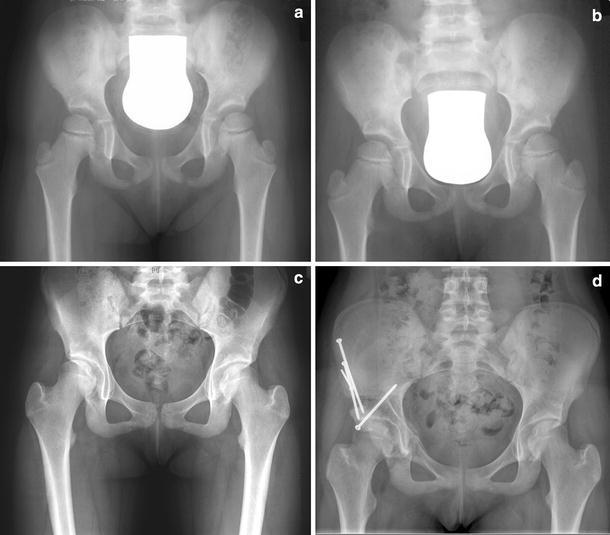
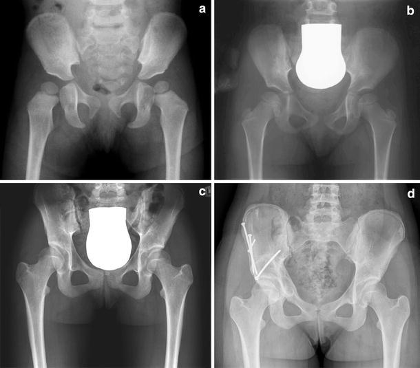
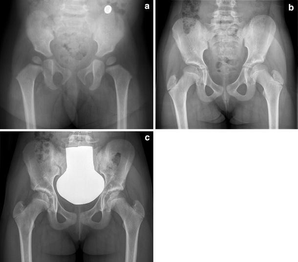
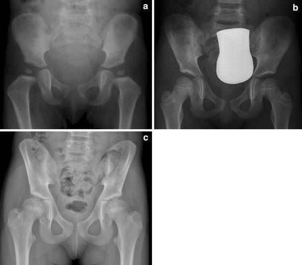
Results: We detected four female subjects among the 150 patients who had been successfully treated for DDH, who had developed dysplastic hips in early adolescence, necessitating acetabular surgery.
Conclusion: The successful treatment of DDH in infancy does not ensure normal hip development; therefore, follow up into maturity may be recommended.
Level of evidence: Level IV.
Keywords: Congenital dysplasia of the hip; Developmental dysplasia of the hip; Pavlik harness.
Figures
Similar articles
-
Late acetabular dysplasia after successful treatment for developmental dysplasia of the hip using the Pavlik method: A systematic literature review.J Orthop. 2018 Dec 4;16(1):5-10. doi: 10.1016/j.jor.2018.11.001. eCollection 2019 Jan-Feb. J Orthop. 2018. PMID: 30765927 Free PMC article. Review.
-
Developmental dysplasia of the hip in neonates: evolution of acetabular dysplasia after hip stabilization by brief Pavlik harness treatment.Orthop Traumatol Surg Res. 2014 Jun;100(4):357-61. doi: 10.1016/j.otsr.2014.03.017. Epub 2014 May 3. Orthop Traumatol Surg Res. 2014. PMID: 24797045
-
Success of Pavlik Harness Treatment Decreases in Patients ≥ 4 Months and in Ultrasonographically Dislocated Hips in Developmental Dysplasia of the Hip.Clin Orthop Relat Res. 2016 May;474(5):1146-52. doi: 10.1007/s11999-015-4388-5. Clin Orthop Relat Res. 2016. PMID: 26047647 Free PMC article.
-
Higher Pavlik Harness Treatment Failure Is Seen in Graf Type IV Ortolani-positive Hips in Males.Clin Orthop Relat Res. 2016 Aug;474(8):1847-54. doi: 10.1007/s11999-016-4776-5. Epub 2016 Mar 14. Clin Orthop Relat Res. 2016. PMID: 26975383 Free PMC article.
-
Cochrane Review: Screening programmes for developmental dysplasia of the hip in newborn infants.Evid Based Child Health. 2013 Jan;8(1):11-54. doi: 10.1002/ebch.1891. Evid Based Child Health. 2013. PMID: 23878122 Review.
Cited by
-
Late acetabular dysplasia after successful treatment for developmental dysplasia of the hip using the Pavlik method: A systematic literature review.J Orthop. 2018 Dec 4;16(1):5-10. doi: 10.1016/j.jor.2018.11.001. eCollection 2019 Jan-Feb. J Orthop. 2018. PMID: 30765927 Free PMC article. Review.
-
Children treated for developmental dysplasia of the hip at birth and with normal acetabular index at 1 year: How many had residual dysplasia at 5 years?J Child Orthop. 2022 Jun;16(3):183-190. doi: 10.1177/18632521221106376. Epub 2022 Jun 30. J Child Orthop. 2022. PMID: 35800653 Free PMC article.
-
Morphological differences between residual childhood hip dysplasia with previous osteotomy and adolescent-onset hip dysplasia.J Orthop Surg Res. 2025 Mar 13;20(1):271. doi: 10.1186/s13018-025-05655-w. J Orthop Surg Res. 2025. PMID: 40075506 Free PMC article.
-
Hip dysplasia in the young adult caused by residual childhood and adolescent-onset dysplasia.Curr Rev Musculoskelet Med. 2016 Dec;9(4):427-434. doi: 10.1007/s12178-016-9369-0. Curr Rev Musculoskelet Med. 2016. PMID: 27613709 Free PMC article. Review.
-
Developmental dysplasia of the hip: What has changed in the last 20 years?World J Orthop. 2015 Dec 18;6(11):886-901. doi: 10.5312/wjo.v6.i11.886. eCollection 2015 Dec 18. World J Orthop. 2015. PMID: 26716085 Free PMC article. Review.
References
-
- Graf R. Hip sonography: 20 years experience and results. Hip Int. 2007;17(Suppl 5):S8–S14. - PubMed
-
- Exner GU, Kern SM. Natural course of mild hip dysplasia from young childhood into adulthood. Orthopade. 1994;23(3):181–184. - PubMed
-
- Exner GU, Schams M (1999) Later development of hip dysplasia after breech presentation with normal hips at birth in otherwise healthy children. Abstract, EPOS, Göteborg
LinkOut - more resources
Full Text Sources
Research Materials
