Review
. 2012 Jul;4(7):1144-68.
doi: 10.3390/v4071144. Epub 2012 Jul 24.

Influenza virus-mediated membrane fusion: determinants of hemagglutinin fusogenic activity and experimental approaches for assessing virus fusion

Affiliations
Review

Influenza virus-mediated membrane fusion: determinants of hemagglutinin fusogenic activity and experimental approaches for assessing virus fusion

Brian S Hamilton et al. Viruses. 2012 Jul.

Abstract

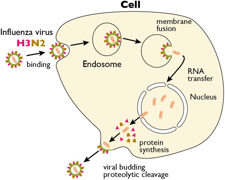
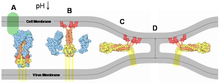
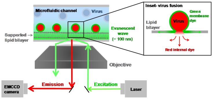
Hemagglutinin (HA) is the viral protein that facilitates the entry of influenza viruses into host cells. This protein controls two critical aspects of entry: virus binding and membrane fusion. In order for HA to carry out these functions, it must first undergo a priming step, proteolytic cleavage, which renders it fusion competent. Membrane fusion commences from inside the endosome after a drop in lumenal pH and an ensuing conformational change in HA that leads to the hemifusion of the outer membrane leaflets of the virus and endosome, the formation of a stalk between them, followed by pore formation. Thus, the fusion machinery is an excellent target for antiviral compounds, especially those that target the conserved stem region of the protein. However, traditional ensemble fusion assays provide a somewhat limited ability to directly quantify fusion partly due to the inherent averaging of individual fusion events resulting from experimental constraints. Inspired by the gains achieved by single molecule experiments and analysis of stochastic events, recently-developed individual virion imaging techniques and analysis of single fusion events has provided critical information about individual virion behavior, discriminated intermediate fusion steps within a single virion, and allowed the study of the overall population dynamics without the loss of discrete, individual information. In this article, we first start by reviewing the determinants of HA fusogenic activity and the viral entry process, highlight some open questions, and then describe the experimental approaches for assaying fusion that will be useful in developing the most effective therapies in the future.

Keywords: hemagglutinin; influenza virus; membrane fusion; protease; virus entry.


Figures

Figure 1
Figure 1
The replication cycle of influenza virus. Influenza virus binds to sialic acid groups present in the glycocalyx of the plasma membrane (glycocalyx omitted for clarity). The bound virus is subsequently endocytosed. Hemagglutinin (HA) is color-coded in red; Neuraminidase is color-coded brown. Ion channels and lipids are color-coded green. During maturation of the endosome, the pH drops, initiating the fusion of the viral envelope (green) with the endosomal membrane (gray) and the release of the viral RNA (orange) and viral proteins into the cytosol. The viral RNA traffics to the nucleus, where replication takes place. Newly formed viral RNAs are exported to the cytosol where they assemble with new virus structural proteins, which are packaged together at the plasma membrane, and bud off to form new virions.
Figure 2
Figure 2
Cleavage of the HA precursor (HA0) primes influenza HA for fusion activation. (A) Structure of the non-cleaved, trimeric A/South Carolina/1/18 HA (H1N1) (PDB code 1RD8) [35]. The cleavage site region is enlarged (inset) with the side chains of the cleavage site residues (IQSR) shown. Equivalent cleavage sites are shown to the right for human H1-H3 and H5 highly pathogenic avian influenza (HPAI), along with the P4-P1’ positions recognized by the protease (B) Schematic diagram of HA. Upon cleavage by a host cell protease, the HA precursor is divided into two functionalsubunits, an HA1(receptor-binding) domain and an HA2 (fusion) domain. The newly created N-terminus of the HA2 subunit is the first residue of the fusion peptide (FP), which is exposed upon subsequent conformational changes and activation of HA triggered by the low pH of the endosome.
Figure 3
Figure 3
HA-mediated membrane binding and fusion between the viral and endosomal membranes. (A) HA1 (blue) binding to a moiety containing sialic acid group on the plasma membrane (light green); (B) After a reduction in pH in the endosome, HA2 undergoes a conformational change that drives the fusion peptides (red) into the host cell membrane; (C) A further conformational change brings the outermost leaflets of the opposing membranes together to form a stalk (D), where it is thought to be the action of several fusogenic HA2 working in concert. The dashed lines divide the upper and lower leaflets of the membranes for clarity. (Figure adapted from the Protein Data Bank [89]). Eventually the stalk collapses to form a pore (not shown).
Figure 4
Figure 4
Total internal reflection fluorescence microscopy integrated with a microfluidic device. A coherent laser source can be steered through an inverted microscope objective at or above the critical angle for total internal reflection as dictated by Snell’s law, generating an evanescent wave at the glass-buffer interface. A supported lipid bilayer adsorbed to the walls of the microfluidic device will reside within this evanescent wave. Fluorescently-labeled viruses bound to the supported bilayer will be excited and emit a red signal. The emitted light is sent back to a sensitive camera for imaging. The addition of a second laser line or more allows multiple fluorophores to be monitored simultaneously, for example, one to mark the viral membrane and another to mark the internal contents. Note that the size of the virus with respect to the bilayer is not drawn to scale. The bilayer is ~4 nm thick, while the virus is typically ~100 nm in diameter. (Inset) Upon acidification of the microfluidic channel, virus fusion commences. A two color virus labeling scheme distinguished the hemifusion step (green) from pore formation (red).

Similar articles

Cited by

References

    1. Horimoto T., Kawaoka Y. Influenza: Lessons from past pandemics, warnings from current incidents. Nat. Rev. Microbiol. 2005;3:591–600. - PubMed
    1. Palese P., Shaw M.L. Orthomyxoviridae: The Viruses and Their Replication. In: Knipe D.M., editor. Fields Virology. Lippincott Williams and Wilkins; Philadelphia, PA, USA: 2007.
    1. Fouchier R.A.M., Munster V., Wallensten A., Bestebroer T.M., Herfst S., Smith D., Rimmelzwaan G.F., Olsen B., Osterhaus A.D.M.E. Characterization of a novel influenza a virus hemagglutinin subtype (H16) obtained from black-headed gulls. J. Virol. 2005;79:2814–2822. - PMC - PubMed
    1. Nelson M.I., Holmes E.C. The evolution of epidemic influenza. Nat. Rev. Genet. 2007;8:196–205. - PubMed
    1. Medina R.A., Garcia-Sastre A. Influenza a viruses: New research developments. Nat. Rev. Microbiol. 2011;9:590–603. - PMC - PubMed

Publication types

MeSH terms

Substances