. 2012:2012:597915.
doi: 10.1155/2012/597915. Epub 2012 Jul 17.

Angiogenic signalling pathways altered in gliomas: selection mechanisms for more aggressive neoplastic subpopulations with invasive phenotype

Affiliations

Angiogenic signalling pathways altered in gliomas: selection mechanisms for more aggressive neoplastic subpopulations with invasive phenotype

Susana Bulnes et al. J Signal Transduct. 2012.

Abstract

The angiogenesis process is a key event for glioma survival, malignancy and growth. The start of angiogenesis is mediated by a cascade of intratumoural events: alteration of the microvasculature network; a hypoxic microenvironment; adaptation of neoplastic cells and synthesis of pro-angiogenic factors. Due to a chaotic blood flow, a consequence of an aberrant microvasculature, tissue hypoxia phenomena are induced. Hypoxia inducible factor 1 is a major regulator in glioma invasiveness and angiogenesis. Clones of neoplastic cells with stem cell characteristics are selected by HIF-1. These cells, called "glioma stem cells" induce the synthesis of vascular endothelial growth factor. This factor is a pivotal mediator of angiogenesis. To elucidate the role of these angiogenic mediators during glioma growth, we have used a rat endogenous glioma model. Gliomas induced by prenatal ENU administration allowed us to study angiogenic events from early to advanced tumour stages. Events such as microvascular aberrations, hypoxia, GSC selection and VEGF synthesis may be studied in depth. Our data showed that for the treatment of gliomas, developing anti-angiogenic therapies could be aimed at GSCs, HIF-1 or VEGF. The ENU-glioma model can be considered to be a useful option to check novel designs of these treatment strategies.


Figures
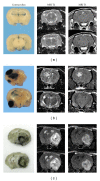
Figure 1
Figure 1
Brain coronal sections of Sprague Dawley rats exposed prenatally to ethylnitrosourea. Two columns show MRI on T1-w and T2-w after gadolinium administration and the left column shows the necropsy thirty minutes after Evans Blue i.v. injection (a, b) or Indian ink (c). (a) On T2-weighted images, tumours in the early development stage show a diminutive proliferation mass growing in association with the subcortical white matter. The blood brain barrier (BBB) remains still intact, shown by the lack of contrast or dye extravasation (microtumours). (b) Multiple tumours on the intermediate development stage. There is a BBB dysfunction indicated by extravasation of Evans Blue and by gadolinium enhancing the contrast on T1 images (macrotumours). (c) A macrotumour in the advanced stage growing over a whole hemisphere. It displays a heterogeneous signal on T2 and on T1 due to the presence of histopathological features of malignancy such as haemorrhages, cysts, and necrosis. With Indian ink, brains show in black a ring of aberrant vessels surrounding the neoplasia. The same typical shape of glioblastoma multiforme may be observed on MRI in T1 with gadolinium.
Figure 2
Figure 2
Confocal microphotographs of the microvascular network during development of an ENU-induced glioma. Histochemistry for tomato lectin (LEA-FITC) performed on 40 μm sections. (a) Similar angioarchitecture to the normal brain during the early stage. (b) Tortuous and dilated vessels during the intermediate development stage. (c) The microvascular network is mainly composed of glomeruloid vessels in the periphery area, (d) Occasional huge dilated vessels in the intratumour region (bar scale of 20 μm).
Figure 3
Figure 3
Expression of stem cell markers: CD133 (red) and Nestin (green), proangiogenic factor VEGF165 (red) and microvasculature markers: LEA lectin (green) and GluT-1 (red) during the ENU-glioma development. (a–f) MRI on T2-w and T1-w after gadolinium injection. (a, d) During the initial stage, the ENU-glioma grows in association with the subcortical white matter. It shows a homogeneous hyperintense signal on T2-w (a) and an isointense signal on T1-w (d). (b) ENU-intermediate stage corresponds with the “angiogenic switch.” (c, f) ENU-Glioblastoma displays heterogeneous hyperintense signal on T2 and on T1-w. (g-h) Overexpression of VEGF165 is shown in the intermediate (h) and advanced stages. (i) Also shown in perivascular cells of glomeruloid vessels. (j–l) CD133 expression is found following the intermediate stage (k). (l) ENU-GBM displays plenty of CD133+ cells in the intratumour hypoxic area and bordering the tumour. (m–o) Nestin+ cells are detected in every ENU development stage. Nestin+ cells are grouped into intratumour niches and around the microvessels. (Immunofluorescence images at 100x amplification, except I, l, and o at 40x).
Figure 4
Figure 4
Intermediate ENU glioma stage showing the expression of Nestin (green) and CD133 (red) by double immunofluorescence. (a–c) Intratumour niches of glioma stem cells positive for Nestin (a) and CD133 (b). (c) Both stem cell markers, Nestin and CD133 are coexpressed in some of these cells. (d–f) Isolated cells in the border of the tumour with a shape similar to astrocytes. Although the majority of these cells are Nestin-CD133 positive, there is a greater density of Nestin+ cells than CD133+ ones. (g–i) Perivascular niche of glioma stem cells displaying colocation of both markers.

Similar articles

Cited by

References

    1. Kleihues P, Burger PC, Aldape KD, et al. Glioblastoma. In: Louis DN, Ohgaki H, Wiestler OD, Cavenee WK, editors. WHO Classification of Tumours of the Central Nervous System. Lyon, France: Agency for Research on Cancer (IARC); 2007. pp. 33–49.
    1. Bergers G, Benjamin LE. Tumorigenesis and the angiogenic switch. Nature Reviews Cancer. 2003;3(6):401–410. - PubMed
    1. Carmeliet P, Jain RK. Angiogenesis in cancer and other diseases. Nature. 2000;407(6801):249–257. - PubMed
    1. Yancopoulos GD, Davis S, Gale NW, Rudge JS, Wiegand SJ, Holash J. Vascular-specific growth factors and blood vessel formation. Nature. 2000;407(6801):242–248. - PubMed
    1. Carmeliet P. Angiogenesis in health and disease. Nature Medicine. 2003;9(6):653–660. - PubMed