

Proteolytic Potential of the MSC Exosome Proteome: Implications for an Exosome-Mediated Delivery of Therapeutic Proteasome
- PMID: 22852084
- PMCID: PMC3407643
- DOI: 10.1155/2012/971907
Proteolytic Potential of the MSC Exosome Proteome: Implications for an Exosome-Mediated Delivery of Therapeutic Proteasome
Abstract
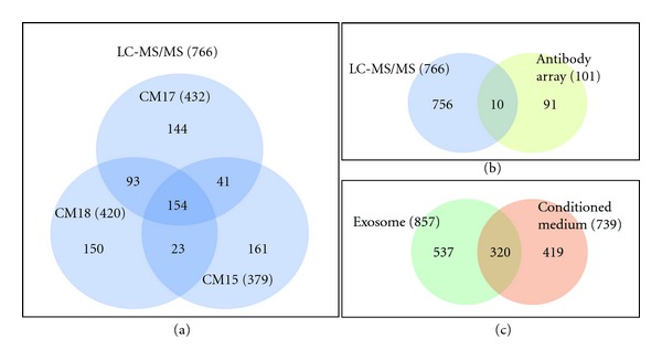
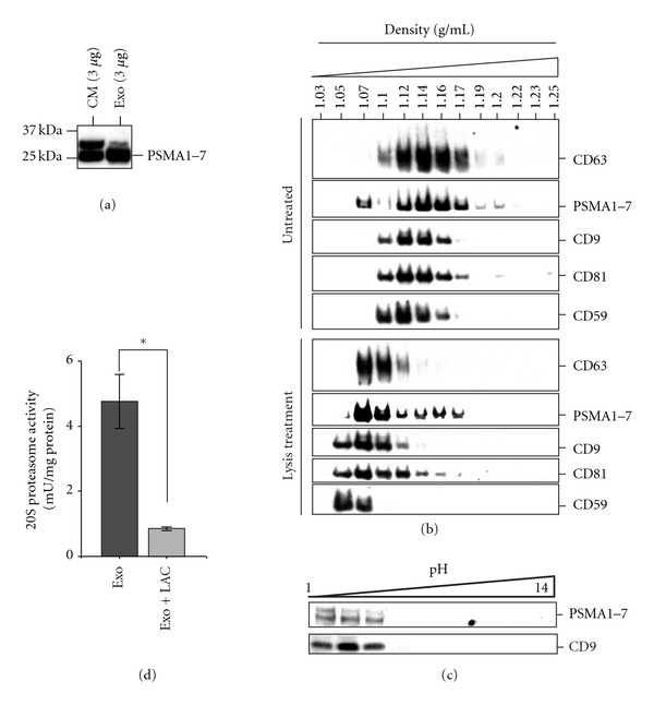
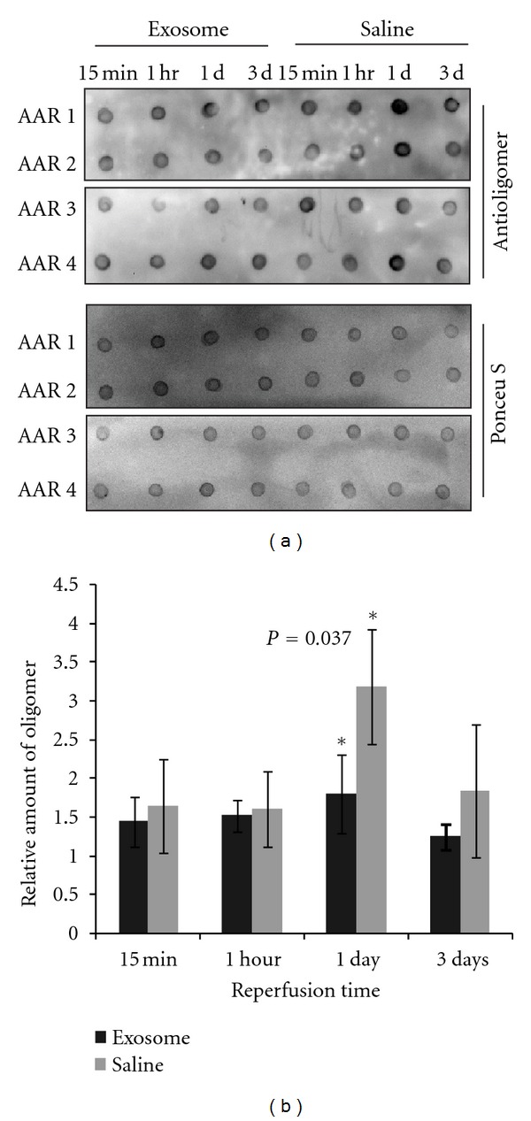
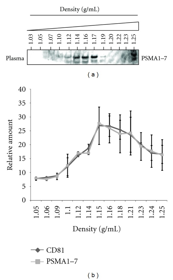
Mesenchymal stem cells (MSCs) are used in many of the current stem cell-based clinical trials and their therapeutic efficacy has increasingly been attributed to secretion of paracrine factors. We have previously demonstrated that a therapeutic constituent of this secretion is exosome, a secreted bilipid membrane vesicle of ~50-100 nm with a complex cargo that is readily internalized by H9C2 cardiomyocytes. It reduces infarct size in a mouse model of myocardial ischemia/reperfusion (MI/R) injury. We postulate that this therapeutic efficacy is derived from the synergy of a select permutation of individual exosome components. To identify protein candidates in this permutation, the proteome was profiled and here we identified 20S proteasome as a protein candidate. Mass spectrometry analysis detected all seven α and seven β chains of the 20S proteasome, and also the three beta subunits of "immunoproteasome" with a very high confidence level. We demonstrated that a functional proteasome copurified with MSC exosomes with a density of 1.10-1.18 g/mL, and its presence correlated with a modest but significant reduction in oligomerized protein in a mouse model of myocardial infarction. Circulating proteasomes in human blood also copurified with exosomes. Therefore, 20S proteasome is a candidate exosome protein that could synergize with other constituents to ameliorate tissue damage.
Figures
Similar articles
-
Self-assembling peptide modified with QHREDGS as a novel delivery system for mesenchymal stem cell transplantation after myocardial infarction.FASEB J. 2019 Jul;33(7):8306-8320. doi: 10.1096/fj.201801768RR. Epub 2019 Apr 10. FASEB J. 2019. PMID: 30970221
-
Exosome secreted by MSC reduces myocardial ischemia/reperfusion injury.Stem Cell Res. 2010 May;4(3):214-22. doi: 10.1016/j.scr.2009.12.003. Epub 2010 Jan 4. Stem Cell Res. 2010. PMID: 20138817
-
MSC exosome works through a protein-based mechanism of action.Biochem Soc Trans. 2018 Aug 20;46(4):843-853. doi: 10.1042/BST20180079. Epub 2018 Jul 9. Biochem Soc Trans. 2018. PMID: 29986939 Free PMC article. Review.
-
Mesenchymal stem cell exosome ameliorates reperfusion injury through proteomic complementation.Regen Med. 2013 Mar;8(2):197-209. doi: 10.2217/rme.13.4. Regen Med. 2013. PMID: 23477399 Review.
-
Therapeutic MSC exosomes are derived from lipid raft microdomains in the plasma membrane.J Extracell Vesicles. 2013 Dec 23;2. doi: 10.3402/jev.v2i0.22614. eCollection 2013. J Extracell Vesicles. 2013. PMID: 24371518 Free PMC article.
Cited by
-
Potential application of mesenchymal stem cells and their exosomes in lung injury: an emerging therapeutic option for COVID-19 patients.Stem Cell Res Ther. 2020 Oct 15;11(1):437. doi: 10.1186/s13287-020-01963-6. Stem Cell Res Ther. 2020. PMID: 33059757 Free PMC article. Review.
-
Stem Cells for Temporomandibular Joint Repair and Regeneration.Stem Cell Rev Rep. 2015 Oct;11(5):728-42. doi: 10.1007/s12015-015-9604-x. Stem Cell Rev Rep. 2015. PMID: 26123357 Review.
-
Extracellular Vesicles: Satellites of Information Transfer in Cancer and Stem Cell Biology.Dev Cell. 2016 May 23;37(4):301-309. doi: 10.1016/j.devcel.2016.04.019. Dev Cell. 2016. PMID: 27219060 Free PMC article. Review.
-
Advances of mesenchymal stem cells and their derived extracellular vesicles as a promising therapy for acute respiratory distress syndrome: from bench to clinic.Front Immunol. 2023 Aug 29;14:1244930. doi: 10.3389/fimmu.2023.1244930. eCollection 2023. Front Immunol. 2023. PMID: 37711624 Free PMC article. Review.
-
Insights of Extracellular Vesicles of Mesenchymal Stem Cells: a Prospective Cell-Free Regenerative Medicine for Neurodegenerative Disorders.Mol Neurobiol. 2022 Jan;59(1):459-474. doi: 10.1007/s12035-021-02603-7. Epub 2021 Oct 29. Mol Neurobiol. 2022. PMID: 34714469 Review.
References
-
- Kern S, Eichler H, Stoeve J, Klüter H, Bieback K. Comparative analysis of mesenchymal stem cells from bone marrow, umbilical cord blood, or adipose tissue. Stem Cells. 2006;24(5):1294–1301. - PubMed
-
- Banas A, Teratani T, Yamamoto Y, et al. Adipose tissue-derived mesenchymal stem cells as a source of human hepatocytes. Hepatology. 2007;46(1):219–228. - PubMed
-
- In ’t Anker PS, Noort WA, Scherjon SA, et al. Mesenchymal stem cells in human second-trimester bone marrow, liver, lung, and spleen exhibit a similar immunophenotype but a heterogeneous multilineage differentiation potential. Haematologica. 2003;88(8):845–852. - PubMed
-
- Young HE, Steele TA, Bray RA, et al. Human reserve pluripotent mesenchymal stem cells are present in the connective tissues of skeletal muscle and dermis derived from fetal, adult, and geriatric donors. Anatomical Record. 2001;264(1):51–62. - PubMed
-
- Roubelakis MG, Pappa KI, Bitsika V, et al. Molecular and proteomic characterization of human mesenchymal stem cells derived from amniotic fluid: comparison to bone marrow mesenchymal stem cells. Stem Cells and Development. 2007;16(6):931–952. - PubMed
LinkOut - more resources
Full Text Sources
Other Literature Sources