

Tuberculosis in Goats and Sheep in Afar Pastoral Region of Ethiopia and Isolation of Mycobacterium tuberculosis from Goat
- PMID: 22852105
- PMCID: PMC3407655
- DOI: 10.1155/2012/869146
Tuberculosis in Goats and Sheep in Afar Pastoral Region of Ethiopia and Isolation of Mycobacterium tuberculosis from Goat
Abstract
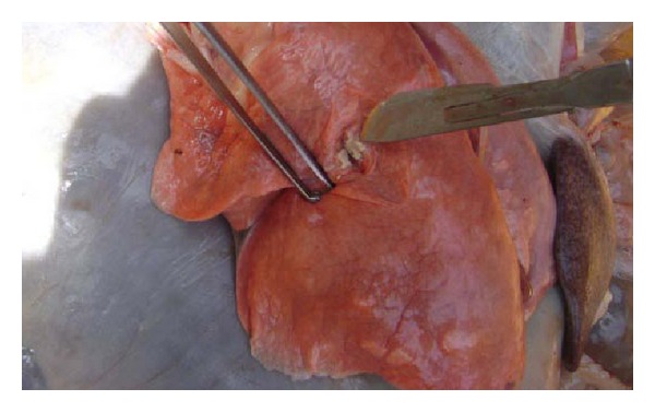
A cross sectional study was conducted on 2231 small ruminants in four districts of the Afar Pastoral Region of Ethiopia to investigate the epidemiology of tuberculosis in goats and sheep using comparative intradermal tuberculin skin test, postmortem examination, mycobacteriological culture and molecular typing methods. The overall animal prevalence of TB in small ruminants was 0.5% (95% CI: 0.2%-0.7%) at ≥4 mm and 3.8% (95% CI: 3%-4.7%) at cutoff ≥2 mm. The herd prevalence was 20% (95% CI: 12-28%) and 47% (95% CI: 37-56%) at ≥4 mm and ≥2 mm cut-off points, respectively. The overall animal prevalence of Mycobacterium avium complex infection was 2.8% (95% CI: 2.1-3.5%) and 6.8% (95% CI: 5.8-7.9%) at ≥4 mm and ≥2 mm cut-off points, respectively. Mycobacteriological culture and molecular characterization of isolates from tissue lesions of tuberculin reactor goats resulted in isolation of Mycobacterium tuberculosis (SIT149) and non-tuberculosis mycobacteria as causative agents of tuberculosis and tuberculosis-like diseases in goats, respectively. The isolation of Mycobacterium tuberculosis in goat suggests a potential transmission of the causative agent from human and warrants further investigation in the role of small ruminants in epidemiology of human tuberculosis in the region.
Figures

Similar articles
-
Bovine tuberculosis at a cattle-small ruminant-human interface in Meskan, Gurage region, Central Ethiopia.BMC Infect Dis. 2011 Nov 15;11:318. doi: 10.1186/1471-2334-11-318. BMC Infect Dis. 2011. PMID: 22085784 Free PMC article.
-
Abattoir-based study on the epidemiology of caprine tuberculosis in Ethiopia using conventional and molecular tools.Acta Vet Scand. 2013 Feb 21;55(1):15. doi: 10.1186/1751-0147-55-15. Acta Vet Scand. 2013. PMID: 23433481 Free PMC article.
-
Low prevalence of bovine tuberculosis in Somali pastoral livestock, southeast Ethiopia.Trop Anim Health Prod. 2012 Oct;44(7):1445-50. doi: 10.1007/s11250-012-0085-5. Trop Anim Health Prod. 2012. PMID: 22286399 Free PMC article.
-
Tuberculosis in small ruminants and dromedary camels in Ethiopia: A systematic review and meta-analysis.Prev Vet Med. 2020 Dec;185:105181. doi: 10.1016/j.prevetmed.2020.105181. Epub 2020 Oct 17. Prev Vet Med. 2020. PMID: 33166824
-
Accuracy of tuberculosis diagnostic tests in small ruminants: A systematic review and meta-analysis.Prev Vet Med. 2020 Sep;182:105102. doi: 10.1016/j.prevetmed.2020.105102. Epub 2020 Jul 26. Prev Vet Med. 2020. PMID: 32739695
Cited by
-
Strain diversity of Mycobacterium tuberculosis isolates from pulmonary tuberculosis patients in Afar pastoral region of Ethiopia.Biomed Res Int. 2014;2014:238532. doi: 10.1155/2014/238532. Epub 2014 Mar 6. Biomed Res Int. 2014. PMID: 24734230 Free PMC article.
-
Identification of peste des petits ruminants virus along with co-infecting diseases of goats in Bangladesh.J Adv Vet Anim Res. 2022 Sep 30;9(3):463-470. doi: 10.5455/javar.2022.i615. eCollection 2022 Sep. J Adv Vet Anim Res. 2022. PMID: 36382033 Free PMC article.
-
Prevalence of bovine tuberculosis in cattle, goats, and camels of traditional livestock raising communities in Eritrea.BMC Vet Res. 2018 Mar 7;14(1):73. doi: 10.1186/s12917-018-1397-0. BMC Vet Res. 2018. PMID: 29514650 Free PMC article.
-
Preliminary investigation of the transmission of tuberculosis between farmers and their cattle in smallholder farms in northwestern Ethiopia: a cross-sectional study.BMC Res Notes. 2017 Jan 7;10(1):31. doi: 10.1186/s13104-016-2349-z. BMC Res Notes. 2017. PMID: 28061860 Free PMC article.
-
Specificity of serological test for detection of tuberculosis in cattle, goats, sheep and pigs under different epidemiological situations.BMC Vet Res. 2019 Mar 1;15(1):70. doi: 10.1186/s12917-019-1814-z. BMC Vet Res. 2019. PMID: 30823881 Free PMC article.
References
-
- FAOSTAT. 2009, http://faostat.fao.org/site/573/DesktopDefault.aspx?PageID=573.
-
- PFE. Pastoralist forum Ethiopia—background to the Ethiopian livestock industry. Proceedings of the 3rd National Conference on Pastoral Development in Ethiopia: Pastoralism and Sustainable Pastoral Development; 2004; Addis Ababa. PFE; pp. 78–79.
-
- Cordes DO, Bullians JA, Lake DE, Carter ME. Observations on tuberculosis caused by Mycobacterium bovis in sheep. New Zealand Veterinary Journal. 1981;29(4):60–62. - PubMed
-
- Tag el Din MH, el Nour Gamaan I. Tuberculosis in sheep in the Sudan. Tropical Animal Health and Production. 1982;14(1):p. 26. - PubMed
-
- Aranaz A, Liébana E, Gómez-Mampaso E, et al. Mycobacterium tuberculosis subsp. caprae subsp. nov.: a taxonomic study of a new member of the Mycobacterium tuberculosis complex isolated from goats in Spain. International Journal of Systematic Bacteriology. 1999;49(3):1263–1273. - PubMed
LinkOut - more resources
Full Text Sources
