. 2012 Aug 1:12:329.
doi: 10.1186/1471-2407-12-329.

Characterization of aldehyde dehydrogenase isozymes in ovarian cancer tissues and sphere cultures

Affiliations

Characterization of aldehyde dehydrogenase isozymes in ovarian cancer tissues and sphere cultures

Yu-Ting Saw et al. BMC Cancer. .

Abstract

Background: Aldehyde dehydrogenases belong to a superfamily of detoxifying enzymes that protect cells from carcinogenic aldehydes. Of the superfamily, ALDH1A1 has gained most attention because current studies have shown that its expression is associated with human cancer stem cells. However, ALDH1A1 is only one of the 19 human ALDH subfamilies currently known. The purpose of the present study was to determine if the expression and activities of other major ALDH isozymes are associated with human ovarian cancer and ovarian cancer sphere cultures.

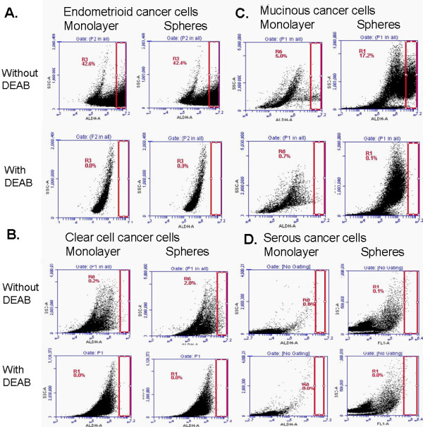
Methods: Immunohistochemistry was used to delineate ALDH isozyme localization in clinical ovarian tissues. Western Blot analyses were performed on lysates prepared from cancer cell lines and ovarian cancer spheres to confirm the immunohistochemistry findings. Quantitative reverse transcription-polymerase chain reactions were used to measure the mRNA expression levels. The Aldefluor® assay was used to measure ALDH activity in cancer cells from the four tumor subtypes.

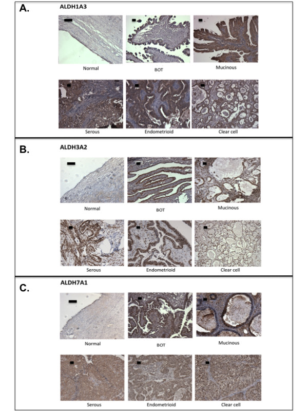
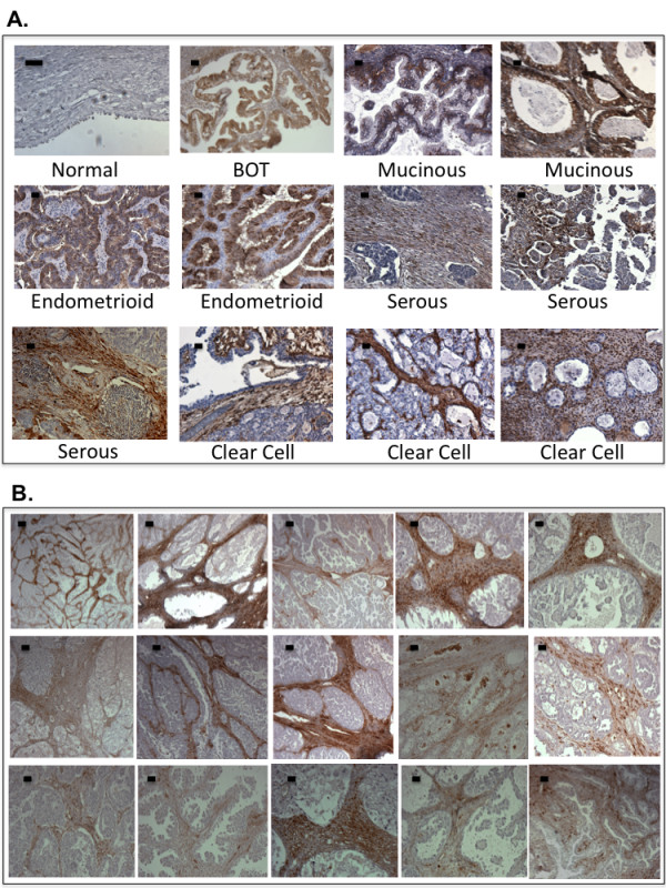
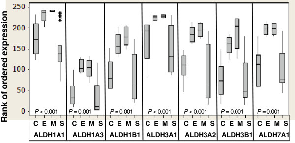
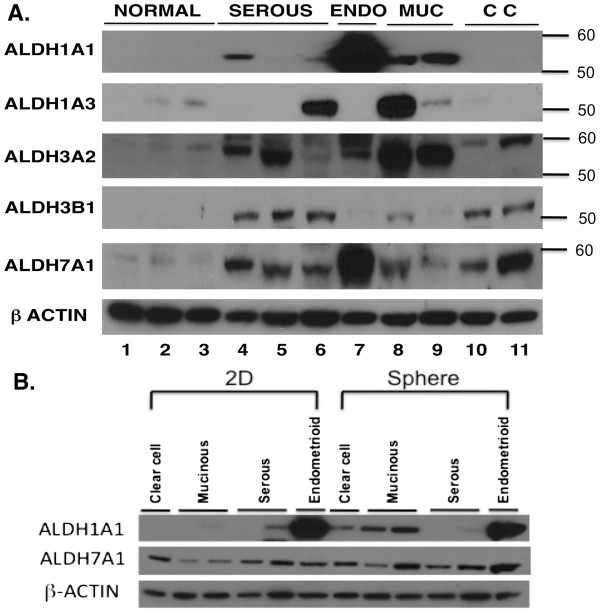
Results: Immunohistochemical staining showed significant overexpression of ALDH1A3, ALDH3A2, and ALDH7A1 isozymes in ovarian tumors relative to normal ovarian tissues. The expression and activity of ALDH1A1 is tumor type-dependent, as seen from immunohistochemisty, Western blot analysis, and the Aldefluor® assay. The expression was elevated in the mucinous and endometrioid ovarian epithelial tumors than in serous and clear cell tumors. In some serous and most clear cell tumors, ALDH1A1 expression was found in the stromal fibroblasts. RNA expression of all studied ALDH isozymes also showed higher expression in endometrioid and mucinous tumors than in the serous and clear cell subtypes. The expression of ALDH enzymes showed tumor type-dependent induction in ovarian cancer cells growing as sphere suspensions in serum-free medium.

Conclusions: The results of our study indicate that ALDH enzyme expression and activity may be associated with specific cell types in ovarian tumor tissues and vary according to cell states. Elucidating the function of the ALDH isozymes in lineage differentiation and pathogenesis may have significant implications for ovarian cancer pathophysiology.


Figures

Figure 1
Figure 1
Expression of ALDH1A3, ALDH3A2, and ALDH7A1 in archived ovarian tissues. Representative figures of immunohistochemical staining of A. ALDH1A3; B. ALDH3A2; and C. ALDH7A1 in ovarian tissues. BOT, borderline tumors. Scale bar represents 50 μm.
Figure 2
Figure 2
Expression of ALDH1A1 in archived ovarian tissues. A. Representative figures of immunohistochemical staining of ALDH1A1 in normal ovaries and different subtypes of ovarian tumor tissues. B. Extended panel of fifteen clear cell ovarian tumor samples stained with ALDH1A1 to demonstrate the predominant staining in the stromal fibroblasts. Scale bar represents 50 μm.
Figure 3
Figure 3
Boxplot to show the quantitative reverse transcription-polymerase chain reaction results of ALDH isozymes in the ovarian cancer cells present in different subtypes of ovarian tumor tissues. RNA was extracted from tumor cells microdissected from 19 high-grade serous, 5 mucinous, 6 clear cell, and 5 endometrioid tumor tissues and quantitative reverse transcription-polymerase chain reactions were performed. Each box covers the middle 50% of ranks of ordered expression of the corresponding ALDH isozyme, and the horizontal line within a box marks the median. The lines extending from a box reach to the minimum and maximum data values, except the presence of outliers that are marked with an asterisk. Kruskal-Wallis P-values are presented to indicate whether the median ranks of the ALDH isozymes are significantly different among the four histologic groups. C, clear cell ovarian tumors; E, endometrioid ovarian tumors; M, mucinous ovarian tumors; S, serous ovarian tumors.
Figure 4
Figure 4
ALDH isozyme protein expression in ovarian cell lines. A. Western blot analysis was used to compare the expression of different ALDH isozymes in normal human ovarian surface epithelial (HOSE) cell lines with cancer cell lines of different subtypes, i.e., serous, endometrioid (ENDO), mucinous (MUC) and clear cell (CC). The cell lines were (starting from left): HOSE1-15, HOSE7, HOSE2170, SKOV3, OVCA432, OVCA433, TOV112D, MCAS, RMUGL, RMG1, and OVCA810. Molecular weights are shown on the right. β-actin served as loading control. B. Western blot analysis was used to compare the levels of ALDH1A1 and ALDH7A1 in ovarian cancer cell lines growing as a monolayer (2D) or sphere culture. The cell lines were (starting from left): RMG1, MCAS, RMUGL, OVCA432, SKOV3, and TOV112D. β-actin served as loading control.
Figure 5
Figure 5
ALDH activity in cancer cells under different culture growing conditions. Aldefluor® was used to estimate ALDH enzyme activity in cells grown as a monolayer culture or in a sphere culture. Flow cytometric graphs show the fluorescence intensity of reacted ALDH substrate in the absence and presence of diethylaminobenzaldehyde, a specific ALDH inhibitor, for A: endometrioid cancer cell line TOV112D; B: Clear cell cancer cell line RMG1; C. Mucinous cancer cell line RMUGL, and D: high-grade serous cancer cells isolated from clinical ascites. Gated regions indicated ALDH+ cells.

References

    1. Altekruse S, Kosary C, Krapcho M, Neyman N, Aminou R, Waldron W, Ruhl J, Howlader N, Tatalovich Z, Cho H, SEER Cancer Statistics Review, 1975–2007. Bethesda, MD, USA :National Cancer Institue; 2010.
    1. Jemal A, Siegel R, Xu J, Ward E. Cancer statistics, 2010. CA Cancer J Clin. 2010;60(5):277–300. doi: 10.3322/caac.20073. - DOI - PubMed
    1. Bast RC, Boyer CM, Olt GJ, Berchuck A, Soper JT, Clarke-Pearson D, Xu FJ, Ramakrishnan S. Identification of marker for early detection of epithelial ovarian cancer. London, England: Chapman and Hall Medical; 1990.
    1. Serov SF, Scullt RE. Histological typing of ovarian tumors. Geneva: World Health Organization; 1993.
    1. Thor AD, Young RH, Clement PB. Pathology of the fallopian tube, broad ligament, peritoneum, and pelvic soft tissues. Hum Pathol. 1991;22(9):856–867. doi: 10.1016/0046-8177(91)90174-N. - DOI - PubMed

Publication types