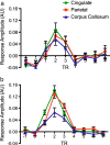
White versus gray matter: fMRI hemodynamic responses show similar characteristics, but differ in peak amplitude
- PMID: 22852798
- PMCID: PMC3469381
- DOI: 10.1186/1471-2202-13-91
White versus gray matter: fMRI hemodynamic responses show similar characteristics, but differ in peak amplitude
Abstract
Background: There is growing evidence for the idea of fMRI activation in white matter. In the current study, we compared hemodynamic response functions (HRF) in white matter and gray matter using 4 T fMRI. White matter fMRI activation was elicited in the isthmus of the corpus callosum at both the group and individual levels (using an established interhemispheric transfer task). Callosal HRFs were compared to HRFs from cingulate and parietal activation.
Results: Examination of the raw HRF revealed similar overall response characteristics. Finite impulse response modeling confirmed that the WM HRF characteristics were comparable to those of the GM HRF, but had significantly decreased peak response amplitudes.
Conclusions: Overall, the results matched a priori expectations of smaller HRF responses in white matter due to the relative drop in cerebral blood flow (CBF) and cerebral blood volume (CBV). Importantly, the findings demonstrate that despite lower CBF and CBV, white matter fMRI activation remained within detectable ranges at 4 T.
Figures


References
-
- Black SE. Imagining white matter and the burden of small vessel disease [abstract] Brain Cogn. 2007;63:191–196. - PubMed
-
- Helenius J, Perkio J, Soinne L, Ostergaard L, Carano RA, Salonen O, Savolainen S, Kaste M, Aronen HJ, Tatlisumak T. Cerebral hemodynamics in a healthy population measured by dynamic susceptibility contrast MR imaging. Acta Radiol. 2003;44:538–546. - PubMed
Publication types
MeSH terms
Substances
Grants and funding
LinkOut - more resources
Full Text Sources
Medical

