. 2012 Sep 10;13(9):2748-59.
doi: 10.1021/bm3007242. Epub 2012 Aug 17.

Low modulus biomimetic microgel particles with high loading of hemoglobin

Affiliations

Low modulus biomimetic microgel particles with high loading of hemoglobin

Kai Chen et al. Biomacromolecules. .

Abstract

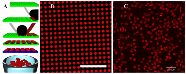
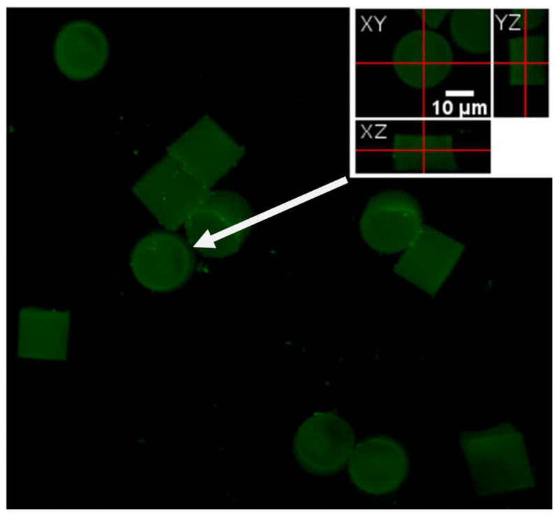
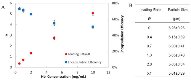
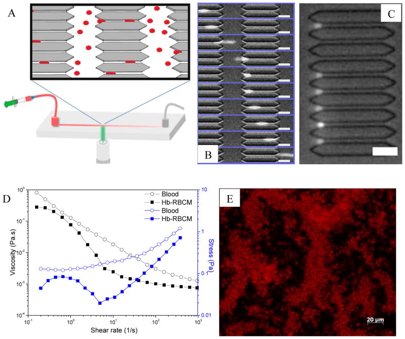
We synthesized extremely deformable red blood cell-like microgel particles and loaded them with bovine hemoglobin (Hb) to potentiate oxygen transport. With similar shape and size as red blood cells (RBCs), the particles were fabricated using the PRINT (particle replication in nonwetting templates) technique. Low cross-linking of the hydrogel resulted in very low mesh density for these particles, allowing passive diffusion of hemoglobin throughout the particles. Hb was secured in the particles through covalent conjugation of the lysine groups of Hb to carboxyl groups in the particles via EDC/NHS coupling. Confocal microscopy of particles bound to fluorescent dye-labeled Hb confirmed the uniform distribution of Hb throughout the particle interior, as opposed to the surface conjugation only. High loading ratios, up to 5 times the amount of Hb to polymer by weight, were obtained without a significant effect on particle stability and shape, though particle diameter decreased slightly with Hb conjugation. Analysis of the protein by circular dichroism (CD) spectroscopy showed that the secondary structure of Hb was unperturbed by conjugation to the particles. Methemoglobin in the particles could be maintained at a low level and the loaded Hb could still bind oxygen, as studied by UV-vis spectroscopy. Hb-loaded particles with moderate loading ratios demonstrated excellent deformability in microfluidic devices, easily deforming to pass through restricted pores half as wide as the diameter of the particles. The suspension of concentrated particles with a Hb concentration of 5.2 g/dL showed comparable viscosity to that of mouse blood, and the particles remained intact even after being sheared at a constant high rate (1000 1/s) for 10 min. Armed with the ability to control size, shape, deformability, and loading of Hb into RBC mimics, we will discuss the implications for artificial blood.


Figures

Figure 1
Figure 1
RBCM particles. (A) Cartoon scheme showing the process of PRINT used to fabricate RBCM microgels. Briefly (Top to bottom), an elastomeric fluoropolymer mold (green) with disc-shaped wells was covered by an aliquot of the pre-polymer liquid (red). The mold was passed through a pressured nip (black) covered by a high-surface-energy sheet (gray), wicking away excess liquid from the mold surface while filling the wells. The filled mold was cured photochemically, yielding crosslinked particles, which were harvested from the mold by laminating a poly(vinyl alcohol) film (blue) on top of the mold through a pressured nip and peeling way the mold. Dissolving the poly(vinyl alcohol) film resulted in a suspension of hydrogel particles. (B) Fluorescent micrograph of polymerized particles (fabricated from 88.85 wt% TEGA, 10 wt% CEA, 0.05 wt% PEG4kDA, 1 wt% HCPK, 0.1 wt% PolyFluor 570) in the mold; and (C) fully hydrated particles free of the mold, suspended in PBS. Scale bars=20 μm.
Figure 2
Figure 2
Conjugation of Hb to RBCM particles. (A) Scheme for the conjugation chemistry. (B) Fluorescent image of particles conjugated with Fluorescein-tagged Hb. Fluorescein-labeled RBCM particles reacted with Rhodamine-tagged Hb were observed in the (C) Fluorescein, (D) Rhodamine channels of fluorescence; (E) overlay of C and D. Scale bars=20 μm. (F) FTIR spectra of blank particles, Hb, and Hb-RBCM conjugate. The arrow pointing to 1579 cm−1 denotes the peak of carboxylate in the particles; the two arrows pointing to 1550 and 1660 cm−1 denote the amide groups in Hb.
Figure 3
Figure 3
A 3D reconstruction view of fluorescein-Hb-conjugated particles observed using xyz scan mode of the confocal laser scanning microscope, with the inset showing orthogonal cross-section views of a representative particle (indicated by the white arrow). The fluorescent protein seems to be homogenously distributed throughout the hydrogel particle.
Figure 4
Figure 4
(A) Loading ratio R and the encapsulation efficiency of Hb into the RBCM particles with respect to different starting concentration of Hb for the conjugation while the particle concentration was maintained at 1mg/mL (n=3). (B) RBCM particle size with respect to different loadings of Hb; the size of the particles were measured by analyzing fluorescent micrographs of the particles (n=50) which had polymerized Rhodamine dye inside.
Figure 5
Figure 5
(A) Far-UV circular dichroism spectra of pure Hb, Hb physically mixed with particles (Hb+particles), and Hb conjugated to particles (Hb-c-particles). For Hb-c-particles, the sample was half diluted as Hb-c-particles Diluted to confirm the isodichroic point as denoted by the arrow. The spectra were all measured in 10 mM K2HPO4 buffer with a path length of 1 mm. (B) Soret CD of the first three samples with Hb concentration of 1 mg/mL in 10 mM K2HPO4 buffer. (C) UV-vis spectra of Hb-particles with the hemes of Hb at different binding states. Particle concentration in the sample was 0.05 mg/mL with Hb concentration of 0.14 mg/mL. The as prepared Hb-particles showed a Soret peak at 405 nm, indicating mostly metHb in the particles. When sodium dithionite was added to the particle suspension, metHb was reduced back to deoxyHb, characterized by the Soret peak at 430 nm. Carbon monoxide purged into the suspension converted deoxyHb into CO-Hb as the Soret peak moved to 419 nm. When exposed to light and air, the Soret peak of the suspension moved to 412 nm, indicating formation of oxyHb. (D) MetHb level in polymer-Hb conjugate over time. The polymer was based on a similar formulation as for the particles but without crosslinker. Overall Hb concentration in the conjugate solution was 5 mg/mL.
Figure 6
Figure 6
(A) Illustration of the microfluidic device. (B) Image sequence (top to bottom) showing how a single particle (R=2.8) passed through a constricted pore (from left to right). The time lapse between the frames was 31 ms. (C) Clogged pore entrance by Hb-RBCM particles with R=5.1. (D) Rheological results showing viscosity and shear stress versus shear rate for mouse blood and Hb-RBCM suspension ([Hb]= 5.2 g/dL) that had been subjected to a rheometer. The shear rate ranged from 0.1 to 1,000 1/s, covering the possible shear rates in blood flow. (E) Microscopic image showing intact Hb-RBCM particles after being sheared at a constant rate of 1,000 1/s for 10 min. Scale bars = 20 μm.
Figure 7
Figure 7
Cytotoxicity of the RBCM particles tested on (A) HUVEC and (B) HeLa cells after 72 h. RBCM particles with 10, 20, and 50 wt% CEA, but unloaded with Hb were studied to assess the biocompatibility of CEA. Hb conjugated particles (10 wt% CEA; R=2.8) with metHb reduced were tested with their un-reduced counterparts. All the particles studied were prepared into 1 mg/mL PBS suspension with regard to the weight of polymers in the particles; therefore, for Hb conjugated particles, at every dose regarding particle concentration, the numbers of particles dosed should be similar to that of unloaded 10 wt% CEA particles.

Similar articles

Cited by

References

    1. Squires JE. Science. 2002;295:1002–5. - PubMed
    1. Riess J. Chem Rev. 2001;101:2720–2797. - PubMed
    1. Winslow RM. Adv Drug Deliv Rev. 2000;40:131–142. - PubMed
    1. Chang TMS. Trends biotechnol. 2006;24:372–377. - PubMed
    1. Alayash AI. Nat Rev Drug Discov. 2004;3:152–9. - PubMed

Publication types

MeSH terms