. 2012 Aug 1:13:41.
doi: 10.1186/1471-2172-13-41.

Multifunctional role of dextran sulfate sodium for in vivo modeling of intestinal diseases

Affiliations

Multifunctional role of dextran sulfate sodium for in vivo modeling of intestinal diseases

William A Rose 2nd et al. BMC Immunol. .

Abstract

Background: Inflammatory bowel diseases (IBDs) are chronic, relapsing disorders that affect the gastrointestinal tract of millions of people and continue to increase in incidence each year. While several factors have been associated with development of IBDs, the exact etiology is unknown. Research using animal models of IBDs is beginning to provide insights into how the different factors contribute to disease development. Oral administration of dextran sulfate sodium (DSS) to mice induces a reproducible experimental colitis that models several intestinal lesions associated with IBDs. The murine DSS colitis model can also be adapted to quantify intestinal repair following injury. Understanding the mechanistic basis behind intestinal repair is critical to development of new therapeutics for IBDs because of their chronic relapsing nature.

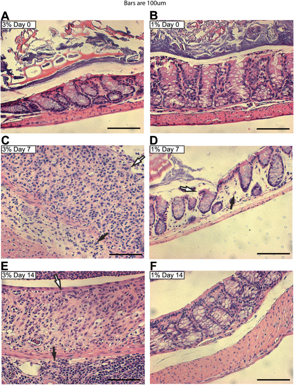
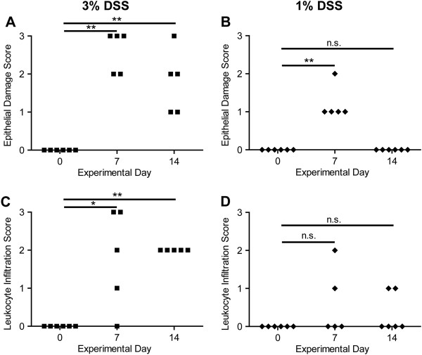
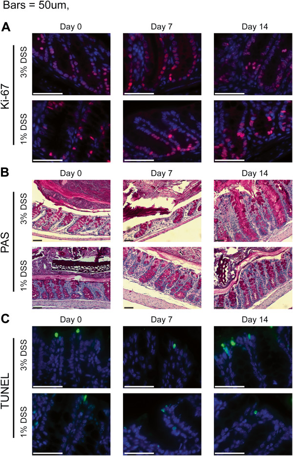
Results: The murine DSS colitis model was adapted to provide a system enabling the quantification of severe intestinal injury with impaired wound healing or mild intestinal injury with rapid restoration of mucosal integrity, by altering DSS concentrations and including a recovery phase. We showed that through a novel format for presentation of the clinical disease data, the temporal progression of intestinal lesions can be quantified on an individual mouse basis. Additionally, parameters for quantification of DSS-induced alterations in epithelial cell populations are included to provide insights into mechanisms underlying the development of these lesions. For example, the use of the two different model systems showed that toll-like receptor 9, a nucleic acid-sensing pattern recognition receptor, is important for protection only following mild intestinal damage and suggests that this model is superior for identifying proteins necessary for intestinal repair.

Conclusions: We showed that using a murine DSS-induced experimental colitis model system, and presenting data in a longitudinal manner on a per mouse basis, enhanced the usefulness of this model, and provided novel insights into the role of an innate immune receptor in intestinal repair. By elucidating the mechanistic basis of intestinal injury and repair, we can begin to understand the etiology of IBDs, enabling development of novel therapeutics or prophylactics.


Figures

Figure 1
Figure 1
Clinical parameter data presented for individual wild-type mice on a daily time scale. Wild-type mice were treated with 3% (A, C, E) or 1% (B, D, F) DSS (n = 5–6 mice/group) in their drinking water for seven days, then supplied with regular drinking water for seven days (closed symbols). Control mice received normal drinking water throughout (open symbols on the X axis) Note that all 1% DSS mice scored 0 and overlap with the control mice on the X axis. Mice were scored for the presence of occult blood (A, B) and stool consistency (C, D) on a daily basis during the fourteen day experiment as described in the materials and methods. (E, F) Mice were weighed on a daily basis to calculate the percent change in weight for the treatment (days 0–7) and recovery (days 7–14) periods. One of two experiments (5–6 mice per condition) with similar results shown. *** p < 0.001; Student’s t-test; n.s., not significant.
Figure 2
Figure 2
Differences in histologic parameters between the two intestinal injury models post cessation of DSS treatments. Wild-type mice were treated with 3% (A, C, E) or 1% (B, D, F) DSS as described in Figure  1. Colons were collected from mice on days 0 (A, B), 7 (C, D), and 14 (E, F). Histopathologic changes in individual crypts (20x dry objective, scale bars = 100 μm) are shown in representative H&E-stained sections. Loss of crypt architecture associated with epithelial damage (white arrows) and leukocyte infiltration (black arrows) is observed following DSS treatment.
Figure 3
Figure 3
Quantification of histologic parameters collected from individual wild-type mice before, during, and after DSS treatment. H&E-stained colon sections were collected as described in Figure  2 and scored in a blinded manner by a board-certified veterinary pathologist. (A, B) Epithelial damage was scored as: 0) no damage; 1) discrete focal lymphoepithelial lesions; 2) mucosal erosion; or 3) mucosal erosion with submucosal damage. (C, D) Leukocyte infiltration was scored as: 0) rare inflammatory cells in the lamina propria; 1) inflammatory cells throughout most of the lamina propria; 2) inflammatory cells extending into the submucosa; or 3) transmural extension of inflammatory cells. * p < 0.05, ** p < 0.01; Mann Whitney test; n.s., not significant.
Figure 4
Figure 4
Representative quantitative histology images. Images from colonic sections of 3% or 1% DSS-treated, wild-type mice collected on days 0, 7, and 14. (A) Sections were stained for Ki-67 (red) and DAPI (blue) and imaged with a 63x oil objective. (B) Sections were stained with PAS for goblet cells and imaged with a 20x dry objective. (C) Sections were TUNEL-stained (green) for apoptotic cells, counterstained for DAPI (blue), and imaged with a 63x oil objective. All scale bars = 50 μm.
Figure 5
Figure 5
DSS treatment induced quantitative changes in colonic epithelial cell populations. Colonic sections were collected from 3 or 1% DSS-treated, wild-type mice on days 0, 7, and 14. 50 crypts from three mice at each time point for each DSS treatment (150 crypts at each treatment time point) were analyzed for: (A) number of Ki-67+ cells per colonic crypt, (B) colonic crypt length, (C) goblet cells per colonic crypt, and (D) number of TUNEL + cells per colonic crypt. Bars represent mean ± standard error of the means. Values were not significant unless indicated: * p < 0.05, ** p < 0.01, *** p < 0.001; ANOVA.
Figure 6
Figure 6
TLR9 is important for protection against mild, but not severe intestinal injury. TLR9-deficient mice were treated with 3% (A, C, E) or 1% (B, D, F) DSS in their drinking water for seven days, then supplied with regular drinking water for seven days. Wild type mice from the same experiment are depicted in Figure  1 for comparison. Mice were scored for the presence of occult blood (A, B) and stool consistency (C, D) on a daily basis during the fourteen day experiment as described in the materials and methods. (E, F) Mice were weighed on a daily basis to calculate the percent change in weight for the treatment (days 0–7) and recovery (days 7–14) periods. One of two experiments (5–6 mice per condition) with similar results shown. *** p < 0.001; Student’s t-test; n.s., not significant.

References

    1. Baumgart DC, Sandborn WJ. Inflammatory bowel disease: clinical aspects and established and evolving therapies. Lancet. 2007;369(9573):1641–1657. doi: 10.1016/S0140-6736(07)60751-X. - DOI - PubMed
    1. Khor B, Gardet A, Xavier RJ. Genetics and pathogenesis of inflammatory bowel disease. Nature. 2011;474(7351):307–317. doi: 10.1038/nature10209. - DOI - PMC - PubMed
    1. Molodecky NA, Soon IS, Rabi DM, Ghali WA, Ferris M, Chernoff G, Benchimol EI, Panaccione R, Ghosh S, Barkema HW. et al. Increasing incidence and prevalence of the inflammatory bowel diseases with time, based on systematic review. Gastroenterology. 2012;142(1):46–54. doi: 10.1053/j.gastro.2011.10.001. e42; quiz e30. - DOI - PubMed
    1. Kaser A, Zeissig S, Blumberg RS. Inflammatory bowel disease. Annu Rev Immunol. 2010;28:573–621. doi: 10.1146/annurev-immunol-030409-101225. - DOI - PMC - PubMed
    1. Baumgart DC, Carding SR. Inflammatory bowel disease: cause and immunobiology. Lancet. 2007;369(9573):1627–1640. doi: 10.1016/S0140-6736(07)60750-8. - DOI - PubMed

Publication types

LinkOut - more resources