. 2012 Aug 1:12:160.
doi: 10.1186/1471-2180-12-160.

Therapeutic concentrations of antibiotics inhibit Shiga toxin release from enterohemorrhagic E. coli O104:H4 from the 2011 German outbreak

Affiliations

Therapeutic concentrations of antibiotics inhibit Shiga toxin release from enterohemorrhagic E. coli O104:H4 from the 2011 German outbreak

Diana Corogeanu et al. BMC Microbiol. .

Abstract

Background: The shiga toxin-producing E. coli (STEC) O104:H4 caused a major outbreak in Germany in spring 2011. STEC are usually susceptible to common antibiotics. However, antibiotic treatment of STEC-infected patients is not recommended because STEC may enhance production and release of shiga toxins (STX) in response to antibiotics, which eventually enhances the frequency and severity of clinical symptoms, including haemolytic uraemic syndrome (HUS) and fatalities.

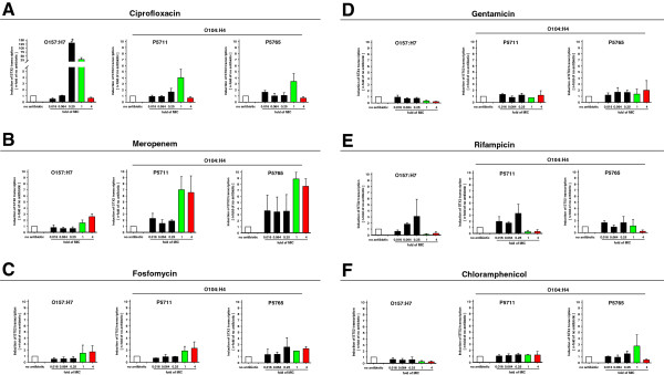
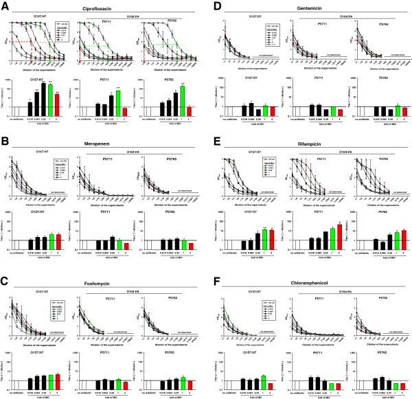
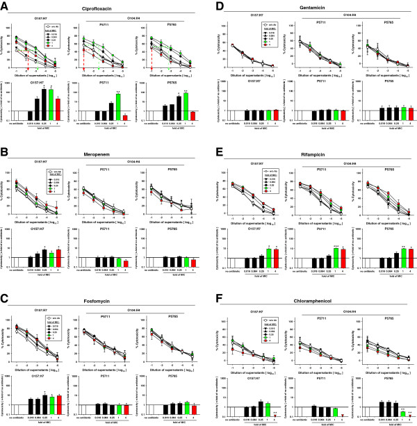
Results: We characterized the response to antibiotics of STEC O104:H4 isolates from two HUS patients during the German STEC outbreak in spring 2011 in comparison to the common STEC O157:H7. Liquid cultures of STEC O157:H7 and O104:H4 were incubated with graded dilutions of the antibiotics ciprofloxacin, meropenem, fosfomycin, gentamicin, rifampicin, and chloramphenicol. At defined times of antibiotic treatment, transcriptional activation of the STX2 gene, contents of STX and STX-activity in the culture supernatants were quantified. Unlike the common serotype O157:H7, STEC O104:H4 does not release STX in response to therapeutic concentrations of ciprofloxacin, meropenem, fosfomycin, and chloramphenicol.

Conclusions: In future outbreaks, the response of the respective epidemiologic STEC strain to antibiotics should be rapidly characterized in order to identify antibiotics that do not enhance the release of STX. This will eventually allow clinical studies tackling the question whether antibiotic treatment impacts on the eradication of STEC, clinical course of disease, and frequency of carriers.


Figures

Figure 1
Figure 1
Transcriptional induction of the STX2 gene in STEC strains O157:H7 and O104:H4 by various antibiotics. STEC strains O157:H7 and O104:H4 were inoculated into L-broth at a density of 1x10⁸ bacteria/ml. The cultures were either left without antibiotics or treated immediately with the indicated n-folds of the MIC of the indicated antibiotics and incubated at 37°C under vigorous shaking. After 2 h, 200 μl of the bacterial suspensions were harvested to prepare total RNA and to determine by qRT-PCR the numbers of STX2-specific transcripts. Green or red columns highlight the values after treatment with the 1-fold or 4-fold MIC, respectively. This colour code is used throughout the manuscript. Shown are the means and standard errors of three independent experiments. Statistical significance is indicated by asterisks: * for p < 0.05.
Figure 2
Figure 2
Quantification of STX in supernatants of STEC strains O157:H7 and O104:H4 treated with various antibiotics. The STEC cultures described in Figure 1 were harvested after 24 h of cultivation and cell-free supernatants were prepared by centrifugation and filtration. The contents of STX were determined with a commercial EIA specific for both STX1 and 2 in two-fold serial dilutions of the supernatants. For each antibiotic, in the upper part of the panel the OD of the STX-specific signal is plotted against the dilution of the supernatants. In the lower part of each panel, the STX-titers are shown which were determined in the plots of the OD as indicated exemplarily for the 1x (green dashed lines) and 4x (red dashed lines) MIC of ciprofloxacin. Briefly, from the OD-value of the undiluted sample of the untreated culture a horizontal dashed line was drawn until it intersected the plot of a given MIC. From this intersection a vertical line was drawn to determine the dilution at which the OD-value of the respective supe rnatant equaled the OD-value of the untreated control. The inverse of this dilution was defined as the STX-titer of the sample. Shown are the means and standard errors of three independent experiments. Statistical significance is indicated by asterisks: * for p < 0.05; ** for p < 0.01.
Figure 3
Figure 3
Cytotoxic activity of supernatants of STEC strains O157:H7 and O104:H4 treated with various antibiotics. The cell free supernatants of STEC cultures described in Figure 2 were 10-fold serially diluted and added to semi-confluent monolayers of Vero cells in microtiter plates. After incubation for 24 h, XTT-labeling reagent was added and cultures were incubated for another 24 h before measuring the viability of the Vero cells as OD450 of the samples. The cytotoxic activity of the supernatants was calculated as described in Methods. For each antibiotic, the cytotoxicity of the supernatants is plotted against the dilution of the supernatants in the upper part of the panel. In these plots, the effect of the antibiotics on the cytotoxicity of the supernatants was determined as the increment of cytotoxicity in comparison to untreated controls, as indicated exemplarily for the 1x and 4x MIC of ciprofloxacin by green dashed lines and red dashed lines, respectively. In the lower part of each panel the increments of the cytotoxicity are plotted for the various MIC of the respective antibiotic. Shown are the means and standard errors of three independent experiments. Statistical significance is indicated by asterisks: * for p < 0.05; ** for p < 0.01.

References

    1. Robert Koch Institute. Report: Final presentation and evaluation of epidemiological findings in the EHEC O104:H4 outbreak, Germany 2011. , Berlin; 2011. www.rki.de.
    1. Serna A, Boedeker EC. Pathogenesis and treatment of Shiga toxin-producing Escherichia coli infections. Curr Opin Gastroenterol. 2008;24(1):38–47. doi: 10.1097/MOG.0b013e3282f2dfb8. - DOI - PubMed
    1. Grif K, Dierich MP, Karch H, Allerberger F. Strain-specific differences in the amount of Shiga toxin released from enterohemorrhagic Escherichia coli O157 following exposure to subinhibitory concentrations of antimicrobial agents. Eur J Clin Microbiol Infect Dis. 1998;17(11):761–766. doi: 10.1007/s100960050181. - DOI - PubMed
    1. Walterspiel JN, Ashkenazi S, Morrow AL, Cleary TG. Effect of subinhibitory concentrations of antibiotics on extracellular Shiga-like toxin I. Infection. 1992;20(1):25–29. doi: 10.1007/BF01704889. - DOI - PubMed
    1. MacConnachie AA, Todd WT. Potential therapeutic agents for the prevention and treatment of haemolytic uraemic syndrome in shiga toxin producing Escherichia coli infection. Curr Opin Infect Dis. 2004;17(5):479–482. doi: 10.1097/00001432-200410000-00013. - DOI - PubMed

MeSH terms