. 2012 Nov 1;125(Pt 21):5061-72.
doi: 10.1242/jcs.105015. Epub 2012 Aug 1.

Polo-like kinase is required for synaptonemal complex disassembly and phosphorylation in mouse spermatocytes

Affiliations

Polo-like kinase is required for synaptonemal complex disassembly and phosphorylation in mouse spermatocytes

Philip W Jordan et al. J Cell Sci. .

Abstract

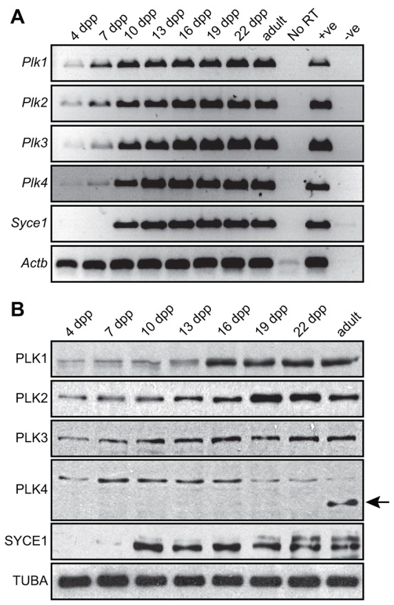
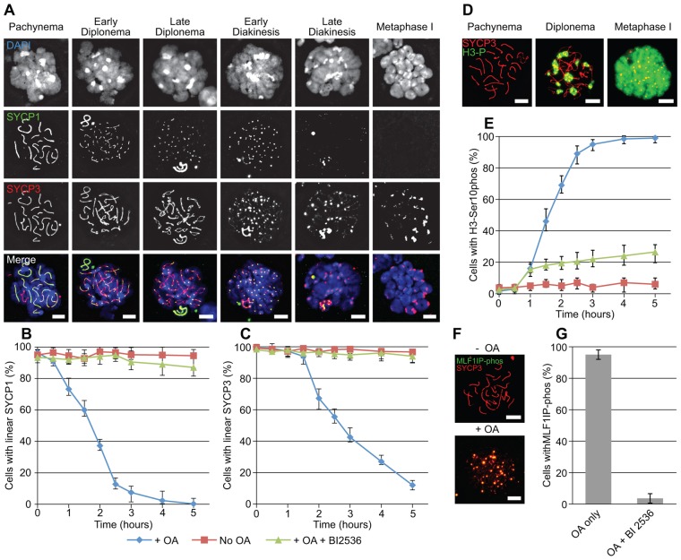
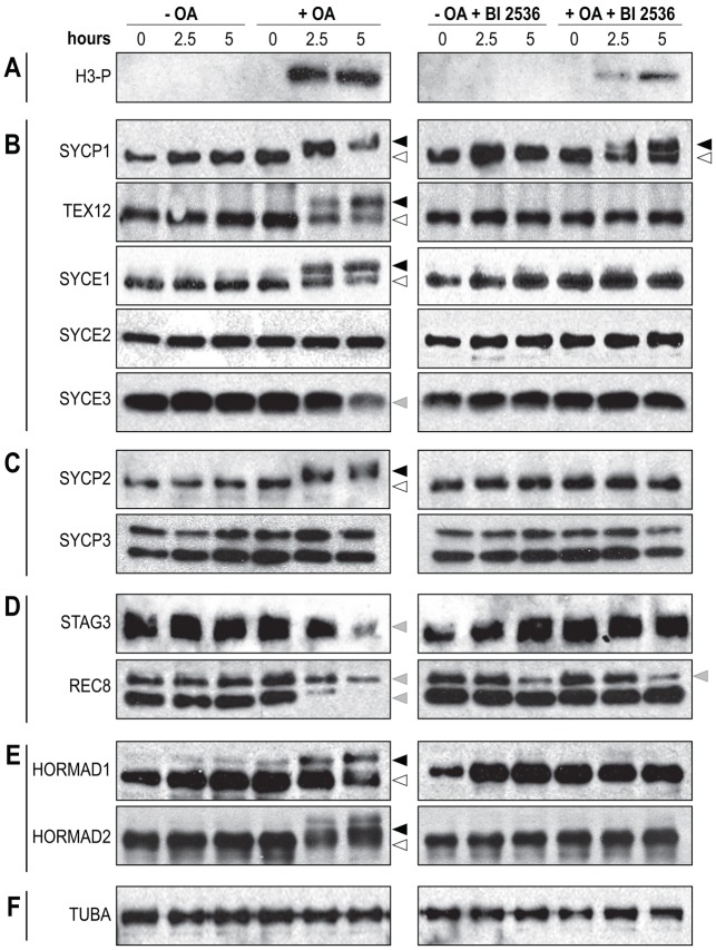
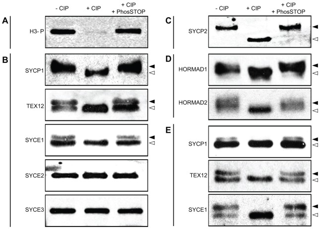
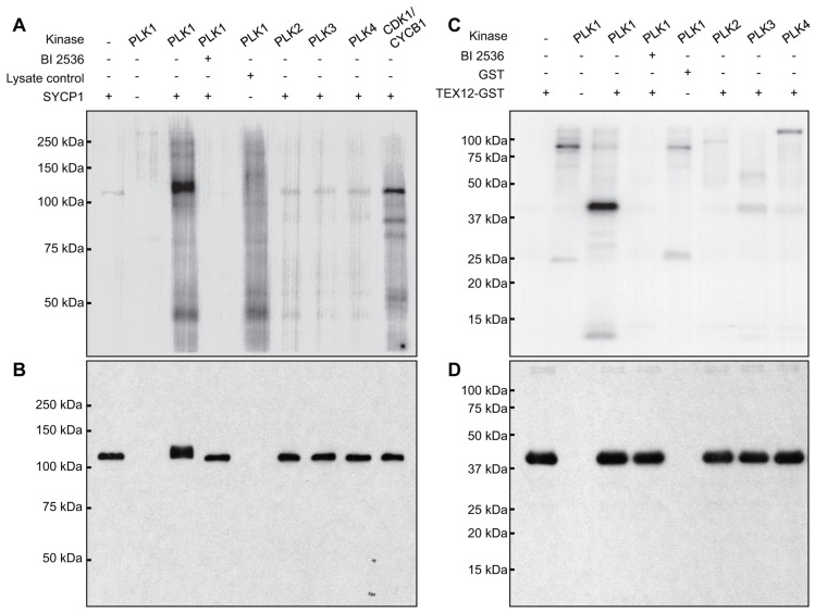
During meiosis, accurate coordination of the completion of homologous recombination and synaptonemal complex (SC) disassembly during the prophase to metaphase I (G2/MI) transition is essential to avoid aneuploid gametes and infertility. Previous studies have shown that kinase activity is required to promote meiotic prophase exit. The first step of the G2/MI transition is the disassembly of the central element components of the SC; however, the kinase(s) required to trigger this process remains unknown. Here we assess roles of polo-like kinases (PLKs) in mouse spermatocytes, both in vivo and during prophase exit induced ex vivo by the phosphatase inhibitor okadaic acid. All four PLKs are expressed during the first wave of spermatogenesis. Only PLK1 (not PLK2-4) localizes to the SC during the G2/MI transition. The SC central element proteins SYCP1, TEX12 and SYCE1 are phosphorylated during the G2/MI transition. However, treatment of pachytene spermatocytes with the PLK inhibitor BI 2536 prevented the okadaic-acid-induced meiotic prophase exit and inhibited phosphorylation of the central element proteins as well as their removal from the SC. Phosphorylation assays in vitro demonstrated that PLK1, but not PLK2-4, phosphorylates central element proteins SYCP1 and TEX12. These findings provide mechanistic details of the first stage of SC disassembly in mammalian spermatocytes, and reveal that PLK-mediated phosphorylation of central element proteins is required for meiotic prophase exit.


Figures

Fig. 1.
Fig. 1.
Expression of PLKs during the first wave of spermatogenesis. This figure provides representative results of analyses of RNA and protein extracted from germ cells of B6SJL F1 male mice aged 4, 7, 10, 13, 16, 19, 22 and 56 (adult) dpp. (A) PCR analysis of mRNA expression of Plk1-4; Syce1 was used as a control for progression of the first wave of spermatogenesis and Actb was used as a mRNA loading control. mRNA extracted from whole testis and RNA-free H2O were used as positive (+ve) and negative (−ve) controls, respectively (35 PCR cycles were used, see Materials and Methods). Each transcript was assayed at least four times. (B) Western blot analysis of PLK1–4 proteins; SYCE1 was used as a control for progression of the first wave of spermatogenesis and TUBA (tubulin) was used as a protein loading control. The faster migrating form of PLK4 detected in adult germ cells is indicated by a black arrow. Each protein was assayed at least two times.
Fig. 2.
Fig. 2.
Localization of PLK1–4 in male germ cells. Nuclear spreads from testicular germ cells of B6SJL F1 mice aged 19–23 dpp were stained with DAPI (blue) and immunolabeled using antibodies against PLKs (green) and the SC lateral element protein SYCP3 (red). (A) PLK1, (B) PLK2, (C) PLK3 and (D) PLK4. In D, the yellow arrowheads point to aggregates of PLK4 and the yellow arrows point to PLK4 that is localized to the X-Y body during pachynema and diplonema. At least 100 nuclear spreads at pachynema, diplonema and metaphase I were scored for each antibody, and three independent rounds of nuclear spread preparations were used for immunolabeling. Scale bars: 10 µm.
Fig. 3.
Fig. 3.
SC disassembly during the OA-initiated G2/MI transition is inhibited by PLK inhibitor BI 2536. (A) Nuclear spreads from STAPUT-purified germ cells depicting steps in post-pachynema prophase exit induced by OA. Nuclear spreads were stained with DAPI (blue) and immunolabeled using antibodies against the SC central element component SYCP1 (green) and the SC lateral element component SYCP3 (red). (B) Time course of the disassembly of the SC central element component SYCP1 during OA-initiated G2/MI transition. STAPUT-purified pachytene spermatocytes were cultured without OA (red line, squares), with 5 µM OA (blue line, diamonds) or with 5 µM OA and 100 nM BI 2536 (green line, triangles). (C) Time course of the disassembly of the SC lateral element component SYCP3 during OA-initiated G2/MI transition. Graph details are as in B. (D) Nuclear spreads from STAPUT-enriched germ cells depicting phosphorylation of the serine 10 residue of histone H3 during the OA-initiated G2/MI transition. Nuclear spreads were immunolabeled using antibodies against phosphorylated H3 (green) and the SC lateral element component SYCP3 (red). Nuclear spreads staged at pachynema (pre-OA treatment), and post-OA diplonema and metaphase I stages are presented. (E) Time course for phosphorylation of the serine 10 residue of histone H3 during OA-initiated G2/MI transition. Graph details are as in B. (F) Nuclear spreads from STAPUT-enriched germ cells depicting phosphorylation of the threonine 78 residue of centromeric protein MLF1IP during OA-initiated G2/MI transition. Nuclear spreads were immunolabeled using antibodies against phosphorylated MLF1IP (green) and the SC lateral element component SYCP3 (red). (G) Bar graph presenting the number of nuclear spreads with phosphorylated MLF1IP after 5 hours in culture with 5 µM OA or with 5 µM OA and 100 nM BI 2536. For all experiments, at least 200 nuclei were counted per time point; experiments were performed in triplicate. Scale bars: 10 µm.
Fig. 4.
Fig. 4.
Modification of SC components during OA-initiated G2/MI transition is inhibited by BI 2536. STAPUT-isolated pachytene spermatocytes were cultured without OA, with 5 µM OA, with 100 nM BI 2536 or with 5 µM OA and 100 nM BI 2536. Protein extracts were made at 0, 2.5 and 5 hours after initiation of treatment and western blot analyses performed for (A) serine 10 histone H3 phosphorylation; (B) SC central element proteins SYCP1, TEX12, SYCE1, SYCE2 and SYCE3; (C) SC lateral element proteins SYCP2 and SYCP3; (D) meiotic cohesin components REC8 and STAG3; and (E) HORMA-domain-containing proteins HORMAD1 and HORMAD2. (F) Tubulin (TUBA) was used as a loading control. White arrowheads point to the faster migrating protein signals; black arrowheads point to the slower migrating protein signals; grey arrowheads point to reduced protein signals (protein degradation). The results presented are typical from experiments repeated at least two times.
Fig. 5.
Fig. 5.
SC components are phosphorylated during the G2/MI transition. STAPUT-enriched pachytene spermatocytes were cultured for 5 hours in 5 µM OA to induce the G2/MI transition, protein was extracted from the spermatocytes and used to assess protein phosphorylation of SC components in a standard calf intestinal phosphatase (CIP) assay. Protein extracts were analyzed in the absence of CIP (−CIP), in the presence of CIP (+CIP) and in the presence of CIP and the phosphatase inhibitor cocktail PhosSTOP (+CIP +PhosSTOP). Product of the CIP assays was used for western analysis of (A) serine 10 histone H3 phosphorylation; (B) central element proteins SYCP1, TEX12, SYCE1, SYCE2 and SYCE3; (C) SC lateral element protein SYCP2; and (D) HORMA-domain-containing proteins HORMAD1 and HORMAD2. (E) Analysis of protein extracted from enriched germ cells of male C57BL/6J mice (22 dpp) in a CIP assay to determine in vivo phosphorylation state of central element proteins SYCP1, TEX12 and SYCE1. White arrowheads point to the faster migrating dephosphorylated protein signals; black arrowheads point to the slower migrating phosphorylated protein signals. Each CIP assay was performed a total of three times.
Fig. 6.
Fig. 6.
In vitro phosphorylation of SC central element proteins by PLK1. (A) Purified SYCP1 protein or control cell lysates were used as substrates for kinase assays with PLK1, 2, 3, 4 or CDK1/CYCB1. The expected molecular mass of unmodified SYCP1 is 114 kDa. (B) Western blot analysis of SYCP1 protein used for the in vitro kinase assay. (C) Purified TEX12–GST protein or control GST protein were used as substrates in kinase assays with PLK1, 2, 3 or 4. The expected molecular mass of unmodified TEX12–GST is 40 kDa. (D) Western blot analysis of TEX12–GST protein used for the in vitro kinase assay. Signals in A and C reflect autoradiographic detection of [γ-32P] incorporation. The in vitro kinase assays were performed a total of three times.

Similar articles

Cited by

References

    1. Abe S., Nagasaka K., Hirayama Y., Kozuka–Hata H., Oyama M., Aoyagi Y., Obuse C., Hirota T. (2011). The initial phase of chromosome condensation requires Cdk1-mediated phosphorylation of the CAP-D3 subunit of condensin II. Genes Dev. 25, 863–874 10.1101/gad.2016411 - DOI - PMC - PubMed
    1. Alexander J., Lim D., Joughin B. A., Hegemann B., Hutchins J. R. A., Ehrenberger T., Ivins F., Sessa F., Hudecz O., Nigg E. A.et al. (2011). Spatial exclusivity combined with positive and negative selection of phosphorylation motifs is the basis for context-dependent mitotic signaling. Sci. Signal. 4, ra42 10.1126/scisignal.2001796 - DOI - PMC - PubMed
    1. Bellvé A. R. (1993). Purification, culture, and fractionation of spermatogenic cells. Methods Enzymol. 225, 84–113 10.1016/0076-6879(93)25009-Q - DOI - PubMed
    1. Bellvé A. R., Millette C. F., Bhatnagar Y. M., O’Brien D. A. (1977). Dissociation of the mouse testis and characterization of isolated spermatogenic cells. J. Histochem. Cytochem. 25, 480–494 10.1177/25.7.893996 - DOI - PubMed
    1. Bolcun–Filas E., Costa Y., Speed R., Taggart M., Benavente R., De Rooij D. G., Cooke H. J. (2007). SYCE2 is required for synaptonemal complex assembly, double strand break repair, and homologous recombination. J. Cell Biol. 176, 741–747 10.1083/jcb.200610027 - DOI - PMC - PubMed

Publication types

MeSH terms