

Metaplastic carcinoma of the right breast and simultaneous giant ovarian teratoma: a case report
- PMID: 22854062
- PMCID: PMC3777448
- DOI: 10.5732/cjc.012.10118
Metaplastic carcinoma of the right breast and simultaneous giant ovarian teratoma: a case report
Abstract
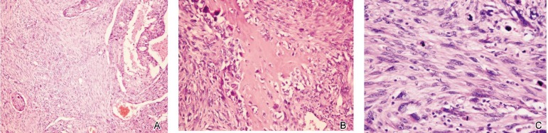
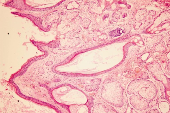
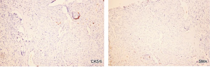
We describe here a female patient who presented with a breast mass and giant abdominal mass. Fine needle aspiration cytology of the breast mass and histological examination after modified radical mastectomy confirmed metaplastic carcinoma of the breast. The epithelial components were formed by infiltrating ductal carcinoma with poor differentiation, and the sarcomatous components were formed by fibrosarcoma and osteosarcoma. Histological examination of the abdominal mass confirmed ovarian teratoma. The patient underwent modified radical mastectomy of the right breast and laparoscopic excision of the abdominal mass in the lower right quadrant. Having underwent six courses of chemotherapy, the patient is now in her tenth month after surgery and under follow-up, and she has no relapsed disease. These two diseases have never seen in one patient before. The case we report here provides some new data for research and clinical experience and it may also provide a new insight into the relationship between metaplastic breast carcinoma and ovarian teratoma.
Figures
References
-
- Lien HC, Hsiao YH, Lin YS, et al. Molecular signatures of metaplastic carcinoma of the breast by large-scale transcriptional profiling: identification of genes potentially related to epithelial-mesenchymal transition. Oncogene. 2007;26:7859–7871. - PubMed
-
- Thiery JP. Epithelial-mesenchymal transitions in tumour progression. Nat Rev Cancer. 2002;2:442–454. - PubMed
Publication types
MeSH terms
Substances
Supplementary concepts
LinkOut - more resources
Full Text Sources
Medical
Research Materials
Miscellaneous