. 2012 Sep 14;47(5):681-93.
doi: 10.1016/j.molcel.2012.06.024. Epub 2012 Jul 30.

PIDD death-domain phosphorylation by ATM controls prodeath versus prosurvival PIDDosome signaling

Affiliations

PIDD death-domain phosphorylation by ATM controls prodeath versus prosurvival PIDDosome signaling

Kiyohiro Ando et al. Mol Cell. .

Abstract

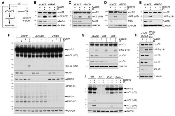
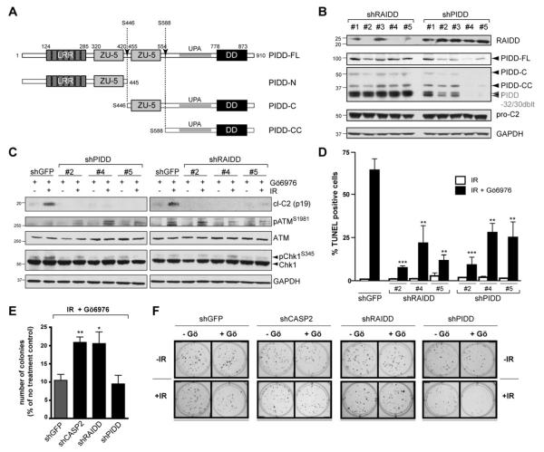
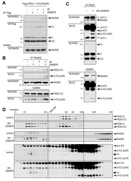
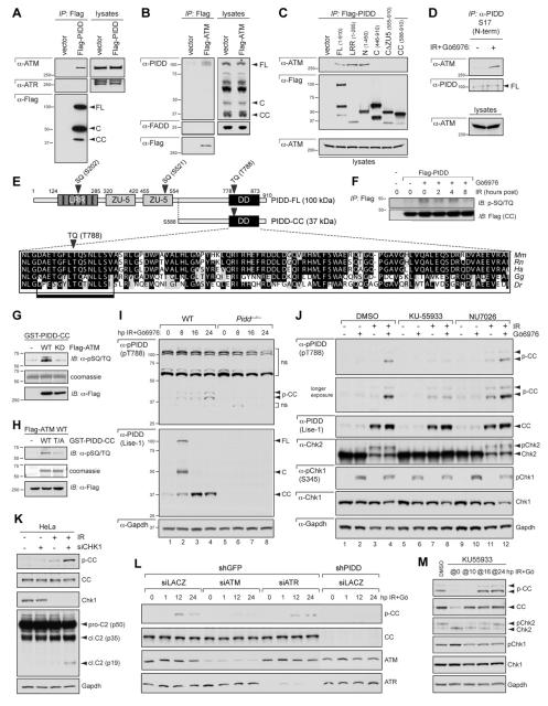
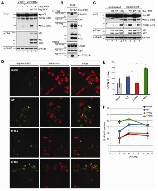
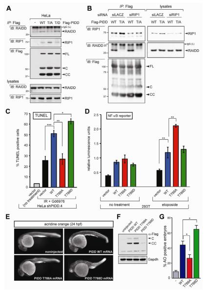
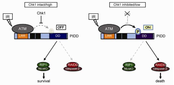
Biochemical evidence implicates the death-domain (DD) protein PIDD as a molecular switch capable of signaling cell survival or death in response to genotoxic stress. PIDD activity is determined by binding-partner selection at its DD: whereas recruitment of RIP1 triggers prosurvival NF-κB signaling, recruitment of RAIDD activates proapoptotic caspase-2 via PIDDosome formation. However, it remains unclear how interactor selection, and thus fate decision, is regulated at the PIDD platform. We show that the PIDDosome functions in the "Chk1-suppressed" apoptotic response to DNA damage, a conserved ATM/ATR-caspase-2 pathway antagonized by Chk1. In this pathway, ATM phosphorylates PIDD on Thr788 within the DD. This phosphorylation is necessary and sufficient for RAIDD binding and caspase-2 activation. Conversely, nonphosphorylatable PIDD fails to bind RAIDD or activate caspase-2, and engages prosurvival RIP1 instead. Thus, ATM phosphorylation of the PIDD DD enables a binary switch through which cells elect to survive or die upon DNA injury.


Figures

Figure 1
Figure 1. PIDD and RAIDD are required for caspase-2 activation in the CS pathway
(A) Diagram of the CS pathway. Pathway activation after IR is restrained by Chk1. Combined delivery of IR and Chk1 inhibitor (Gö6976) or siRNA activates the pathway. IR and Chk1 inhibition are individually insufficient for pathway activation (Sidi et al., 2008). (B-E) HeLa cells transfected with the indicated siRNAs were treated with Gö6976 (1 μM) with or without IR (10 Gy) and harvested 24 hr post IR. Lysates were analyzed by western blot. pro-C2, procaspase-2; cl-C2 (p19), cleaved caspase-2, p19 fragment (mature cleavage product). cl-C2 images are longer exposures of the same membrane. (F) HeLa cells stably expressing the indicated shRNAs were transfected with CHK1 siRNA (+) or LACZ siRNA (-), treated with or without IR (10 Gy) 16 hr after transfection, and harvested 16 hr post IR. Lysates were analyzed by western blot. (G) HeLa cells transfected with the indicated siRNAs were treated with Gö6976 (1 μM) with or without IR (10 Gy) and harvested 24 hr post IR. Lysates were analyzed by western blot. C8, caspase-8; C9, caspase-9. (H) HeLa cells stably expressing the indicated shRNAs and transfected with the indicated siRNAs were treated with Gö6976 (1 μM) with or without IR (10 Gy), and harvested 24 hr post IR. Lysates were analyzed by western blot. C3, caspase-3; C6, caspase-6; C7, caspase-7. (I) SV40-transformed MEFs of indicated genotypes were treated with Gö6976 (1 μM) with or without IR (10 Gy) and harvested 24 hr post IR. Lysates were analyzed by western blot.
Figure 2
Figure 2. PIDD and RAIDD are required for CS apoptosis
(A) Schematic diagram of PIDD maturation. Autoproteolytic cleavage sites are indicated (arrows). (B) HeLa cells stably expressing the indicated shRNAs were analyzed by western blot. PIDD-32/30 doublet: PIDD specific bands running at ~30 kDa, presumably resulting from autoproteolysis of shorter PIDD isoforms (Cuenin et al., 2008). (C) HeLa cells stably expressing the indicated shRNAs were treated with Gö6976 (1 μM) with or without IR (10 Gy), and harvested 24 hr post IR. Lysates were analyzed by western blot. (D) HeLa cells stably expressing the indicated shRNAs were treated with 10 Gy IR with or without Gö6976 (1 μM) (black and white bars, respectively), and were analyzed by TUNEL staining at 48 hr post IR. Data are means +/− SEM. **p < 0.01; ***p < 0.001 two-tailed Student’s t-test. (E) HeLa cells stably expressing the indicated shRNAs were treated with Gö6976 (1 μM) and IR (5 Gy) and colony numbers were recorded 14 days post IR. Data are means +/− SEM. *p < 0.05; **p < 0.01 two-tailed Student’s t-test. (F) Representative images of a clonogenic assay. HeLa cells stably expressing the indicated shRNAs were treated with or without Gö6976 (1 μM) or IR (2 Gy) and stained with crystal violet 14 days after IR.
Figure 3
Figure 3. The CS pathway triggers PIDDosome assembly
(A) HeLa cells transfected with the indicated constructs were treated with or without Gö6976 (1 μM) or 10 Gy IR and harvested 24 hr post IR. Anti-Flag immunoprecipitates were analyzed by western blot. PIDD-FL is constitutively autoprocessed in cells into -C and -CC fragments (Tinel et al., 2007). (B) HeLa cells treated with or without IR (10 Gy) or Gö6976 (1 μM) were harvested 24 hr post IR. Lysates were immunoprecipated with anti-RAIDD antibody and analyzed by western blot. (C) HeLa cells treated with or without IR (10 Gy) or Gö6976 (1 μM) were harvested 24 hr post IR. Lysates were immunoprecipated with anti-PIDD antibody and analyzed by western blot. (D) HeLa cells treated with or without IR (10 Gy) and Gö6976 (1 μM) were lysed 24 hr post IR and loaded on a S400 HiPrep 16/60 Sephacryl column (1 ml/min). An aliquot of each fraction was analyzed by SDS-PAGE with the indicated antibodies. Empty arrowheads indicate non-specific bands.
Figure 4
Figure 4. ATM phosphorylates PIDD on T788 during CS apoptosis
(A) HeLa cells transfected with Flag-tagged PIDD were lysed, immunoprecipitated with anti-Flag antibodies and analyzed by western blot. (B) HeLa cells transfected with Flag-ATM were lysed, immunoprecipitated with anti-Flag antibodies and analyzed by western blot. (C) HeLa cells transfected with the indicated Flag-tagged PIDD deletion constructs were lysed 24 hr post transfection, immunoprecipitated with anti-Flag antibodies and analyzed by western blot. (D) HeLa cells either untreated or treated with IR (10 Gy) and Gö6976 (1 μM) were harvested 24 hr post IR. Nuclear extracts were immunoprecipated with an anti-PIDD antibody targeted to the PIDD N-terminus. Immunoprecipitates were analyzed by western blot. (E) Diagram of PIDD-FL highlighting three candidate ATM phosphorylation sites (bold arrowheads). Blow up shows a clustal alignment of the DD amino acid sequences of PIDD proteins from the indicated species. Mm, Mus musculus; Rn, Rattus norvergicus; Hs, Homo sapiens; Gg, Gallus gallus (chicken); Dr, Danio rerio (zebrafish). The predicted full ATM target sequence (box) and the target TQ motif (bold arrowhead) are indicated. (F) HeLa cells transfected with empty vector or C-terminally Flag-tagged PIDD were treated with IR (10 Gy) with or without Gö6976 (1 μM) and harvested at the indicated time points after IR. Protein extracts were immunoprecipitated with anti-Flag antibodies and analyzed by western blot. (G) Recombinant GST-PIDD-CC proteins were incubated with wild-type (WT) or kinase-dead (KD) Flag-ATM for an in vitro kinase assay (IVK). Reactions were analyzed by coomassie staining and western blot. (H) Recombinant wild-type (WT) or T788A mutant (T/A) GST-PIDD-CC proteins were incubated with Flag-ATMWT for an IVK. Reactions were analyzed by coomassie staining and western blot. Residual pSQ/TQ immunoreactivity in the T/A sample suggests additional specific or non-specific phosphorylation events occurring in the reaction. (I) SV40 MEFs of indicated genotypes were treated with IR (10 Gy) and Gö6976 (1 μM) and harvested at the indicated time points after IR. Extracts were analyzed by western blot. p-CC, phospho-PIDD-CC; upper arrowhead marks p-CC harboring an additional post-translational modification (see text). ns, non-specific bands. (J) SV40 WT MEFs treated with or without IR (10 Gy) or Gö6976 (1 μM) were additionally treated with ATM inhibitor KU55933 or DNA-PKcs inhibitor NU7026 (10 μM each) at 8 hpIR. Cells were harvested at 24 hpIR and analyzed by western blot. (K) HeLa cells transfected with CHK1 siRNA (+) or LACZ siRNA (-) were treated with or without IR (10 Gy) or Gö6976 (1 μM) at 16 hr post-transfection. Cells were harvested at 16 hpIR and analyzed by western blot. (L) HeLa cells stably expressing the indicated shRNAs were transfected with the indicated siRNAs and treated with or without Gö6976 (1 μM) or IR (10 Gy) 36 hr post-transfection. Cells were harvested at the indicated time points after IR and analyzed by western blot. (M) SV40 WT MEFs treated with IR (10 Gy) and Gö6976 (1 μM) were exposed to ATM inhibitor KU55933 added at (‘@’) the indicated time points after stimulus. Cells were harvested at 24 hr after IR+Gö6976 treatment and analyzed by western blot.
Figure 5
Figure 5. PIDD phosphorylation on T788 is necessary and sufficient for caspase-2 activation
(A) HeLa cells stably expressing the indicated shRNAs were transfected with the indicated C-terminally Flag-tagged, shRNA-resistant PIDD constructs, treated with or without Gö6976 (1 μM) or IR (10 Gy), and harvested 24 hr post IR. Lysates were analyzed by western blot. WT, wild-type PIDD; T/A, T788A mutant. (B) 293T cells transfected with the indicated C-terminally Flag-tagged PIDD constructs were harvested at 24 hr post transfection. Protein extracts were immunoprecipitated with anti-Flag antibodies. Immunoprecipitates and whole-cell lysates were analyzed by western blot. Asterisk indicates IgG light chain. (C) HeLa cells transfected with the indicated C-terminally Flag-tagged PIDD constructs were either left untreated or treated with Gö6976 (1 μM) and IR (10 Gy), and harvested 24 hr post IR. Lysates were analyzed by western blot. T/D, T788D mutant. (D) HeLa-Bcl-XL cells were transfected with 5 ng of C2-CARD-VC and C2-CARD-VN pBiFC plasmids, encoding the C- and N-terminal moieties of the Venus protein, respectively, fused to the caspase recruitment domain (CARD) of caspase-2. Cells were co-transfected with the indicated PIDD constructs (10 ng) and dsRed-mito (10 ng, used as transfection reporter), and imaged 24 hr post-transfection by confocal microscopy. Caspase-2 bifluorescence complementation (BiFC; left column) is the Venus fluorescence signal that occurs when N- and C-terminal Venus moieties are brought in proximity as a result of caspase-2 recruitment to the PIDDosome. (E) HeLa cells transfected and imaged as in (D) were scored for Venus positivity. Data from 3 independent experiments (>100 cells each) are shown as means +/− SEM. **p < 0.01, two-tailed Student’s t-test. (F) HeLa cells transfected as in (D) but with incremental amounts of the indicated PIDD constructs (10, 20, or 40 ng) were imaged as in (D) and scored for Venus positivity. Data from 3 independent experiments (>100 cells each) are shown as means +/− SEM.
Figure 6
Figure 6. T788 phosphorylation determines RAIDD versus RIP1 selection and fate decision by PIDD
(A) HeLa cells transfected with the indicated C-terminally Flag-tagged PIDD constructs were harvested at 24 hr post transfection. Protein extracts were immunoprecipitated with anti-Flag antibodies and analyzed by western blot. (B) HeLa cells co-transfected with the indicated siRNAs and C-terminally Flag-tagged PIDD constructs were harvested at 36 hr post siRNA transfection. Protein extracts were immunoprecipitated with anti-Flag antibodies and analyzed by western blot. Asterisk indicates IgG light chain. (C) shPIDD.4 HeLa cells transfected with the indicated PIDD constructs were treated with or without 10 Gy IR and Gö6976 (1 μM) and analyzed by TUNEL staining at 48 hr post IR. Data are means +/− SEM. *p < 0.05; **p < 0.01; ***p < 0.001, two-tailed Student’s t-test. (D) HEK293T cells transiently transfected with the indicated PIDD constructs were treated with or without etoposide (40 μM) and analyzed with a NF-κB reporter. **p < 0.01, two-tailed Student’s t-test. (E) One-cell stage wild-type zebrafish embryos were injected with the indicated synthetic Flag-PIDD mRNAs (100 pg each), stained with the cell-death marker acridine orange at 24 hr post-fertilization (hpf), and imaged with a fluorescence microscope. Representative images are shown, anterior to the left. (F) Whole-body protein extracts from 24-hpf embryos as in (E) were analyzed by western blot. (G) Quantification of acridine-orange stains such as in (E). Data collected from six independent experiments (at least 30 embryos per construct in each) are represented as means +/− SEM. *p < 0.05, two-tailed Student’s t-test.
Figure 7
Figure 7. Mechanism of life vs. death decisions at the PIDD docking site
See text for details.

Comment in

References

    1. Baptiste-Okoh N, Barsotti AM, Prives C. A role for caspase 2 and PIDD in the process of p53-mediated apoptosis. Proc Natl Acad Sci U S A. 2008;105:1937–1942. - PMC - PubMed
    1. Berube C, Boucher LM, Ma W, Wakeham A, Salmena L, Hakem R, Yeh WC, Mak TW, Benchimol S. Apoptosis caused by p53-induced protein with death domain (PIDD) depends on the death adapter protein RAIDD. Proc Natl Acad Sci U S A. 2005;102:14314–14320. - PMC - PubMed
    1. Bouchier-Hayes L, Oberst A, McStay GP, Connell S, Tait SW, Dillon CP, Flanagan JM, Beere HM, Green DR. Characterization of cytoplasmic caspase-2 activation by induced proximity. Mol Cell. 2009;35:830–840. - PMC - PubMed
    1. Ciccia A, Elledge SJ. The DNA damage response: making it safe to play with knives. Mol Cell. 2010;40:179–204. - PMC - PubMed
    1. Cuenin S, Tinel A, Janssens S, Tschopp J. p53-induced protein with a death domain (PIDD) isoforms differentially activate nuclear factor-kappaB and caspase-2 in response to genotoxic stress. Oncogene. 2008;27:387–396. - PubMed

Publication types