. 2012 Sep;4(9):1049-58.
doi: 10.1039/c2ib20083j. Epub 2012 Aug 2.

Soft microenvironments promote the early neurogenic differentiation but not self-renewal of human pluripotent stem cells

Affiliations

Soft microenvironments promote the early neurogenic differentiation but not self-renewal of human pluripotent stem cells

Albert J Keung et al. Integr Biol (Camb). 2012 Sep.

Abstract

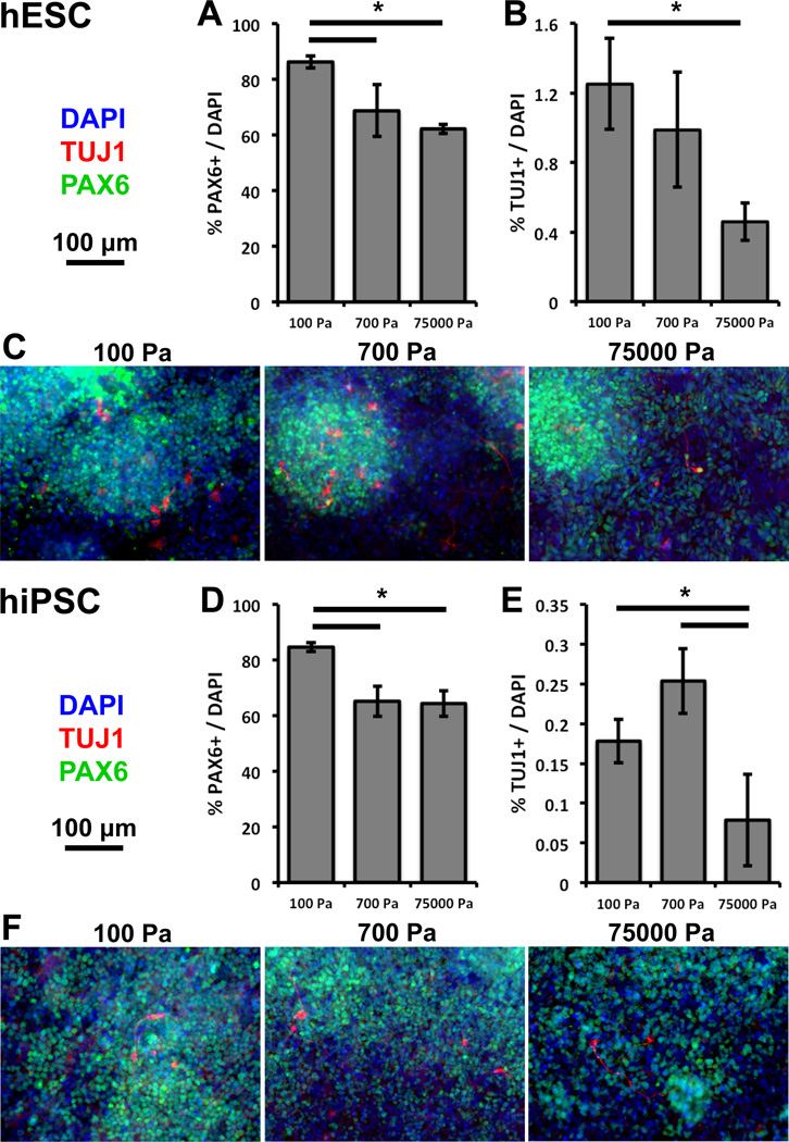
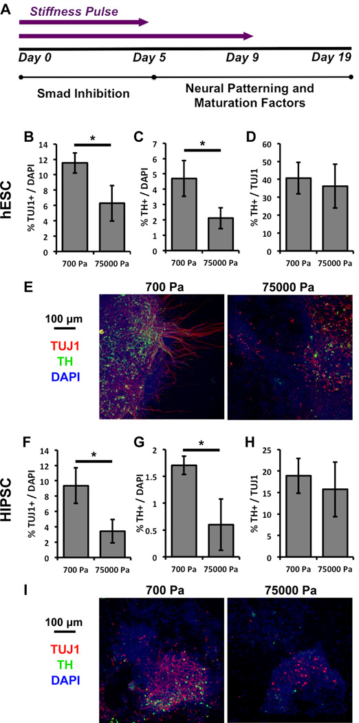
Human pluripotent stem cells (hPSCs) are of great interest in biology and medicine due to their ability to self-renew and differentiate into any adult or fetal cell type. Important efforts have identified biochemical factors, signaling pathways, and transcriptional networks that regulate hPSC biology. However, recent work investigating the effect of biophysical cues on mammalian cells and adult stem cells suggests that the mechanical properties of the microenvironment, such as stiffness, may also regulate hPSC behavior. While several studies have explored this mechanoregulation in mouse embryonic stem cells (mESCs), it has been challenging to extrapolate these findings and thereby explore their biomedical implications in hPSCs. For example, it remains unclear whether hPSCs can be driven down a given tissue lineage by providing tissue-mimetic stiffness cues. Here we address this open question by investigating the regulation of hPSC neurogenesis by microenvironmental stiffness. We find that increasing extracellular matrix (ECM) stiffness in vitro increases hPSC cell and colony spread area but does not alter self-renewal, in contrast to past studies with mESCs. However, softer ECMs with stiffnesses similar to that of neural tissue promote the generation of early neural ectoderm. This mechanosensitive increase in neural ectoderm requires only a short 5-day soft stiffness "pulse", which translates into downstream increases in both total neurons as well as therapeutically relevant dopaminergic neurons. These findings further highlight important differences between mESCs and hPSCs and have implications for both the design of future biomaterials as well as our understanding of early embryonic development.


Figures

Figure 1
Figure 1
hPSC colony area increases with substrate stiffness. Quantification of colony area and phase contrast images of human (A) embryonic H1 and (B) induced pluripotent MSC-iPS stem cells cultured on 100, 700, and 75000 Pa polyacrylamide substrates after 3 days in self-renewal conditions. Error bars are 95% confidence intervals, n = 220–300 colonies. *p < .05 (ANOVA, Tukey-Kramer). Abbreviations: ANOVA, analysis of variance; MSC-iPS, mesencymal stem cell-induced pluripotent stem cell; hPSC, human pluripotent stem cell; hESC, human embryonic stem cell; hiPSC, human induced pluripotent stem cell.
Figure 2
Figure 2
hPSC pluripotency marker expression is not altered by substrate stiffness. hESCs (left) and hiPSCs (right) show similar (A) cell numbers per cell culture surface area and (B) expression of pluripotency markers after 3 days in self-renewal conditions. (C) Substrate stiffness does not alter the expression of pluripotency marker Tra-1-60 even with the withdrawal of growth factors. Error bars are 95% confidence intervals, n = 3. *p < .05 (ANOVA, Tukey-Kramer). Abbreviations: ANOVA, analysis of variance; hESC, human embryonic stem cell; hiPSC, human induced pluripotent stem cell.
Figure 3
Figure 3
Softer substrates promote neural ectodermal and neuronal differentiation from hPSCs after 9 days of differentiation. Compared to 700 and 75000 Pa substrates, 100 Pa substrates promote PAX6 gene expression in cultures derived from (A) hESCs and (D) hiPSCs. Compared to 75000 Pa substrates, 100 and 700 Pa substrates promote neuronal marker TUJ1 gene expression from cultures derived from (B) hESCs and (E) hiPSCs. Representative immunofluorescence images of cultures derived from (C) hESCs and (F) hiPSCs. Red-TUJ1, Green-PAX6, Blue-DAPI. Error bars are 95% confidence intervals, n = 3. *p < .05 (ANOVA, Tukey-Kramer). Abbreviations: ANOVA, analysis of variance; hPSC, human pluripotent stem cell; hESC, human embryonic stem cell; hiPSC, human induced pluripotent stem cell.
Figure 4
Figure 4
Softer substrates promote neuronal and early ectodermal differentiation from hPSCs after 9 days in culture. (A) 100 Pa substrates promote PAX6 gene expression in (left) hESCs and (right) hiPSCs. (B) 100 and 700 Pa substrates promote TUJ1 gene expression in hPSCs. (C) 100 and 700 Pa substrates promote SOX1 gene expression in hPSCs. Error bars are 95% confidence intervals, n = 4. *p < .05 (ANOVA, Tukey-Kramer). Abbreviations: ANOVA, analysis of variance; hPSC, human pluripotent stem cell; hESC, human embryonic stem cell; hiPSC, human induced pluripotent stem cell; PS, polystyrene.
Figure 5
Figure 5
Softer substrates promote the differentiation of hPSCs into neurons and dopaminergic neurons but do not change the proportion of dopaminergic to total neurons after 19 days of differentiation. The percentage of cells (DAPI+) that are TUJ1+ (A-hESC, E-hiPSC) and TH+ (B-hESC, F-hiPSC) decreases with increasing substrate stiffness. The percentage of TUJ1+ cells that are also TH+ does not change with substrate stiffness (C-hESC, G-hiPSC). Representative immunofluorescence images of cultures derived from (D) hESCs and (H) hiPSCs. Red-TUJ1, Green-TH, Blue-DAPI. Error bars are 95% confidence intervals, n = 4. *p < .05 (ANOVA, Tukey-Kramer). Abbreviations: ANOVA, analysis of variance; hPSC, human pluripotent stem cell; hESC, human embryonic stem cell; hiPSC, human induced pluripotent stem cell; PS, polystyrene.
Figure 6
Figure 6
An early substrate stiffness signal is sufficient to recapitulate the full differentiation phenotypes of hPSCs. (A) A shortened substrate stiffness pulse (from 9 to 5 days) prior to neural patterning is sufficient to modulate neuronal and dopaminergic differentiation to the same extent as a 9 day pulse. The percentage of cells (DAPI+) that are TUJ1+ (B-hESC, F-hiPSC) and TH+ (C-hESC, G-hiPSC) decreases with increasing substrate stiffness. The ratio of TH+ to TUJ1+ cells does not change with substrate stiffness (D-hESC, H-hiPSC). Representative immunofluorescence images of cultures derived from (E) hESCs and (I) hiPSCs. Red-TUJ1, Green-TH, Blue-DAPI. Error bars are 95% confidence intervals, n = 4. *p < .05 (ANOVA, Tukey-Kramer). Abbreviations: ANOVA, analysis of variance; hESC, human embryonic stem cell; hiPSC, human induced pluripotent stem cell; PS, polystyrene.

Similar articles

Cited by

References

    1. Chambers SM, Fasano Ca, Papapetrou EP, Tomishima M, Sadelain M, Studer L. Highly efficient neural conversion of human ES and iPS cells by dual inhibition of SMAD signaling. Nature biotechnology. 2009;27:275–280. - PMC - PubMed
    2. Kriks S, Shim J-W, Piao J, Ganat YM, Wakeman DR, Xie Z, Carrillo-Reid L, Auyeung G, Antonacci C, Buch A, Yang L, Beal MF, Surmeier DJ, Kordower JH, Tabar V, Studer L. Dopamine neurons derived from human ES cells efficiently engraft in animal models of Parkinson,Äôs disease. Nature. 2011;480:547–551. - PMC - PubMed
    1. Takahashi K, Tanabe K, Ohnuki M, Narita M, Ichisaka T, Tomoda K, Yamanaka S. Induction of pluripotent stem cells from adult human fibroblasts by defined factors. Cell. 2007;131:861–872. - PubMed
    2. Yu J, Vodyanik Ma, Smuga-Otto K, Antosiewicz-Bourget J, Frane JL, Tian S, Nie J, Jonsdottir Ga, Ruotti V, Stewart R, Slukvin II, Thomson Ja. Induced pluripotent stem cell lines derived from human somatic cells. Science (New York, N.Y.) 2007;318:1917–1920. - PubMed
    1. Yan Y, Yang D, Zarnowska ED, Du Z, Werbel B, Valliere C, Pearce Ra, Thomson Ja, Zhang S-C. Directed differentiation of dopaminergic neuronal subtypes from human embryonic stem cells. Stem cells (Dayton, Ohio) 2005;23:781–790. - PMC - PubMed
    1. Chen X, Xu H, Yuan P, Fang F, Huss M, Vega VB, Wong E, Orlov YL, Zhang W, Jiang J, Loh Y-H, Yeo HC, Yeo ZX, Narang V, Govindarajan KR, Leong B, Shahab A, Ruan Y, Bourque G, Sung W-K, Clarke ND, Wei C-L, Ng H-H. Integration of external signaling pathways with the core transcriptional network in embryonic stem cells. Cell. 2008;133:1106–1117. - PubMed
    2. Desbordes SC, Placantonakis DG, Ciro A, Socci ND, Lee G, Djaballah H, Studer L. High-throughput screening assay for the identification of compounds regulating self-renewal and differentiation in human embryonic stem cells. Cell stem cell. 2008;2:602–612. - PMC - PubMed
    3. Loh Y-H, Yang L, Yang JC, Li H, Collins JJ, Daley GQ. Genomic Approaches to Deconstruct Pluripotency. Annual review of genomics and human genetics. 2010 - PMC - PubMed
    1. Engler AJ, Sen S, Sweeney HL, Discher DE. Matrix elasticity directs stem cell lineage specification. Cell. 2006;126:677–689. - PubMed
    2. Fu J, Wang Y-k, Yang MT, Desai Ra, Yu X, Liu Z, Chen CS. Mechanical regulation of cell function with geometrically modulated elastomeric substrates. Nature methods. 2010;7:733–736. - PMC - PubMed
    3. Keung AJ, de Juan-Pardo EM, Schaffer DV, Kumar S. Rho GTPases Mediate the Mechanosensitive Lineage Commitment of Neural Stem Cells. Stem cells (Dayton, Ohio) 2011:1886–1897. - PMC - PubMed

Publication types