. 2012 Aug 23;488(7412):527-30.
doi: 10.1038/nature11344.

Defining the mode of tumour growth by clonal analysis

Affiliations

Defining the mode of tumour growth by clonal analysis

Gregory Driessens et al. Nature. .

Abstract

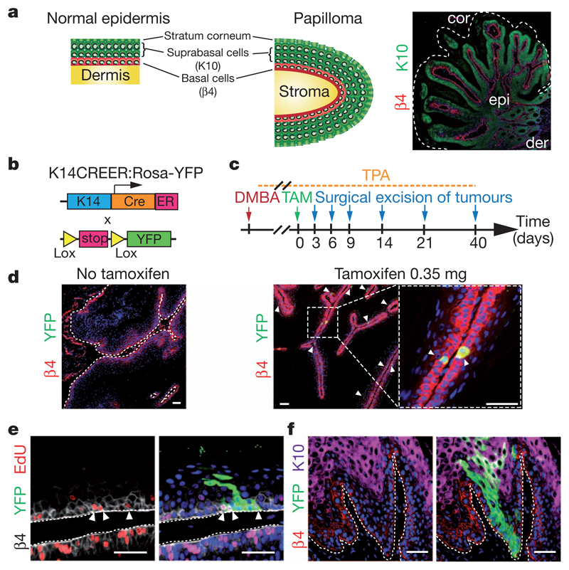
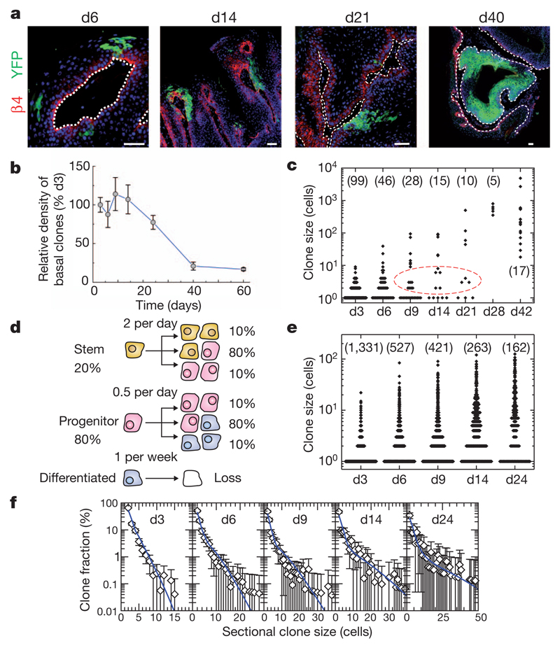
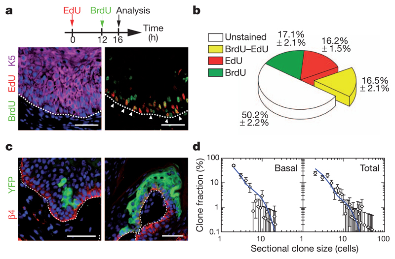
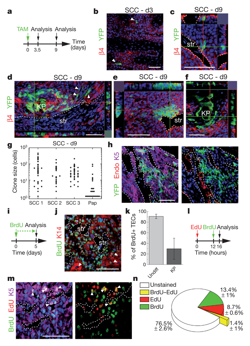
Recent studies using the isolation of a subpopulation of tumour cells followed by their transplantation into immunodeficient mice provide evidence that certain tumours, including squamous skin tumours, contain cells with high clonogenic potential that have been referred to as cancer stem cells (CSCs). Until now, CSC properties have only been investigated by transplantation assays, and their existence in unperturbed tumour growth is unproven. Here we make use of clonal analysis of squamous skin tumours using genetic lineage tracing to unravel the mode of tumour growth in vivo in its native environment. To this end, we used a genetic labelling strategy that allows individual tumour cells to be marked and traced over time at different stages of tumour progression. Surprisingly, we found that the majority of labelled tumour cells in benign papilloma have only limited proliferative potential, whereas a fraction has the capacity to persist long term, giving rise to progeny that occupy a significant part of the tumour. As well as confirming the presence of two distinct proliferative cell compartments within the papilloma, mirroring the composition, hierarchy and fate behaviour of normal tissue, quantitative analysis of clonal fate data indicates that the more persistent population has stem-cell-like characteristics and cycles twice per day, whereas the second represents a slower cycling transient population that gives rise to terminally differentiated tumour cells. Such behaviour is shown to be consistent with double-labelling experiments and detailed clonal fate characteristics. By contrast, measurements of clone size and proliferative potential in invasive squamous cell carcinoma show a different pattern of behaviour, consistent with geometric expansion of a single CSC population with limited potential for terminal differentiation. This study presents the first experimental evidence for the existence of CSCs during unperturbed solid tumour growth.


Conflict of interest statement

The authors declare no competing financial interests.

Figures

Figure 1
Figure 1. Genetic tracing of skin papilloma at clonal density.
a, Scheme of the stereotypical architecture of papilloma similar to normal epidermis. cor, stratum corneum; der, dermis; epi, epithelial cells. b, Scheme of genetic strategy to induce YFP expression in papilloma. ER, tamoxifen-responsive hormone-binding domain of the oestrogen receptor; K14, human keratin 14 promoter. c, Protocol of DMBA/TPA and tamoxifen (TAM) administration. d, Immunostainings for β4-integrin (β4) and YFP in papilloma with or without TAM. e, Immunostaining for β4-integrin, YFP and 5-ethynyl-2′-deoxyuridine (EdU) in a papilloma 6 days post-labelling. f, Immunostaining for β4 integrin, YFP and K10 in a papilloma 14 days post-labelling. Scale bars, 50 μm.
Figure 2
Figure 2. Quantitative clonal analysis in skin papilloma.
a, Immunostainings for β4-integrin and YFP in papilloma after TAM administration. b, Quantification of the relative density of basal clones over time (n = 666, 383, 328, 194, 162, 35 and 33 clones from at least 5 different tumours at day 3, 6, 9, 14, 24, 40 and 60, respectively). c, Total size of clones derived from 3D serial reconstruction over time. The n value is shown. d, Tumour growth model showing the proliferative stem/progenitor cells. e, Clone sizes derived from single sections over time. The n value is shown. f, Fit of the hierarchical model (d) to the corresponding cumulative sectional clonal size distribution. Points show data (e) and the blue lines show the model prediction. Scale bars, 50 μm. Error bars denote s.e.m.
Figure 3
Figure 3. Challenging the stem/progenitor cell model in papilloma.
a, Immunostaining for BrdU, EdU and K5 in papilloma from mice treated following the protocol schemed above. b, Quantification of the proportion of unmarked cells, BrdU, EdU and EdU–BrdU double-labelled cells within the basal layer of papilloma (n = 5,512 cells from 8 papilloma). c, Immunostaining for YFP and β4-integrin on papilloma 9 days after TAM administration. d, Comparison between the model prediction (blue lines), with parameters defined in Fig. 2d and in the text, and the clone size distribution (n = 105 clones from 7 tumours). Scale bars, 50 μm. Error bars denote s.e.m.
Figure 4
Figure 4. Quantitative clonal analysis of squamous cell carcinoma.
a, Protocol of TAM administration and clonal analysis in SCC. b, Immunostaining for YFP and β4-integrin in SCC 3 days after TAM administration. c–f, Same as in b 9 days after TAM administration. KP, keratin pearl; str, stroma. g, Total size of clones quantified from serial 30 μm sections 9 days after TAM administration (n = 37, 25, 42 and 28 clones in SCC 1, 2, 3 and papilloma (Pap), respectively). h, Immunostaining for YFP, K5 and endoglin (Endo) in SCC 9 days after TAM administration. i, Protocol of BrdU administration. j, Immunostaining for BrdU and K14 in SCC. k, Quantification of the BrdU+ tumour epithelial cells (TECs) within SCC after 5 days of continuous BrdU administration (n = 676 undifferentiated tumour cells and 282 KP cells). Error bars denote s.d. Undiff, undifferentiated. l, Protocol of BrdU and EdU administration. m, Immunostaining for BrdU, EdU and K5 in SCC from mice treated following the protocol shown in l. n, Quantification of the proportion of unmarked cells, BrdU, EdU and EdU–BrdU double-labelled cells within SCC (n = 9,010 cells from 2 SCC). Scale bars, 50 μm.

Comment in

Similar articles

  • SOX2 controls tumour initiation and cancer stem-cell functions in squamous-cell carcinoma.
    Boumahdi S, Driessens G, Lapouge G, Rorive S, Nassar D, Le Mercier M, Delatte B, Caauwe A, Lenglez S, Nkusi E, Brohée S, Salmon I, Dubois C, del Marmol V, Fuks F, Beck B, Blanpain C. Boumahdi S, et al. Nature. 2014 Jul 10;511(7508):246-50. doi: 10.1038/nature13305. Epub 2014 Jun 8. Nature. 2014. PMID: 24909994
  • Fate mapping of human glioblastoma reveals an invariant stem cell hierarchy.
    Lan X, Jörg DJ, Cavalli FMG, Richards LM, Nguyen LV, Vanner RJ, Guilhamon P, Lee L, Kushida MM, Pellacani D, Park NI, Coutinho FJ, Whetstone H, Selvadurai HJ, Che C, Luu B, Carles A, Moksa M, Rastegar N, Head R, Dolma S, Prinos P, Cusimano MD, Das S, Bernstein M, Arrowsmith CH, Mungall AJ, Moore RA, Ma Y, Gallo M, Lupien M, Pugh TJ, Taylor MD, Hirst M, Eaves CJ, Simons BD, Dirks PB. Lan X, et al. Nature. 2017 Sep 14;549(7671):227-232. doi: 10.1038/nature23666. Epub 2017 Aug 30. Nature. 2017. PMID: 28854171 Free PMC article.
  • A vascular niche and a VEGF-Nrp1 loop regulate the initiation and stemness of skin tumours.
    Beck B, Driessens G, Goossens S, Youssef KK, Kuchnio A, Caauwe A, Sotiropoulou PA, Loges S, Lapouge G, Candi A, Mascre G, Drogat B, Dekoninck S, Haigh JJ, Carmeliet P, Blanpain C. Beck B, et al. Nature. 2011 Oct 19;478(7369):399-403. doi: 10.1038/nature10525. Nature. 2011. PMID: 22012397
  • Skin precancer.
    Brash DE, Pontén J. Brash DE, et al. Cancer Surv. 1998;32:69-113. Cancer Surv. 1998. PMID: 10489624 Review.
  • Breast-cancer stem cells-beyond semantics.
    Badve S, Nakshatri H. Badve S, et al. Lancet Oncol. 2012 Jan;13(1):e43-8. doi: 10.1016/S1470-2045(11)70191-7. Lancet Oncol. 2012. PMID: 22225725 Review.

Cited by

References

    1. Shackleton M, Quintana E, Fearon ER, Morrison SJ. Heterogeneity in cancer: cancer stem cells versus clonal evolution. Cell. 2009;138:822–829. - PubMed
    1. Lobo NA, Shimono Y, Qian D, Clarke MF. The biology of cancer stem cells. Annu Rev Cell Dev Biol. 2007;23:675–699. - PubMed
    1. Malanchi I, et al. Cutaneous cancer stem cell maintenance is dependent on β-catenin signalling. Nature. 2008;452:650–653. - PubMed
    1. Beck B, et al. A vascular niche and a VEGF–Nrp1 loop regulate the initiation and stemness of skin tumours. Nature. 2011;478:399–403. - PubMed
    1. Schober M, Fuchs E. Tumor-initiating stem cells of squamous cell carcinomas and their control by TGF-β and integrin/focal adhesion kinase (FAK) signaling. Proc Natl Acad Sci USA. 2011;108:10544–10549. - PMC - PubMed

Publication types

MeSH terms