. 2012 Oct 11;490(7419):283-7.
doi: 10.1038/nature11398. Epub 2012 Aug 1.

HIV-infected T cells are migratory vehicles for viral dissemination

Affiliations

HIV-infected T cells are migratory vehicles for viral dissemination

Thomas T Murooka et al. Nature. .

Abstract

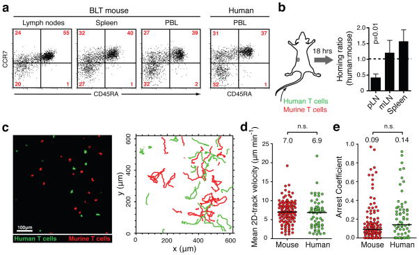
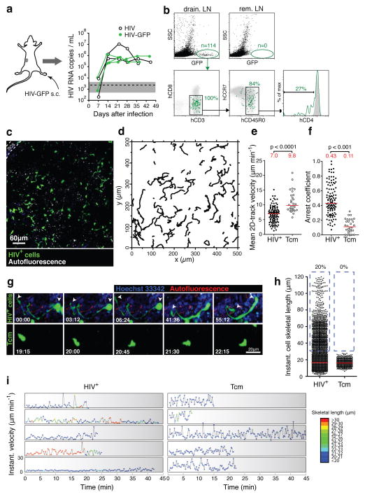
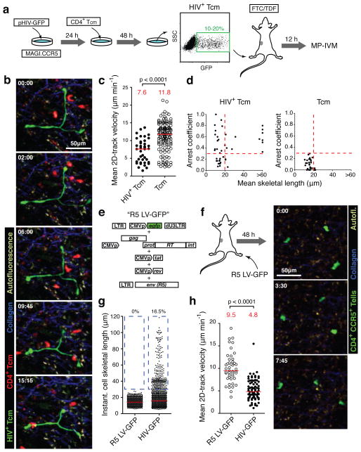
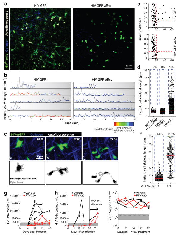
After host entry through mucosal surfaces, human immunodeficiency virus-1 (HIV-1) disseminates to lymphoid tissues to establish a generalized infection of the immune system. The mechanisms by which this virus spreads among permissive target cells locally during the early stages of transmission and systemically during subsequent dissemination are not known. In vitro studies suggest that the formation of virological synapses during stable contacts between infected and uninfected T cells greatly increases the efficiency of viral transfer. It is unclear, however, whether T-cell contacts are sufficiently stable in vivo to allow for functional synapse formation under the conditions of perpetual cell motility in epithelial and lymphoid tissues. Here, using multiphoton intravital microscopy, we examine the dynamic behaviour of HIV-infected T cells in the lymph nodes of humanized mice. We find that most productively infected T cells migrate robustly, resulting in their even distribution throughout the lymph node cortex. A subset of infected cells formed multinucleated syncytia through HIV envelope-dependent cell fusion. Both uncoordinated motility of syncytia and adhesion to CD4(+) lymph node cells led to the formation of long membrane tethers, increasing cell lengths to up to ten times that of migrating uninfected T cells. Blocking the egress of migratory T cells from the lymph nodes into efferent lymph vessels, and thus interrupting T-cell recirculation, limited HIV dissemination and strongly reduced plasma viraemia. Thus, we have found that HIV-infected T cells are motile, form syncytia and establish tethering interactions that may facilitate cell-to-cell transmission through virological synapses. Migration of T cells in lymph nodes therefore spreads infection locally, whereas their recirculation through tissues is important for efficient systemic viral spread, suggesting new molecular targets to antagonize HIV infection.


Figures

FIGURE 1
FIGURE 1. Human T cell migration in lymph nodes of BLT mice
a. Human CD4+T cells with naïve (CD45RA+ CCR7+), central memory (CD45RA, CCR7+) and effector memory (CD45RA, CCR7) phenotype are represented in lymphoid organs and peripheral blood of BLT mice. Relative frequencies in blood are similar to humans. Plots are gated on hCD4+, hCD3+ cells. b. Homing of human and murine CD4+ T cells into lymphoid tissues (pLN=peripheral, mLN=mesenteric LNs). Data pooled from three independent experiments. Values are means±s.e.m. c. Multiphoton intravital micrograph of a BLT popliteal LN 24 hours after adoptive transfer of human (green) and murine (red) T cells. Graph on the right shows migratory tracks of each population during a 30-minute recording. d, e. Mean 2D-track velocities (d) and arrest coefficients (e) of murine and human T cells in BLT LNs. Lines and numbers indicate medians. Data pooled from three recordings/two independent experiments.
FIGURE 2
FIGURE 2. In vivo dynamics and phenotype of HIV-infected LN cells
a. Footpad injection of BLT mice with HIV-GFP produces robust and sustained viremia. ‘HIV’ is identical to HIV-GFP but lacks an IRES-GFP cassette. Similar results as shown here for 5 mice were obtained with other routes of infection (Supplementary Fig. 2d). Dashed line and grey-shaded area indicate mean and 95% confidence interval of background signals obtained from plasma of uninfected mice. b. Draining and non-draining LN cells two days after footpad infection with HIV-GFP. Grey dot plots and histograms show GFPSSClow LN cells. rem. LN: remote LNs. c. An intravital micrograph recorded from a popLN two days after footpad infection with HIV-GFP. d. Migratory tracks of GFP+ LN cells during a 30-minute recording. e, f. Mean 2D-track velocities (e) and arrest coefficients (f) of HIV+ LN cells compared to uninfected, GFP-expressing Tcm, recorded in LNs of uninfected BLT mice. Lines and numbers indicate medians. Data on HIV-infected LN cells and Tcm are representative of four and two independent experiments, resp. g. MP-IVM time-lapse recordings of an HIV-infected LN cell (top) and an uninfected Tcm (bottom) in BLT LNs. Arrows indicate leading and trailing edge of the infected cell. Elapsed time in min:sec. h. Instantaneous cell skeletal length of HIV-infected LN cells and Tcm from recordings as shown in (g). Lines indicate medians. Percentages indicate events>30 μm, highlighted by dashed blue box. i. Representative traces of infected LN cells and Tcm showing instantaneous cell skeletal length (color-coded) and instantaneous migratory velocity over time. Traces selected from 142 recorded in 4 movies/3 independent experiments.
FIGURE 3
FIGURE 3. HIV induces an elongated phenotype in infected T cells
a. Analysis of in vitro HIV-infected Tcm in BLT LNs. FTC: Emtricitabine; TDF: Tenofovir. b. MP-IVM time-lapse series of uninfected (red) and HIV+ (green) Tcm in a BLT LN. Elapsed time in min:sec. c. Mean 2D-track velocities. Red lines and numbers indicate medians. d. Correlation of mean cell skeletal length and arrest coefficient of individual cells. Dashed lines indicate threshold values based on measurements of uninfected Tcm. Data pooled from two independent experiments. e. Production of an R5-tropic, GFP-expressing lentiviral vector. f. 48 hours after footpad injection of 1.4 × 10⁵ infectious units, GFP+ cells are found in draining LNs. g, h. These cells do not elongate (g) and do not show the reduction of cell motility of HIV-infected LN cells (h). Percentages in (g) refer to cells >30 μm, highlighted by dashed blue box. The red lines and numbers in (h) indicate medians. Data are representative of two independent experiments/three mice.
FIGURE 4
FIGURE 4. HIV-infected T cells tether to other LN cells and form syncytia through Env, and migrate to distant tissues to disseminate infection
a. Intravital micrographs from LNs of BLT mice injected 48 hours earlier with HIV-GFP (left) in one, and HIV-GFP ΔEnv (right) into the other footpad. b. Representative traces of infected T cells depicting instantaneous cell skeletal length (color-coded) and migratory velocity over time. Traces selected from 281 recorded in 14 movies/3 independent experiments. c. Mean cell skeletal length and arrest coefficient of individual cells infected with HIV-GFP or HIV-GFP ΔEnv. Data are pooled from six and eight recordings, respectively, from three independent experiments. d. Cell lengths of HIV-GFP-infected Tcm injected into NK cell-depleted, antibody-deficient DHLMP2A mice, and of BLT LN cells infected in situ with HIV-GFP D368R or HIV-GFP. e. Recording of LN cells infected with HIV-nGFP. Bottom panels indicate border between cytoplasm and nuclei, based on a 80% of maximum fluorescence intensity (FI) threshold. f. Cell lengths of mono- and multinucleated, HIV-infected cells. g, h. Viremia in NSG BLT mice treated with FTY720 or vehicle starting at the day of s.c. HIV-infection via the cheek skin. g. Infection with HIV-GFP. h. Infection with clone SF162R3. FTY720 treatment was ended at day 56. i. Plasma viremia under FTY-treatment started four weeks after in i.p-infection, when virema had stabilized. One representative experiment of two is shown.

Comment in

Similar articles

Cited by

References

    1. Haase AT. Early events in sexual transmission of HIV and SIV and opportunities for interventions. Annu Rev Med. 2011;62:127–39. - PubMed
    1. Sattentau Q. Avoiding the void: cell-to-cell spread of human viruses. Nat Rev Microbiol. 2008;6:815–26. - PubMed
    1. Gebhardt T, et al. Different patterns of peripheral migration by memory CD4+ and CD8+ T cells. Nature. 2011;477:216–9. - PubMed
    1. Mempel TR, Henrickson SE, von Andrian UH. T-cell priming by dendritic cells in lymph nodes occurs in three distinct phases. Nature. 2004;427:154–9. - PubMed
    1. Melkus MW, et al. Humanized mice mount specific adaptive and innate immune responses to EBV and TSST-1. Nat Med. 2006;12:1316–22. - PubMed

Publication types