Specific capture and temperature-mediated release of cells in an aptamer-based microfluidic device
- PMID: 22854859
- PMCID: PMC3976991
- DOI: 10.1039/c2lc40411g
Specific capture and temperature-mediated release of cells in an aptamer-based microfluidic device
Abstract
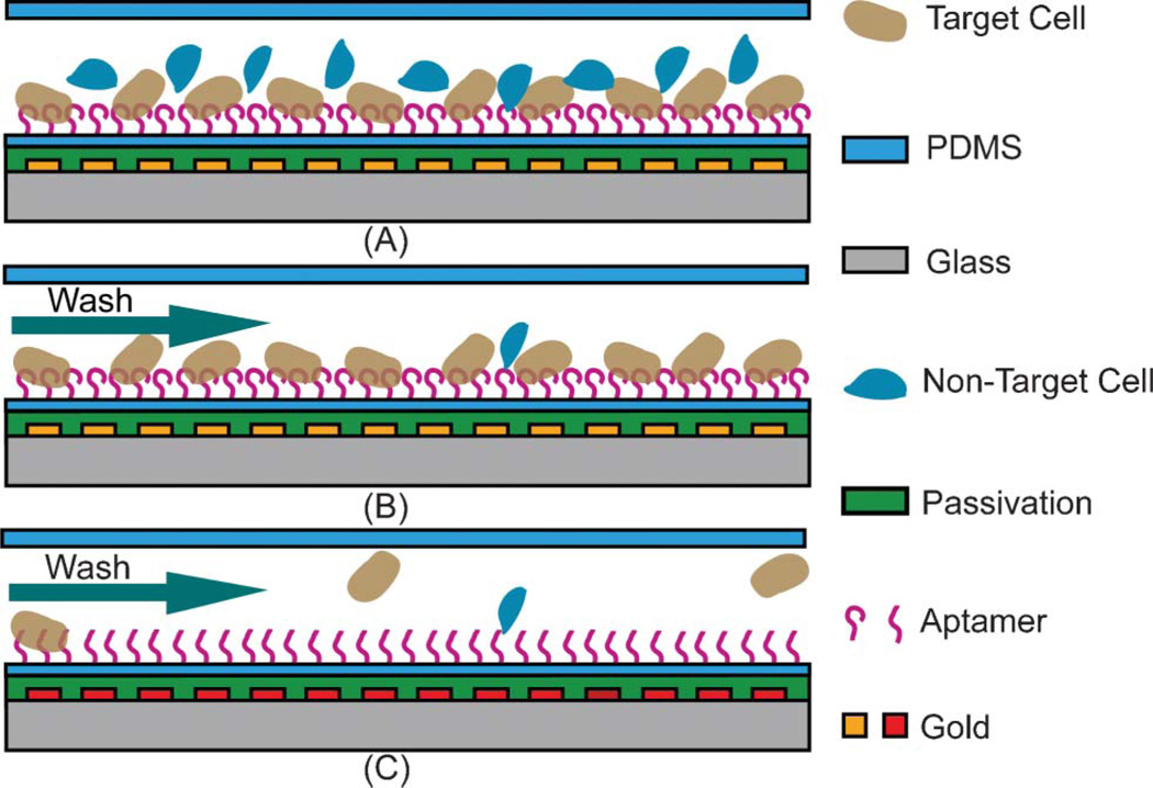
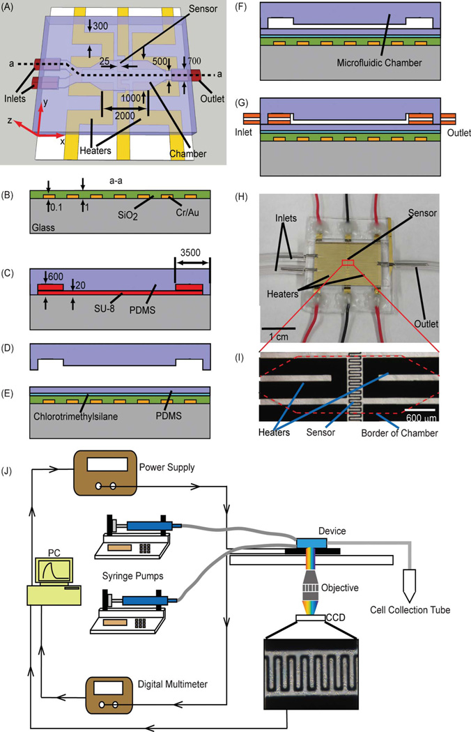
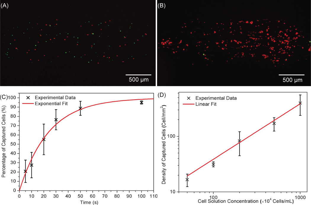
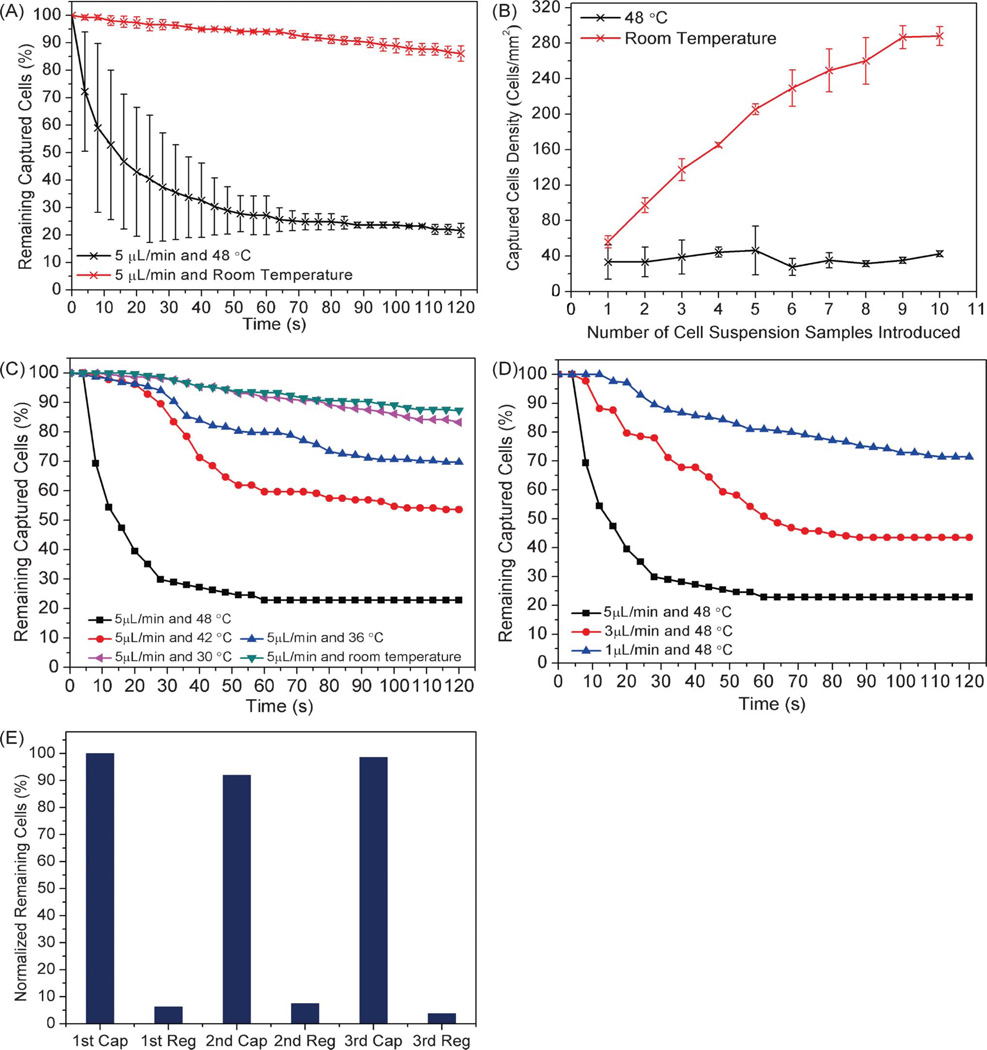
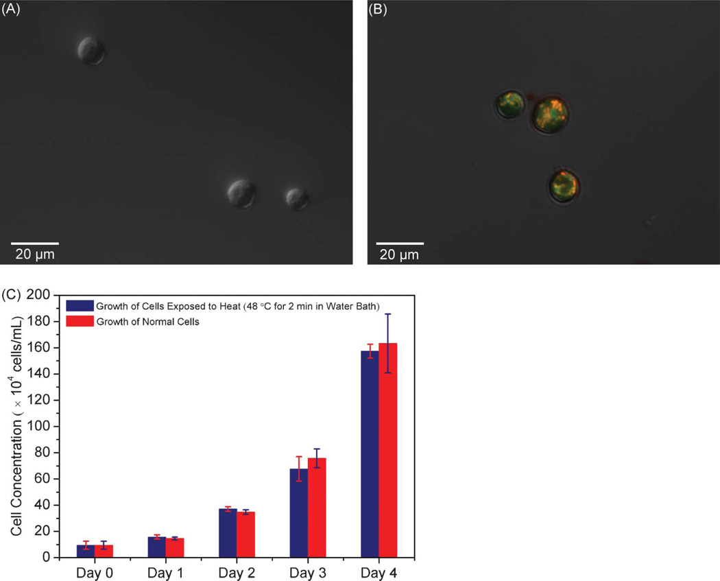
Isolation of cells from heterogeneous mixtures is critically important in both basic cell biology studies and clinical diagnostics. Cell isolation can be realized based on physical properties such as size, density and electrical properties. Alternatively, affinity binding of target cells by surface-immobilized ligands, such as antibodies, can be used to achieve specific cell isolation. Microfluidics technology has recently been used in conjunction with antibody-based affinity isolation methods to capture, purify and isolate cells with higher yield rates, better efficiencies and lower costs. However, a method that allows easy release and collection of live cells from affinity surfaces for subsequent analysis and detection has yet to be developed. This paper presents a microfluidic device that not only achieves specific affinity capture and enrichment, but also enables non-destructive, temperature-mediated release and retrieval of cells. Specific cell capture is achieved using surface-immobilized aptamers in a microchamber. Release of the captured cells is realized by a moderate temperature change, effected via integrated heaters and a temperature sensor, to reversibly disrupt the cell-aptamer interaction. Experimental results with CCRF-CEM cells have demonstrated that the device is capable of specific capture and temperature-mediated release of cells, that the released cells remain viable and that the aptamer-functionalized surface is regenerable.
Figures


Similar articles
-
Spatially selective release of aptamer-captured cells by temperature mediation.IET Nanobiotechnol. 2014 Mar;8(1):2-9. doi: 10.1049/iet-nbt.2013.0028. IET Nanobiotechnol. 2014. PMID: 24888185 Free PMC article.
-
Enrichment of cancer cells using aptamers immobilized on a microfluidic channel.Anal Chem. 2009 Feb 1;81(3):1033-9. doi: 10.1021/ac802092j. Anal Chem. 2009. PMID: 19115856 Free PMC article.
-
Aptamer-enabled efficient isolation of cancer cells from whole blood using a microfluidic device.Anal Chem. 2012 May 1;84(9):4199-206. doi: 10.1021/ac3005633. Epub 2012 Apr 17. Anal Chem. 2012. PMID: 22482734 Free PMC article.
-
Fundamentals of affinity cell separations.Electrophoresis. 2018 Mar;39(5-6):732-741. doi: 10.1002/elps.201700311. Epub 2017 Oct 25. Electrophoresis. 2018. PMID: 28960354 Review.
-
Screening of aptamers on microfluidic systems for clinical applications.Sensors (Basel). 2012;12(7):9514-29. doi: 10.3390/s120709514. Epub 2012 Jul 11. Sensors (Basel). 2012. PMID: 23012556 Free PMC article. Review.
Cited by
-
Nanotechnology-Assisted Isolation and Analysis of Circulating Tumor Cells on Microfluidic Devices.Micromachines (Basel). 2020 Aug 14;11(8):774. doi: 10.3390/mi11080774. Micromachines (Basel). 2020. PMID: 32823926 Free PMC article. Review.
-
Nucleic acid aptamers in orthopedic diseases: promising therapeutic agents for bone disorders.Bone Res. 2025 Jul 24;13(1):71. doi: 10.1038/s41413-025-00447-8. Bone Res. 2025. PMID: 40707473 Free PMC article. Review.
-
Combined use of EpCAM and FRα enables the high-efficiency capture of circulating tumor cells in non-small cell lung cancer.Sci Rep. 2018 Jan 19;8(1):1188. doi: 10.1038/s41598-018-19391-1. Sci Rep. 2018. PMID: 29352248 Free PMC article.
-
Multivalent Aptamer Approach: Designs, Strategies, and Applications.Micromachines (Basel). 2022 Mar 12;13(3):436. doi: 10.3390/mi13030436. Micromachines (Basel). 2022. PMID: 35334728 Free PMC article. Review.
-
Fundamentals and application of magnetic particles in cell isolation and enrichment: a review.Rep Prog Phys. 2015 Jan;78(1):016601. doi: 10.1088/0034-4885/78/1/016601. Epub 2014 Dec 4. Rep Prog Phys. 2015. PMID: 25471081 Free PMC article. Review.
References
Publication types
MeSH terms
Substances
Grants and funding
LinkOut - more resources
Full Text Sources
Other Literature Sources