

Epithelial machines of morphogenesis and their potential application in organ assembly and tissue engineering
- PMID: 22854913
- PMCID: PMC3664917
- DOI: 10.1007/s10237-012-0423-6
Epithelial machines of morphogenesis and their potential application in organ assembly and tissue engineering
Abstract
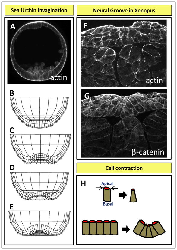
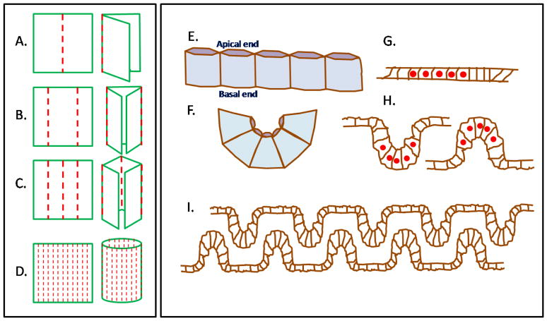
Sheets of embryonic epithelial cells coordinate their efforts to create diverse tissue structures such as pits, grooves, tubes, and capsules that lead to organ formation. Such cells can use a number of cell behaviors including contractility, proliferation, and directed movement to create these structures. By contrast, tissue engineers and researchers in regenerative medicine seeking to produce organs for repair or replacement therapy can combine cells with synthetic polymeric scaffolds. Tissue engineers try to achieve these goals by shaping scaffold geometry in such a way that cells embedded within these scaffold self-assemble to form a tissue, for instance aligning to synthetic fibers, and assembling native extracellular matrix to form the desired tissue-like structure. Although self-assembly is a dominant process that guides tissue assembly both within the embryo and within artificial tissue constructs, we know little about these critical processes. Here, we compare and contrast strategies of tissue assembly used by embryos to those used by engineers during epithelial morphogenesis and highlight opportunities for future applications of developmental biology in the field of tissue engineering.
Figures

Similar articles
-
Concise review: can the intrinsic power of branching morphogenesis be used for engineering epithelial tissues and organs?Stem Cells Transl Med. 2013 Dec;2(12):993-1000. doi: 10.5966/sctm.2013-0076. Epub 2013 Nov 4. Stem Cells Transl Med. 2013. PMID: 24191267 Free PMC article. Review.
-
Tubular collagen scaffolds with radial elasticity for hollow organ regeneration.Acta Biomater. 2017 Apr 1;52:1-8. doi: 10.1016/j.actbio.2017.02.005. Epub 2017 Feb 5. Acta Biomater. 2017. PMID: 28179160
-
Self-organization and branching morphogenesis of primary salivary epithelial cells.Tissue Eng. 2007 Apr;13(4):721-35. doi: 10.1089/ten.2006.0123. Tissue Eng. 2007. PMID: 17341161
-
The development of a bioengineered organ germ method.Nat Methods. 2007 Mar;4(3):227-30. doi: 10.1038/nmeth1012. Epub 2007 Feb 18. Nat Methods. 2007. PMID: 17322892
-
Calcium as a signal integrator in developing epithelial tissues.Phys Biol. 2018 May 16;15(5):051001. doi: 10.1088/1478-3975/aabb18. Phys Biol. 2018. PMID: 29611534 Free PMC article. Review.
Cited by
-
Tissue-specific roles of Fgfr2 in development of the external genitalia.Development. 2015 Jun 15;142(12):2203-12. doi: 10.1242/dev.119891. Development. 2015. PMID: 26081573 Free PMC article.
-
Tubular organ epithelialisation.J Tissue Eng. 2016 Dec 19;7:2041731416683950. doi: 10.1177/2041731416683950. eCollection 2016 Jan-Dec. J Tissue Eng. 2016. PMID: 28228931 Free PMC article. Review.
References
Publication types
MeSH terms
Grants and funding
LinkOut - more resources
Full Text Sources
