. 2012 Sep 21;287(39):32430-9.
doi: 10.1074/jbc.M112.397067. Epub 2012 Jul 31.

Alternative modes of binding of poly(ADP-ribose) polymerase 1 to free DNA and nucleosomes

Affiliations

Alternative modes of binding of poly(ADP-ribose) polymerase 1 to free DNA and nucleosomes

Nicholas J Clark et al. J Biol Chem. .

Abstract

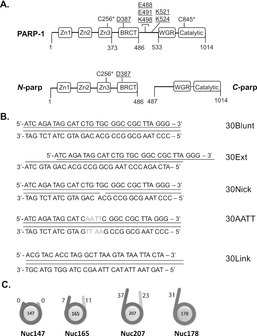
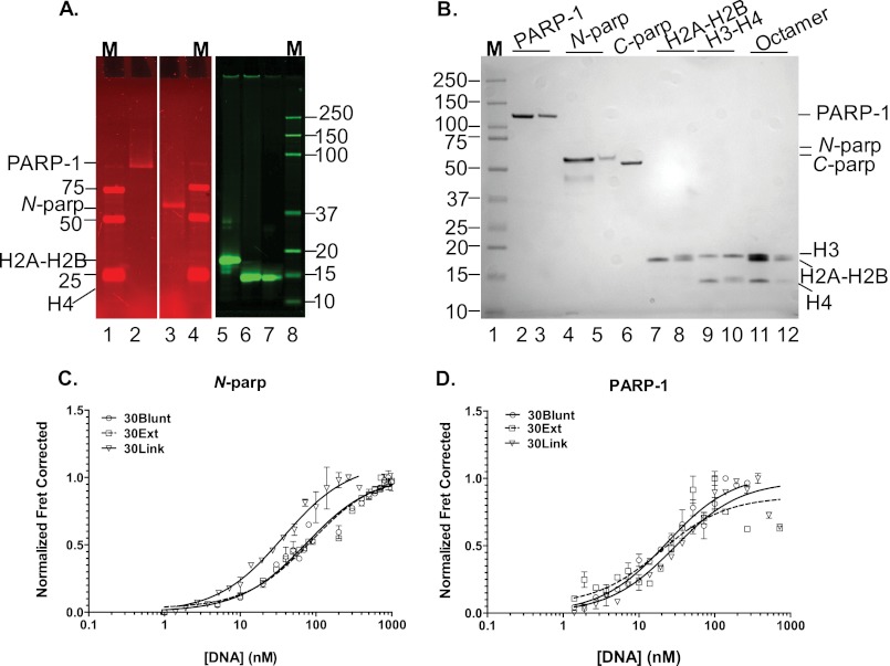
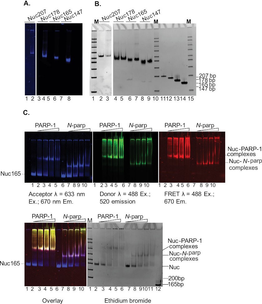
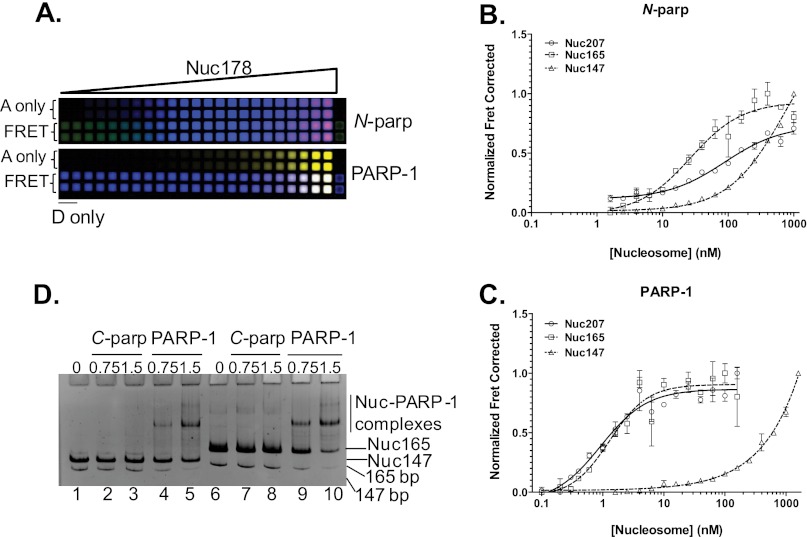
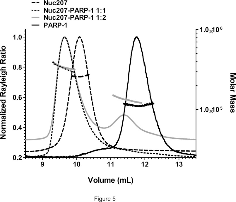
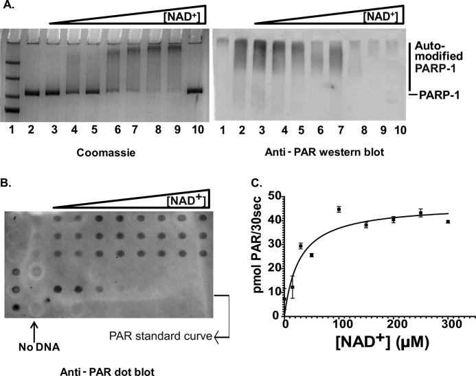
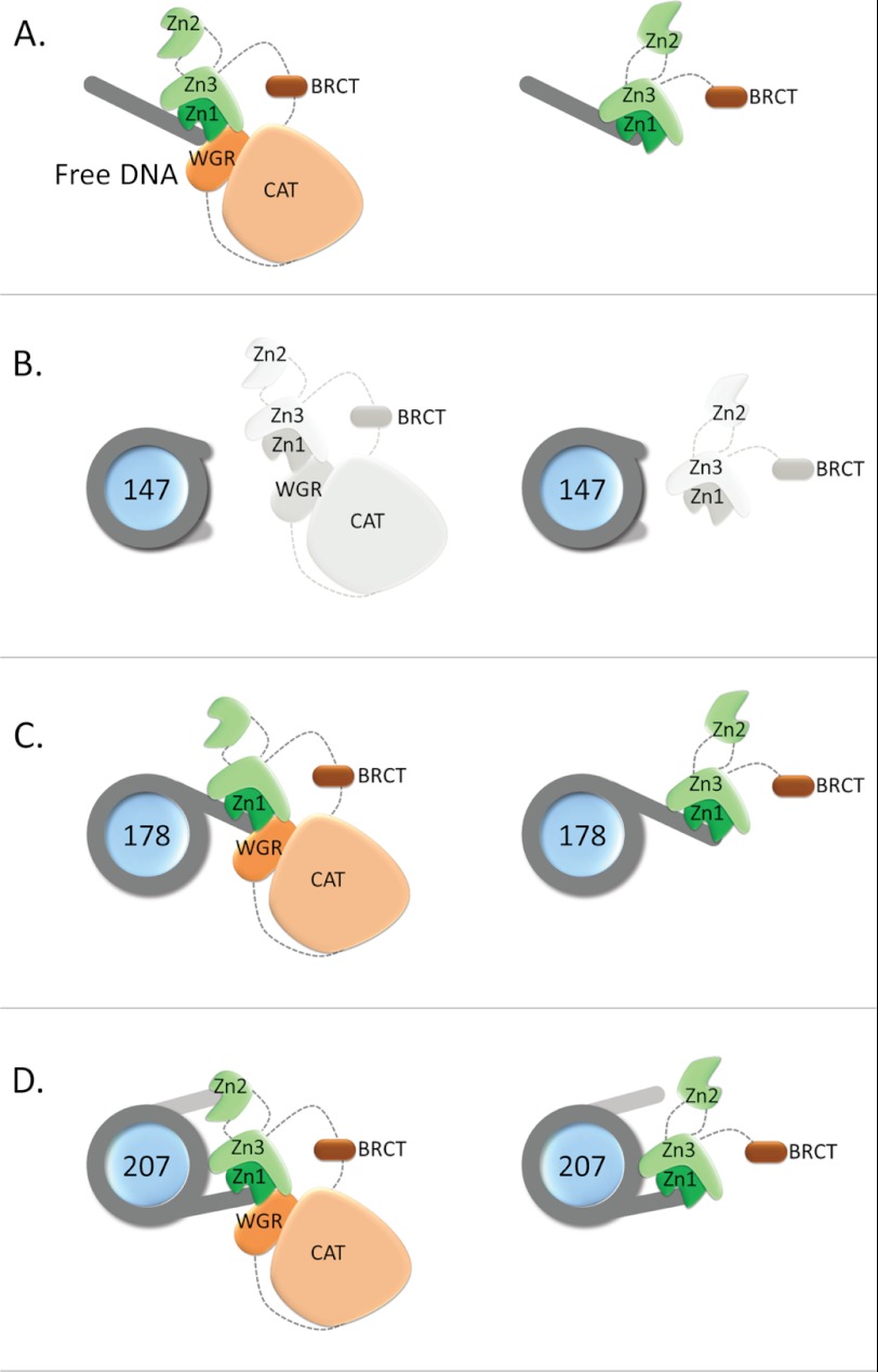
Poly(ADP-ribose) polymerase 1 (PARP-1) is an abundant nuclear protein that binds chromatin and catalyzes the transfer of ADP-ribose groups to itself and to numerous target proteins upon interacting with damaged DNA. The molecular basis for the dual role of PARP-1 as a chromatin architectural protein and a first responder in DNA repair pathways remains unclear. Here, we quantified the interactions of full-length PARP-1 and its N-terminal half with different types of DNA damage and with defined nucleosome substrates. We found that full-length PARP-1 prefers nucleosomes with two linker DNA extensions over any other substrate (including several free DNA models) and that the C-terminal half of PARP-1 is necessary for this selectivity. We also measured the ability of various substrates to activate PARP-1 activity and found that the most important feature for activation is one free DNA end rather than tight interaction with the activating nucleic acid. Our data provide insight into the different modes of interaction of this multidomain protein with nucleosomes and free DNA.


Figures

FIGURE 1.
FIGURE 1.
PARP-1 constructs and substrates assayed in this study. A, full-length PARP-1 contains all six domains; N-parp encompasses zinc fingers Zn1–Zn3 and the BRCT domain (amino acids 1–486); and C-parp spans residues 487-1014 and includes the WGR and CAT domains. Surface-exposed native cysteine residues (positions 256 and 845; indicated by asterisks) were labeled with Alexa Fluor 488. Underlined residues denote auto-PARylation sites (8). B, DNA models used for PARP-1 binding and activity assays. 30Blunt, 30Ext, and 30Nick are identical in sequence. 30AATT replaces 4 central bp with AATT. 30Link is identical in sequence to the linker in Nuc178. All DNA models were labeled at the 5′-end with Cy5 or ATTO 647N. C, nucleosome substrates were labeled with ATTO 647N at histone H4 E63C on the histone octamer (21). All nucleosomal DNA is based on the 601 positioning sequence (34). The length of the linker DNA in each particle is indicated in base pairs.
FIGURE 2.
FIGURE 2.
The CAT domain of PARP-1 contributes moderately to the interaction with DNA. A, fluorescently labeled PARP-1 constructs and histones. All samples were run on a Criterion XT 4-12% gradient SDS-polyacrylamide gel that was scanned on a Typhoon Imager at wavelengths appropriate for measuring donor (488 nm excitation and 520 nm emission) for the left panels depicting PARP-1 constructs and for measuring acceptor (633 nm excitation and 670 nm emission) for the right panel with labeled histones. Lanes 1, 4, and 8, molecular weight markers (M); lane 2, Alexa Fluor 488-labeled PARP-1 (at Cys-256 and Cys-845); lane 3, Alexa Fluor 488-labeled N-parp (at Cys-256); lane 5, H2A-H2B dimer (with ATTO 647N-labeled at histone H2B T112C); lane 6, (H3-H4)2 tetramer (labeled with ATTO 647N at histone H4 E63C); lane 7, histone octamer (labeled with ATTO 647N at histone H4 E36C). B, the same gel was visualized with Imperial stain. Lane 1, protein size marker; lane 2, unlabeled PARP-1; lane 3, labeled PARP-1; lane 4, unlabeled N-parp; lane 5, labeled N-parp; lane 6, C-parp; lane 7, unlabeled H2A-H2B; lane 8, labeled H2A-H2B; lane 9, unlabeled H3-H4; lane 10, labeled H3-H4; lane 11, unlabeled histone octamer; lane 12, labeled histone octamer. C, N-parp (labeled with Alexa Fluor 488 at Cys-256) binding to selected free DNA models shown in B measured by HI-FI FRET. D, PARP-1 binding curves for the same DNA fragments. The concentrations of the titrated acceptor species are plotted on the x axis, and normalized FRET-corrected values are plotted on the y axis (19, 24). Affinities from this and similar experiments are listed in Table 1. Error bars shown here were obtained from duplicates from individual representative experiments.
FIGURE 3.
FIGURE 3.
PARP-1 interacts with nucleosomes. A, fluorescently labeled nucleosome substrates. DNA fragments 207, 178, 165, and 147 bp in length, all containing the 601 positioning sequence, were assembled into nucleosomes with histone octamers labeled at histone H4 E63C with ATTO 647N. Nucleosomes were run on 5% native polyacrylamide gel and scanned on a Typhoon Imager at an emission wavelength of 670 nm. Lanes 2, 4, 6, and 8 are nucleosomes assembled on 207-, 178-, 165-, and 147-bp DNAs, respectively. Lanes 1, 3, 5, and 7 are unlabeled nucleosomes assembled on 207, 178, 165, and 147 bp DNA respectively. B, the same gel stained with ethidium bromide. Lanes 1 and 2 are labeled and unlabeled Nuc207, respectively. Lanes 3 and 4 are labeled and unlabeled Nuc178, respectively. Lanes 5 and 6 are labeled and unlabeled Nuc165, respectively. Lanes 7 and 8 are labeled and unlabeled Nuc147, respectively. Lanes 9–12 are 207-, 178-, 165-, and 147-bp DNA fragments, respectively. Note the absence of free DNA (<1%) in the nucleosome samples. C, ATTO 647N (acceptor)-labeled nucleosomes (Nuc165) were incubated with increasing amounts of Alexa Fluor 488-labeled PARP-1 or N-parp and analyzed by native PAGE. Gels were scanned on a Typhoon Imager at the indicated wavelengths. Lower left panel, acceptor, donor, and FRET channels are overlaid. Lanes 1 and 6, Nuc165; lanes 2–5, nucleosomes incubated with increasing molar ratios of PARP-1 (0.5-, 1-, 1.5-, and 2-fold excess); lanes 7–10, nucleosomes incubated with increasing molar ratios of N-parp (0.5-, 1-, 1.5-, and 2-fold excess); lane 11 in the lower right panel, free 165-bp DNA.
FIGURE 4.
FIGURE 4.
Quantification of interactions between PARP-1 and nucleosomes. A, HI-FI FRET plate assay. A portion of a typical 384-well plate is shown for Nuc178 and N-parp (upper panel) and full-length PARP-1 (lower panel). Increasing amounts of Nuc178 labeled with ATTO 647N at histone H4 E63C were titrated with a constant amount of either N-parp or PARP-1 labeled with Alexa Fluor 488. The upper two rows in each panel represent acceptor-only (A only) controls. The first two wells in the lower two rows in each panel are donor-only (D only) wells. FRET between the interacting partners is shown in the lower two rows in each panel (pink/purple). The plate was scanned using a Typhoon Imager as described for the gel in Fig. 2. Data from experiments were normalized, and the resulting curves were fit as described (19). Results from this plate are shown in Table 1. B and C, N-parp and PARP-1 interactions, respectively, with the various mononucleosome substrates. All values from this and similar experiments are summarized in Table 1. D, C-parp does not bind nucleosomes. Nucleosomes were incubated with C-parp or PARP-1 at increasing molar excess (as indicated) and loaded on a prerun 5% native TBE gel. Gels were stained with ethidium Bromide. C-parp did not interact with Nuc147 (lanes 2 and 3) or Nuc165 (lanes 7 and 8), whereas PARP-1 caused an upshift in both (lanes 4 and 5 for Nuc147 and lanes 9 and 10 for Nuc165).
FIGURE 5.
FIGURE 5.
One PARP-1 molecule binds per nucleosome. Shown are SEC-MALS profiles for Nuc207 and its complexes with PARP-1. Nuc207 formed a 1:1 complex with PARP-1 even when excess PARP-1 was added to the reaction mixture. The molecular weights for the various complexes derived from this and similar SEC-MALS experiments are listed in Table 2.
FIGURE 6.
FIGURE 6.
PARP-1 is activated by DNA. A, SDS-PAGE showing a shift in PARP-1 mobility as it undergoes auto-PARylation in the presence of 1 μm 30Blunt DNA and increasing concentrations (0, 10, 20, 50, 100, 200, and 400 μm in lanes 3–9, respectively) of NAD+ (left panel). The right panel is a Western blot of an identical gel probed with anti-PAR antibodies. Lane 1, protein size marker; lane 2, no NAD+; lanes 3–9, increasing amounts of NAD+; lane 10, no DNA in the presence of 400 μm NAD+. B, a slot blot of the above reaction was probed with anti-PAR antibody and ATTO 647N-conjugated secondary antibodies and visualized on a Typhoon scanner at 633-nm excitation and 670-nm emission wavelengths. C, the data were quantified using ImageQuant TL and analyzed in a Michaelis-Menten plot. A complete list of all parameters for this and other activators is listed in Table 3.
FIGURE 7.
FIGURE 7.
Model for N-parp and PARP-1 interactions with DNA and nucleosomes. A, interaction with free DNA as shown in Ref. . (The schematic is adapted from Ref. .) B, nucleosomes without linker do not interact with either PARP-1 construct. C, nucleosomes with one asymmetric linker arm likely interact similarly with PARP-1 as they do with free DNA, with some steric inhibition from the nucleosome, as indicated by the weaker binding affinities. D, interaction with nucleosomes with two linker arms. Both linker arms are engaged in PARP-1 binding, with Zn2 binding the second DNA linker. Regions from C-parp are required for correct orientation of Zn2.

Similar articles

Cited by

References

    1. Kraus W. L. (2008) Transcriptional control by PARP-1: chromatin modulation, enhancer binding, coregulation, and insulation. Curr. Opin. Cell Biol. 20, 294–302 - PMC - PubMed
    1. Lord C. J., Ashworth A. (2012) The DNA damage response and cancer therapy. Nature 481, 287–294 - PubMed
    1. Krishnakumar R., Kraus W. L. (2010) The PARP side of the nucleus: molecular actions, physiological outcomes, and clinical targets. Mol. Cell 39, 8–24 - PMC - PubMed
    1. Ji Y., Tulin A. V. (2010) The roles of PARP-1 in gene control and cell differentiation. Curr. Opin. Genet. Dev. 20, 512–518 - PMC - PubMed
    1. Krishnakumar R., Gamble M. J., Frizzell K. M., Berrocal J. G., Kininis M., Kraus W. L. (2008) Reciprocal binding of PARP-1 and histone H1 at promoters specifies transcriptional outcomes. Science 319, 819–821 - PubMed

Publication types

LinkOut - more resources