. 2012 Sep 21;287(39):32494-511.
doi: 10.1074/jbc.M112.368282. Epub 2012 Jul 31.

Human telomerase reverse transcriptase (hTERT) is a novel target of the Wnt/β-catenin pathway in human cancer

Affiliations

Human telomerase reverse transcriptase (hTERT) is a novel target of the Wnt/β-catenin pathway in human cancer

Yong Zhang et al. J Biol Chem. .

Abstract

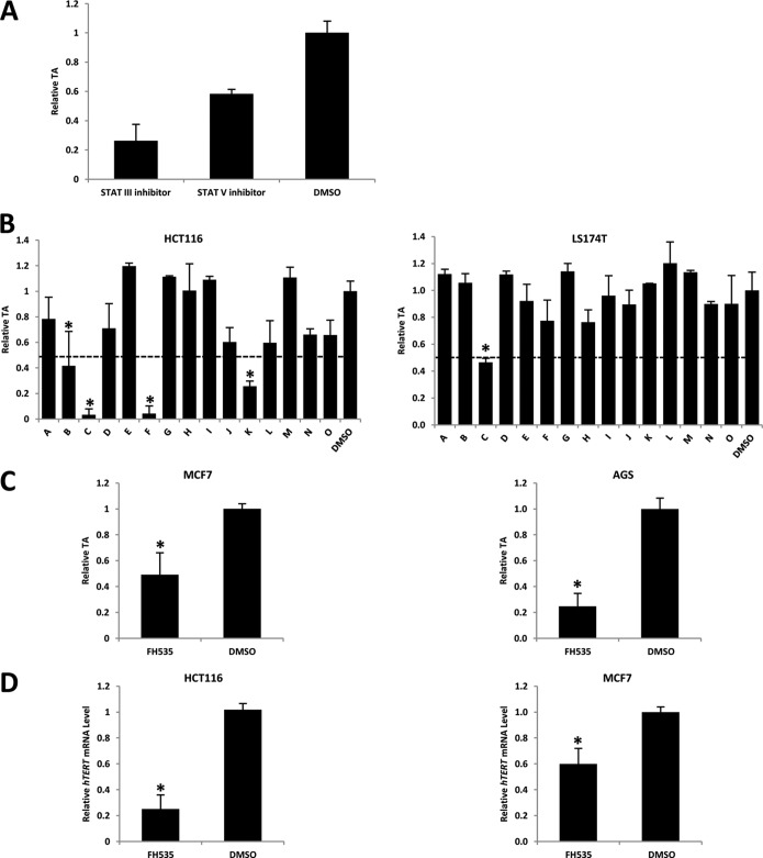
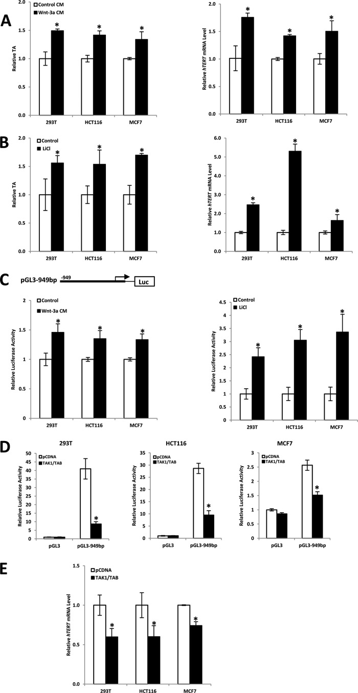
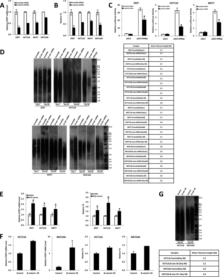
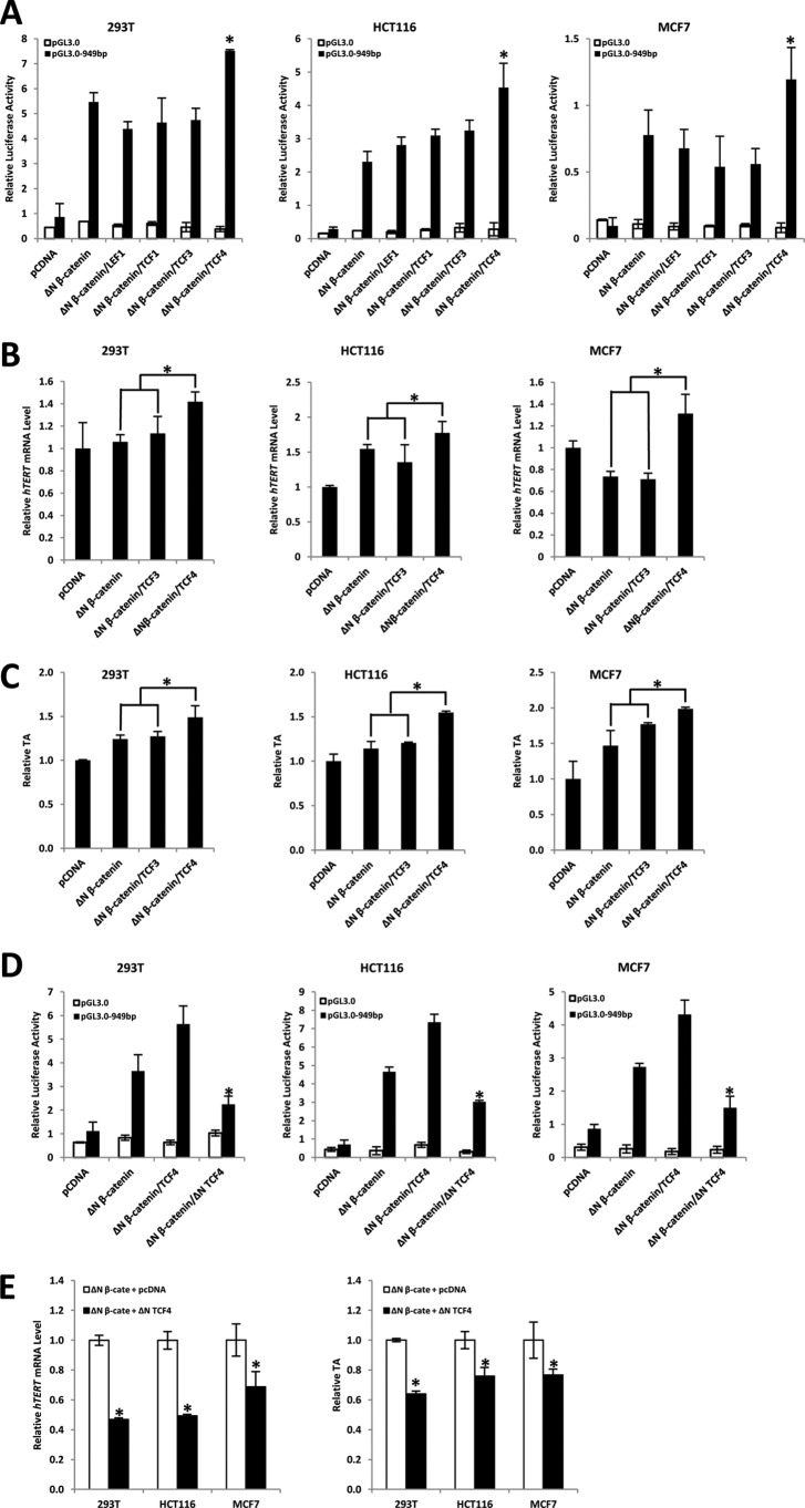
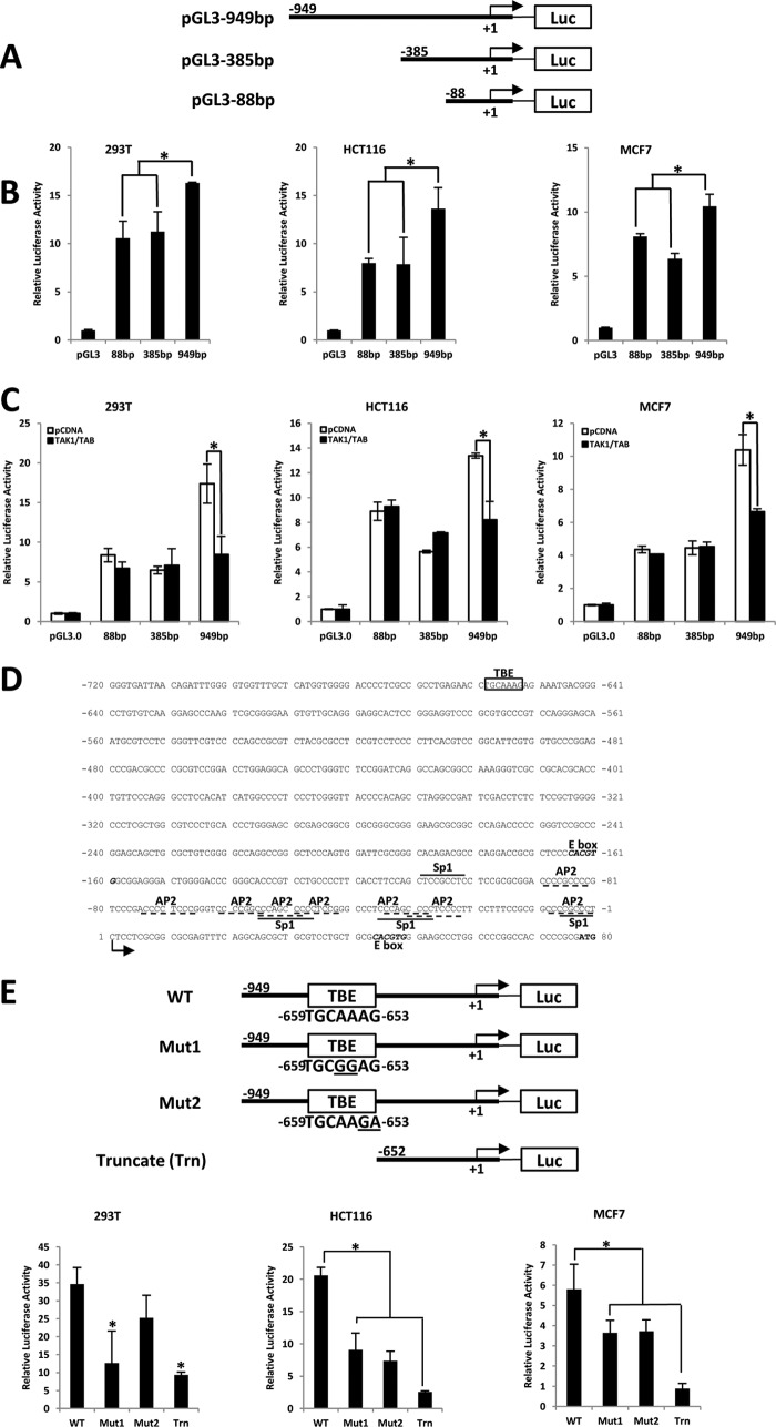
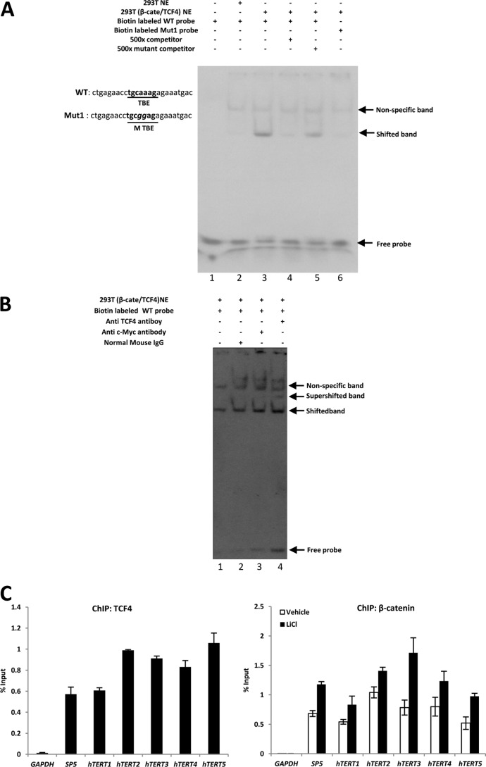
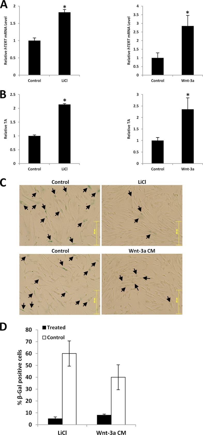
Telomerase activation plays a critical role in human carcinogenesis through the maintenance of telomeres, but the activation mechanism during carcinogenesis remains unclear. The human telomerase reverse transcriptase (hTERT) promoter has been shown to promote hTERT gene expression selectively in tumor cells but not in normal cells. Deregulation of the Wnt/β-catenin signaling pathway is reported to be associated with human carcinogenesis. However, little is known about whether the Wnt/β-catenin pathway is involved in activating hTERT transcription and inducing telomerase activity (TA). In this study, we report that hTERT is a novel target of the Wnt/β-catenin pathway. Transient activation of the Wnt/β-catenin pathway either by transfection of a constitutively active form of β-catenin or by LiCl or Wnt-3a conditioned medium treatment induced hTERT mRNA expression and elevated TA in different cell lines. Furthermore, we found that silencing endogenous β-catenin expression by β-catenin gene-specific shRNA effectively decreased hTERT expression, suppressed TA, and accelerated telomere shortening. Of the four members of the lymphoid-enhancing factor (LEF)/T-cell factor (TCF) family, only TCF4 showed more effective stimulation on the hTERT promoter. Ectopic expression of a dominant negative form of TCF4 inhibited hTERT expression in cancer cells. Through promoter mapping, electrophoretic mobility shift assay, and chromatin immunoprecipitation assay, we found that hTERT is a direct target of β-catenin·TCF4-mediated transcription and that the TCF4 binding site at the hTERT promoter is critical for β-catenin·TCF4-dependent expression regulation. Given the pivotal role of telomerase in carcinogenesis, these results may offer insight into the regulation of telomerase in human cancer.


Figures

FIGURE 1.
FIGURE 1.
Screening telomerase inhibitor from well known signaling pathway inhibitor libraries. A, STAT III/V inhibitors inhibit TA in HCT116 cells. 24 h before drug treatment, 4 × 10⁵ HCT116 cells were seeded into 12-well plates. The seeding density was chosen to obtain the optimum 60% confluence prior to drug treatment. Cells were treated with inhibitors or DMSO (vehicle control) and incubated for 48 h. After treatment, cells were harvested for qTRAP to measure TA. B, effect of Wnt pathway inhibitors on TA in HCT116 and LS174T cells. FH535, inhibitor C, is the most effective TA inhibitor. Inhibitors named A–O are listed in supplemental Table S1. The dotted line marks 50% TA inhibition, which was normalized with DMSO control. C, FH535 inhibitory effect on TA is validated in MCF7 and AGS cell lines. D, FH535 treatment leads to reduction of hTERT expression. Data were normalized against DMSO treatment. Data are the average of three independent experiments. *, p < 0.05. Error bars, S.D.
FIGURE 2.
FIGURE 2.
Activated Wnt/β-catenin directly affects hTERT gene expression and TA. A and B, Wnt-3a and LiCl stimulation of 293T, HCT116, and MCF7 cells increases hTERT gene expression and TA. Cells were treated with Wnt-3a CM or control CM (A) for 48 h or 15 mm LiCl for 24 h (B), respectively, and assayed for real-time PCR and qTRAP to measure TA (left) and hTERT expression level (right). C, activated Wnt pathway elevates hTERT promoter activities. A luciferase reporter construct, containing 0.95 kb upstream of the hTERT gene transcription initiation site, was transiently transfected into 293T, HCT116, and MCF7 cells. 24 h after transfection, cells were treated with Wnt-3a CM (left) or 15 mm LiCl (right), respectively. Wnt-3a CM or 15 mm LiCl could significantly activate hTERT promoter compared with controls. D and E, Wnt pathway negative regulator TAK1 represses hTERT promoter activities. 293T (Wnt-3a CM-treated), HCT116, and MCF7 cells were co-transfected with a 949-bp hTERT promoter-driven reporter construct and the indicated expression constructs for TAK1 and TAB (D). In another assay, after transfection with TAK1, cells were subjected to real-time PCR to measure hTERT expression (E). Empty expression vector pCDNA was used as a control. Expression of TAK1/TAB abolished hTERT promoter activities compared with control. Relative luciferase activity was standardized to Renilla luciferase activities. Data are the average of three independent experiments. *, p < 0.05. Error bars, S.D.
FIGURE 3.
FIGURE 3.
Effects of β-catenin knockdown on the hTERT gene and TA. A, real-time PCR analysis of hTERT expression in β-catenin knockdown stable 293T (Wnt-3a CM-treated), HCT116, MCF7, and MCF10A cells. hTERT mRNA levels were normalized by comparison with GAPDH as internal controls. -Fold change was calculated relative to the control. B, TRAP was carried out to measure TA in β-catenin knockdown stable cells and control cells. C, 949-bp hTERT promoter-driven luciferase reporter construct was transiently transfected into β-catenin knockdown stable cells: 293T (Wnt-3a CM-treated), HCT116, and MCF7. 24 h after transfection, cells were collected for luciferase activity measurement. β-Catenin knockdown could significantly inhibit hTERT promoter activity compared with controls. Relative luciferase activity was standardized to Renilla luciferase activities. Data are the average of three independent experiments. *, p < 0.05. D, telomere length was measured by Southern blotting in β-catenin knockdown stable cells and control cells. The table beside the Southern blot indicates the mean telomere length measured. E, expression construct containing the constitutively active form of β-catenin (ΔN β-catenin) was transiently transfected into 293T, HCT116, and MCF7 cells. 48 h after transfection, cells were harvested and subjected to real-time PCR or qTRAP to measure hTERT mRNA expression level (left) or TA (right). Empty expression vector pCDNA was used as a control. Data are the average of three independent experiments. *, p < 0.05. F, β-catenin overexpression (OE) stable HCT116 and MCF10A cell lines were obtained as described under “Experimental Procedures.” These stable cells were harvested and subjected to real-time PCR or qTRAP to measure hTERT mRNA expression level (top) or TA (bottom). Empty retroviral expression vector was used as a control. G, telomere length was measured by Southern blotting in β-catenin overexpression stable HCT116 and MCF10A cell lines. The table beside the Southern blot indicates the mean telomere length measured. Error bars, S.D.
FIGURE 4.
FIGURE 4.
β-Catenin·TCF4 specifically up-regulates promoter activity of hTERT. A, 293T, HCT116, and MCF7 cells were co-transfected with a 949-bp hTERT promoter-driven reporter construct, the indicated expression construct for ΔN β-catenin, and/or LEF1, TCF1, TCF3, and TCF4 expression constructs. pGL3.0 reporter plasmid was used as a control. Co-transfection of β-catenin and TCF4 specifically activates the hTERT promoter. B, 293T, HCT116, and MCF7 cells were transfected with the expression construct for ΔN β-catenin alone or with TCF3 or TCF4 expression constructs, respectively. Empty expression vector pCDNA was used as a control. 48 h after transfection, cells were harvested and subjected to real-time PCR to measure hTERT mRNA. Co-transfection of β-catenin and TCF4 could more efficiently increase hTERT expression. C, 293T, HCT116, and MCF7 cells were transfected with expression construct for ΔN β-catenin alone or with TCF3 or TCF4 expression constructs, respectively. Empty expression vector pCDNA was used as a control. 48 h after transfection, cells were harvested and subjected for qTRAP to measure TA. Co-transfection of β-catenin and TCF4 could more efficiently elevate TA. D, 293T, HCT116, and MCF7 cells were co-transfected with a 949-bp hTERT promoter-driven reporter construct or the indicated expression constructs for ΔN β-catenin, TCF4, or ΔN TCF4. pGL3.0 reporter plasmid was used as a control. Co-transfection of ΔN TCF4 significantly inhibited hTERT promoter activation. Relative luciferase activity was standardized to Renilla luciferase activities. E, 293T, HCT116, and MCF7 cells were co-transfected with expression construct for ΔN β-catenin and ΔN TCF4. pcDNA plasmid was used as a control. Co-transfection of ΔN TCF4 was able to reduce β-catenin-induced hTERT expression (left) and TA (right). Data are the average of three independent experiments. *, p < 0.05. Error bars, S.D.
FIGURE 5.
FIGURE 5.
Identification of TBE in the hTERT promoter region. A, schematic representation of various luciferase reporter plasmids of hTERT promoter (88, 385, and 949 bp). B, 949-bp hTERT promoter is most responsive to β-catenin·TCF4. 293T, HCT116, and MCF7 cells were transiently transfected with various luciferase reporter constructs containing 88, 385, and 949 bp (upstream of the transcriptional start site) of the hTERT promoter and β-catenin·TCF4 expression constructs. C, TAK1/TAB reduces promoter activity of the hTERT by β-catenin·TCF4. 293T, HCT116, and MCF7 cells were co-transfected with the indicated various hTERT promoter luciferase reporters and β-catenin·TCF4 expression constructs. TAK1/TAB overexpression leads to a significant reduction in 949-bp hTERT promoter activity only. D, the sequence of the distal and proximal promoter of hTERT. Putative protein binding sites for various transcription factors in the first 181 bp are indicated. The +1 indicates the first nucleotide of the hTERT mRNA. Consensus motifs for Sp1 and AP2 are underlined by solid and broken lines, respectively, and the E box (c-Myc binding site) consensus motif is in italic type. The putative TBE consensus motif is boxed. The initiating ATG codon is shown in boldface type. E, TBE is important for β-catenin·TCF4 function on the hTERT promoter. Schematic representation of various luciferase reporter plasmids of the hTERT promoter. The putative TCF4 binding element is located between −659 and −653 bp from the transcription initiation site. Construct WT contains the wild-type binding element; Mut1 has a 2-bp substitution (AA → GG, underlined); Mut2 has another 2-bp substitution (AG → GA, underlined); and Truncate (Trn) only consists of the 652-bp sequence upstream of the transcriptional start site of the hTERT promoter, which does not contain the putative TBE. 293T, HCT116, and MCF7 cells were transiently transfected with various luciferase reporter constructs (WT, Mut1, Mut2, and Truncate) and β-catenin·TCF4 expression constructs. The promoter reporter activity was normalized with Renilla luciferase reporter. Data are the average of three independent experiments. *, p < 0.05. Error bars, S.D.
FIGURE 6.
FIGURE 6.
Binding of TCF-4 and β-catenin to a putative TBE in the hTERT promoter. A, TBE is important and is the minimal sequence for TCF4 binding. Biotin-labeled wild-type TBE probe (lanes 1–5) and mutant TBE probe (lane 6) were incubated with 10 μg of 293T nuclear extracts or 293T nuclear extracts with β-catenin·TCF4 overexpression for 30 min. Competition experiments were performed by preincubating with a 500-fold molar excess of the unlabeled TBE probes (competitor; lane 4), or 500-fold molar excess of the unlabeled mutant TBE (mutant competitor; lane 5). The arrow indicates the position of specific transcription factor complexes. B, biotin-labeled TBE (lanes 1–4) were incubated with 10 μg of 293T cell nuclear extracts with β-catenin·TCF4 overexpression for 30 min. Antibody binding experiments were carried out following the vendor's instruction with anti-TCF4 antibody (lane 4), normal mouse IgG as control (lane 2), or anti-c-Myc antibody (lane 3). The arrows indicate the position of shifted and supershifted bands. C, TCF4 binds to the hTERT promoter in vivo. A ChIP assay was performed using anti-TCF4 or β-catenin antibodies and analyzed by real-time PCR. GAPDH was used as a negative control, and SP5 was used as a positive control. Error bars, S.D.
FIGURE 7.
FIGURE 7.
The Wnt/β-catenin signaling pathway is involved in telomerase reactivation in somatic cells. A and B, LiCl or Wnt-3a CM stimulation of BJ cells increases hTERT gene expression and reactivates telomerase. BJ cells were treated with LiCl or Wnt-3a and subjected to real-time PCR or TRAP to measure hTERT expression level (A) or TA (B), respectively. C, activation of the Wnt pathway could extend the life span of BJ cells. BJ cells were treated with LiCl or Wnt-3a CM for 20 population doublings, and the morphological changes were monitored under a microscope. Blue coloration in cells indicates senescence, indicated by arrows. Bar, 251 μm. D, bars represent the percentage of β-galactosidase-positive cells. Data are mean ± S.D. (error bars) from five images each.

Similar articles

Cited by

References

    1. Blackburn E. H. (1991) Telomeres. Trends Biochem. Sci. 16, 378–381 - PubMed
    1. Perrem K., Bryan T. M., Englezou A., Hackl T., Moy E. L., Reddel R. R. (1999) Repression of an alternative mechanism for lengthening of telomeres in somatic cell hybrids. Oncogene 18, 3383–3390 - PubMed
    1. Greider C. W., Blackburn E. H. (1989) A telomeric sequence in the RNA of Tetrahymena telomerase required for telomere repeat synthesis. Nature 337, 331–337 - PubMed
    1. Blasco M. A., Hahn W. C. (2003) Evolving views of telomerase and cancer. Trends Cell Biol. 13, 289–294 - PubMed
    1. Kim N. W., Piatyszek M. A., Prowse K. R., Harley C. B., West M. D., Ho P. L., Coviello G. M., Wright W. E., Weinrich S. L., Shay J. W. (1994) Specific association of human telomerase activity with immortal cells and cancer. Science 266, 2011–2015 - PubMed

Publication types

MeSH terms

LinkOut - more resources