. 2012 Sep 21;287(39):32535-45.
doi: 10.1074/jbc.M112.402727. Epub 2012 Aug 1.

DERP6 (ELP5) and C3ORF75 (ELP6) regulate tumorigenicity and migration of melanoma cells as subunits of Elongator

Affiliations

DERP6 (ELP5) and C3ORF75 (ELP6) regulate tumorigenicity and migration of melanoma cells as subunits of Elongator

Pierre Close et al. J Biol Chem. .

Abstract

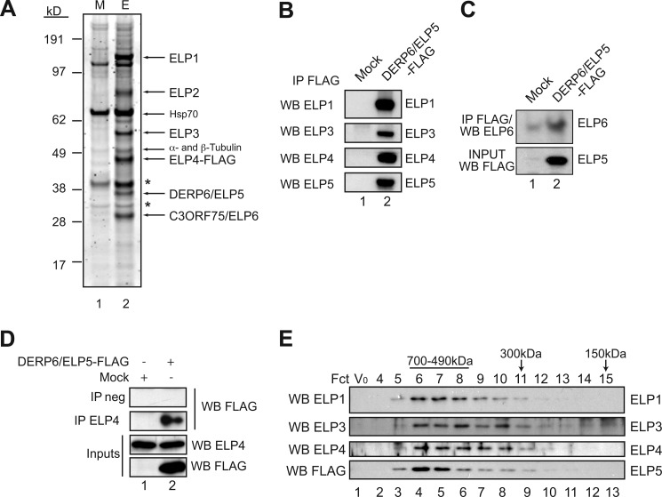
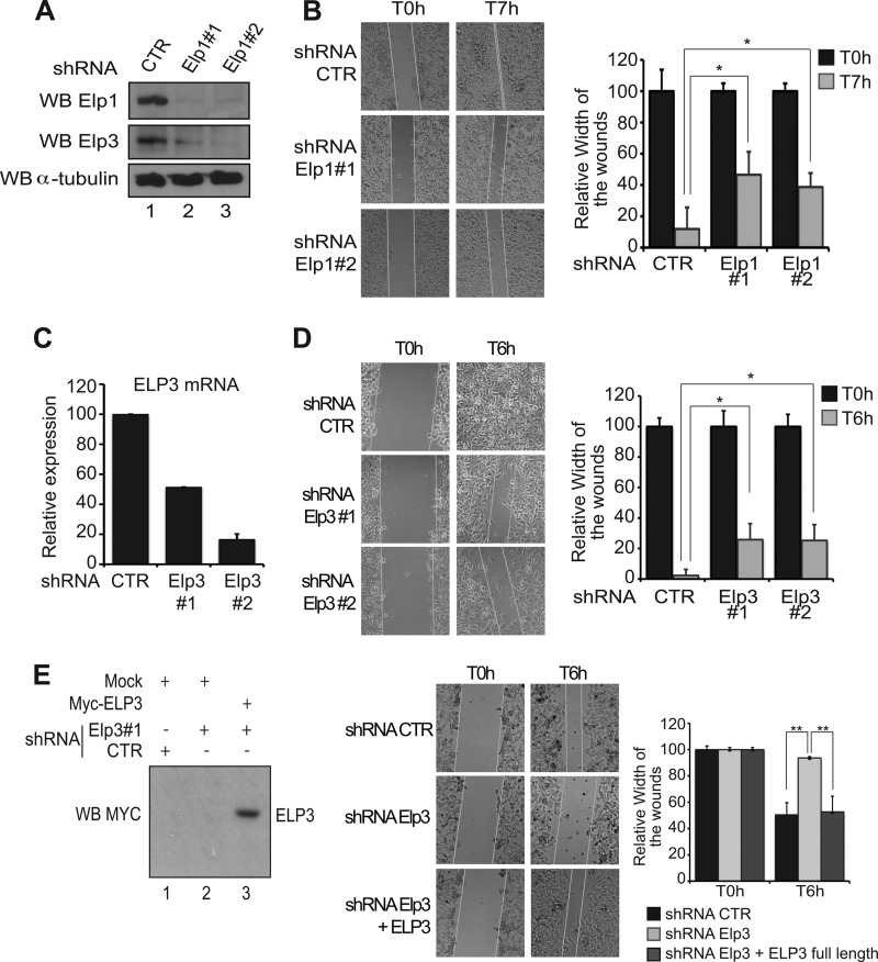
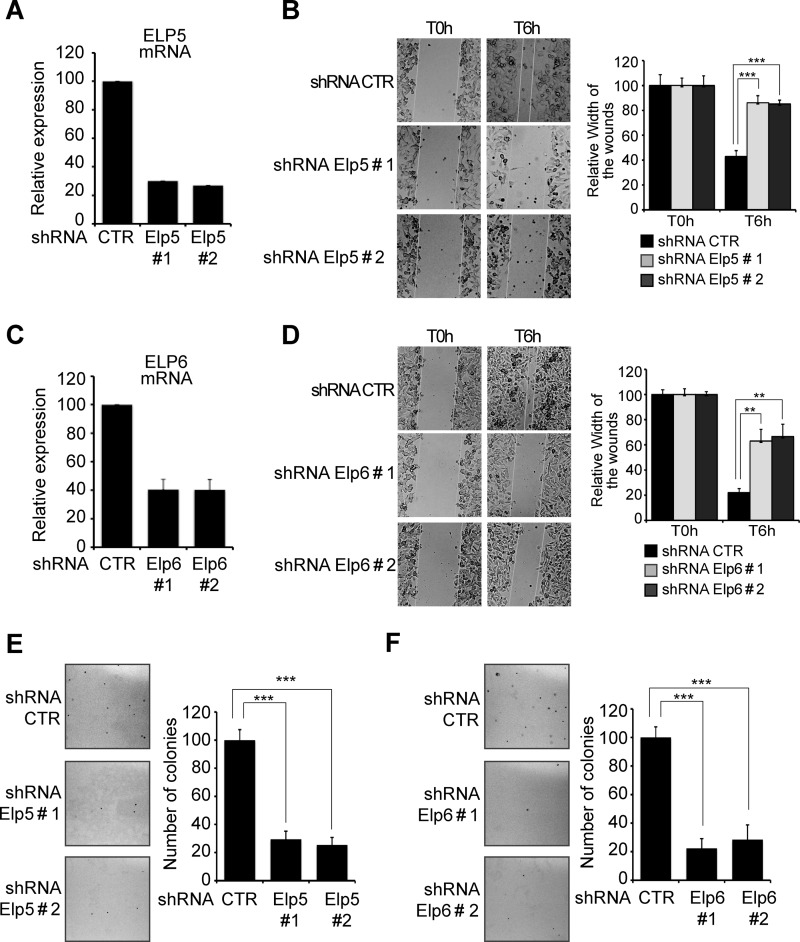
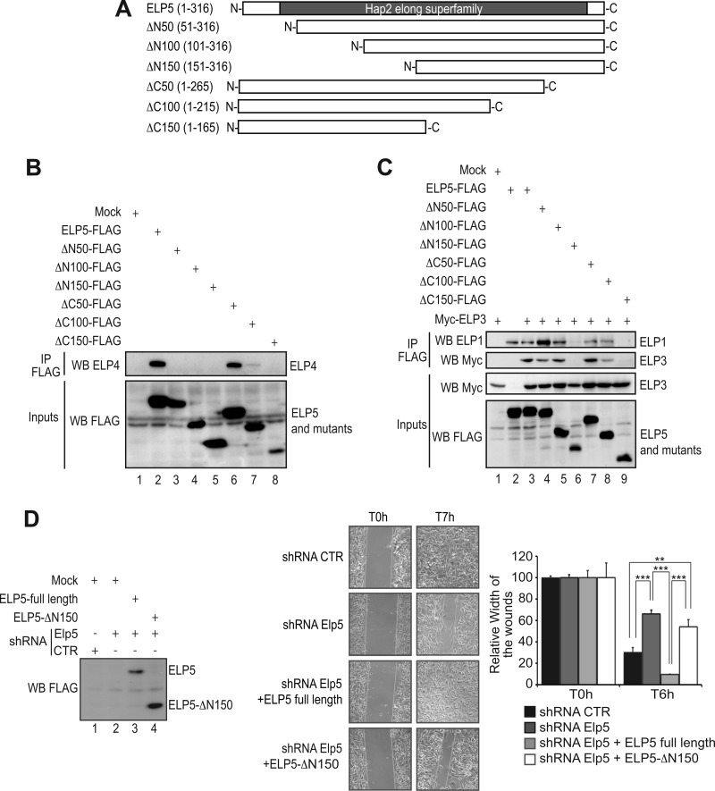
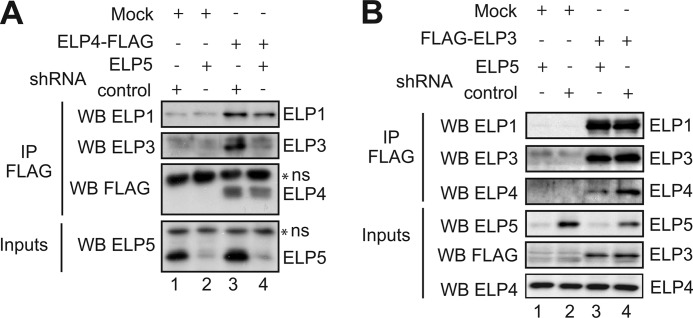
The Elongator complex is composed of 6 subunits (Elp1-Elp6) and promotes RNAPII transcript elongation through histone acetylation in the nucleus as well as tRNA modification in the cytoplasm. This acetyltransferase complex directly or indirectly regulates numerous biological processes ranging from exocytosis and resistance to heat shock in yeast to cell migration and neuronal differentiation in higher eukaryotes. The identity of human ELP1 through ELP4 has been reported but human ELP5 and ELP6 have remained uncharacterized. Here, we report that DERP6 (ELP5) and C3ORF75 (ELP6) encode these subunits of human Elongator. We further investigated the importance and function of these two subunits by a combination of biochemical analysis and cellular assays. Our results show that DERP6/ELP5 is required for the integrity of Elongator and directly connects ELP3 to ELP4. Importantly, the migration and tumorigenicity of melanoma-derived cells are significantly decreased upon Elongator depletion through ELP1 or ELP3. Strikingly, DERP6/ELP5 and C3ORF75/ELP6-depleted melanoma cells have similar defects, further supporting the idea that DERP6/ELP5 and C3ORF75/ELP6 are essential for Elongator function. Together, our data identify DERP6/ELP5 and C3ORF75/ELP6 as key players for migration, invasion and tumorigenicity of melanoma cells, as integral subunits of Elongator.


Figures

FIGURE 1.
FIGURE 1.
Identification of DERP6 and C3ORF75 as Elongator subunits. A, Elongator was purified from the cytoplasmic fraction of ELP4-FLAG expressing cells. Equal amount of the M2 chromatography eluates from Mock (M) and ELP4-FLAG (E) were separated by 4–12% SDS-PAGE and stained with Sypro Ruby. Bands were extracted and analyzed by mass spectrometry. Arrows indicate the co-eluting Elongator subunits as well as Hsp70, α- and β-tubulin (* shows nonspecific bands). B, Western blot analysis of M2 eluates from control (Mock) or DERP6/ELP5-FLAG expressing cells. Proteins were detected with antibodies as shown on the left. C, Anti-ELP6 Western blot analysis on anti-FLAG immunoprecipitates from control or DERP6/ELP5-FLAG-expressing cells (top panel). At the bottom, anti-ELP5 Western blot performed on the crude cell extracts (input). D, endogenous ELP4 was immunoprecipitated from Mock or DERP6/ELP5-FLAG expressing cells and proteins detected with antibodies listed on the right. E, M2-purified Elongator complex from B was analyzed by size exclusion chromatography. Vo is the void volume fraction. Proteins were detected by Western blotting using the antibodies indicated on the left.
FIGURE 2.
FIGURE 2.
ELP1, ELP3, ELP4, and ELP5 are mainly cytoplasmic. A, HEK293 cells were transfected with the indicated expression plasmids and immunofluorescence analysis were conducted using anti-Myc or -FLAG antibodies (left column). To-Pro (blue) was used to visualize the nuclei (middle and right columns). B, DERP6/ELP5-FLAG was transfected in HEK293 cells and cytoplasmic versus nuclear extracts (C and N, respectively) were isolated from the resulting cells. Those extracts were subjected to anti-FLAG, -α-tubulin, and -Histone H3 (cytoplasmic and nuclear markers, respectively), as indicated. A quantification of the cellular distribution of DERP6/ELP5 is illustrated as well.
FIGURE 3.
FIGURE 3.
Elp1 and Elp3 regulate cell motility of melanoma-derived B16-F10 cells. A, generation and characterization of Elp1-deficient B16-F10 cells. Anti-Elp1, -Elp3, and -α-tubulin (loading control) Western blot analysis were carried out using cell extracts from B16-F10 infected with lentiviral constructs delivering small hairpin RNAs targeting two distinct sequences of the Elp1 transcript, or a control sequence as a negative control (“shRNA Elp1#1,” “shRNA Elp1#2,” and “shRNA control (CTR),” respectively). B, migration of control (CTR) or Elp1-depleted (Elp1#1 or Elp1#2) melanoma-derived cells was measured by wound healing assay. Pictures were taken at the indicated times after the wound. A quantification of the data obtained is illustrated on the right. For each experimental condition, the width of the wound was set to 100% at time 0 and the width in other time points expressed relative to that. The figure shows the data from a representative experiment performed in triplicates (mean values + S.D.). C, generation and characterization of Elp3-depleted melanoma-derived cells. mRNA levels from B16-F10 cells infected with lentiviral constructs delivering small hairpin RNAs targeting two distinct sequences of the Elp3 transcript, or a control sequence (shRNA Elp3#1, shRNA Elp3#2, and shRNA CTR, respectively), were assessed by qRT-PCR. Elp3 mRNA levels in control B16-F10 cells were set to 100%, and mRNA levels in other experimental conditions are relative to that. The figure shows the data from a representative experiment performed in triplicates (mean values + S.D.). D, same as B, but using Elp3-depleted melanoma-derived generated in C. E, wound healing assays were conducted with shRNA control, shRNA Elp3 B16-F10 cells or with shRNA Elp3 B16-F10 cells transfected with full-length Myc-ELP3. A quantification of the data obtained is illustrated on the right. For each experimental condition, the width of the wound was set to 100% at time 0 and the width in other time points expressed relative to that. The figure shows the data from a representative experiment performed in triplicates (mean values + S.D.). On the left, anti-Myc Western blots were carried out with protein extracts from the indicated cells collected at the end of the wound healing assay. “Mock” denotes experimental conditions in which cells were transfected with a control plasmid.
FIGURE 4.
FIGURE 4.
Elongator affects tumorigenic potential of melanoma cells. A and B, ability of Elp1 (A) or Elp3 (B)-depleted B16-F10 cells to form colonies in soft agar was examined. The indicated cells were seeded in agar-containing media, as described under “Experimental Procedures,” and pictures were taken 2 weeks after seeding. The number of colonies observed in control B16-F10 cells was set to 100%, and the number of colonies obtained in other experimental conditions expressed relative to that. The figures show the data from a representative experiment performed in triplicates (mean values + S.D.). C, invasion of control or Elp1-depleted cells was evaluated by using a 3D cell culture system (described under “Experimental Procedures”). On the top, pictures were taken 3 weeks after seeding. At the bottom, the number of colonies obtained in control cells was set to 100%, and the number of colonies observed in other experimental conditions expressed relative to that. The figures show the data from a representative experiment performed in triplicates (mean values + S.D.).
FIGURE 5.
FIGURE 5.
Elp5 and Elp6 are essential for migration and tumorigenicity of melanoma-derived cells. A and C, Elp5 (A) or Elp6 (C) were depleted in B16-F10 cells by infection with two specific shRNAs (#1 and #2), as judged by qRT-PCR analysis. B and D, migration of control, Elp5 (B) or Elp6 (D)-depleted cells was assessed by wound healing assay. Pictures were taken at the indicated times. The quantification of the data is plotted. For each experimental condition, the width of the wound was set to 100% at time 0 and the width in other time points expressed relative to that. The figure shows the data from a representative experiment performed in triplicates (mean values + S.D.). E and F, number of colonies formed in soft agar was dramatically reduced in Elp5 (E) or Elp6 (F)-deficient versus control B16-F10 cells. The indicated cells were seeded in agar-containing media, as described under “Experimental Procedures.” Pictures were taken 2 weeks after seeding. The number of colonies observed in control B16-F10 cells was set to 100%, and the number of colonies obtained in Elp5 or Elp6-depleted cells expressed relative to that. The figure show the data from a representative experiment performed in triplicates (mean values + S.D.).
FIGURE 6.
FIGURE 6.
Elp5 regulates cell migration of melanoma-derived cells as part of the Elongator complex. A, schematic representation of full-length ELP5 and mutants tested for interaction with ELP1, ELP3, and ELP4. The Hap2 elong superfamily domain (described in Ref. 12) is schematically illustrated. B, HEK293 cells were transfected with the indicated expression plasmids, and cell extracts were subjected to anti-FLAG immunoprecipitations followed by an anti-ELP4 Western blot (top panel). As inputs, FLAG expression constructs were detected in crude cell extracts by an anti-FLAG Western blot analysis (bottom panel). C, HEK293 cells were transfected with the indicated expression plasmids and cell extracts were subjected to anti-FLAG immunoprecipitations followed by anti-ELP1 or -Myc Western blots (top and second panel from the top). As inputs, FLAG- or Myc-expression constructs were detected in crude cell extracts by an anti-FLAG or anti-Myc Western blot analysis (bottom panel). D, wound healing assays were conducted with control, shRNA Elp5 B16-F10 cells or with shRNA Elp5 B16-F10 cells transfected with full-length ELP5 or with the ELP5 mutant lacking the first 150 N-terminal amino acids (“ELP5-ΔN150”). A quantification of the data obtained is illustrated on the right. For each experimental condition, the width of the wound was set to 100% at time 0 and the width in other time points expressed relative to that. The figure shows the data from a representative experiment performed in triplicates (mean values + S.D.). On the left, anti-FLAG Western blots were carried out with protein extracts from the indicated cells collected at the end of the wound healing assay. “Mock” denotes experimental conditions in which cells were transfected with a control plasmid.
FIGURE 7.
FIGURE 7.
ELP5 connects ELP3 to ELP4. A and B, control or shRNA ELP5 HEK293 cells were transfected with a control plasmid (“Mock”) or a ELP4-FLAG (A) or FLAG-ELP3 (B) expression plasmid, as indicated. Cell extracts from the resulting cells were subjected to anti-FLAG immunoprecipitations followed by anti-ELP1 (A and B), -ELP3 (A and B), -FLAG (A), and -ELP4 (B) Western blots. As inputs, ELP5 (A and B), ELP4 (B) or FLAG-expressing constructs (B) were detected in crude cell extracts by Western blotting (bottom panels).

Similar articles

Cited by

References

    1. Otero G., Fellows J., Li Y., de Bizemont T., Dirac A. M., Gustafsson C. M., Erdjument-Bromage H., Tempst P., Svejstrup J. Q. (1999) Elongator, a multisubunit component of a novel RNA polymerase II holoenzyme for transcriptional elongation. Mol. Cell 3, 109–118 - PubMed
    1. Kim J. H., Lane W. S., Reinberg D. (2002) Human Elongator facilitates RNA polymerase II transcription through chromatin. Proc. Natl. Acad. Sci. U.S.A. 99, 1241–1246 - PMC - PubMed
    1. Hawkes N. A., Otero G., Winkler G. S., Marshall N., Dahmus M. E., Krappmann D., Scheidereit C., Thomas C. L., Schiavo G., Erdjument-Bromage H., Tempst P., Svejstrup J. Q. (2002) Purification and characterization of the human elongator complex. J. Biol. Chem. 277, 3047–3052 - PubMed
    1. Krogan N. J., Greenblatt J. F. (2001) Characterization of a six-subunit holo-elongator complex required for the regulated expression of a group of genes in Saccharomyces cerevisiae. Mol. Cell Biol. 21, 8203–8212 - PMC - PubMed
    1. Wittschieben B. O., Otero G., de Bizemont T., Fellows J., Erdjument-Bromage H., Ohba R., Li Y., Allis C. D., Tempst P., Svejstrup J. Q. (1999) A novel histone acetyltransferase is an integral subunit of elongating RNA polymerase II holoenzyme. Mol. Cell 4, 123–128 - PubMed

Publication types

MeSH terms