. 2013 Jun;26(3):472-82.
doi: 10.1007/s10278-012-9520-4.

Region-based nasopharyngeal carcinoma lesion segmentation from MRI using clustering- and classification-based methods with learning

Affiliations

Region-based nasopharyngeal carcinoma lesion segmentation from MRI using clustering- and classification-based methods with learning

Wei Huang et al. J Digit Imaging. 2013 Jun.

Abstract

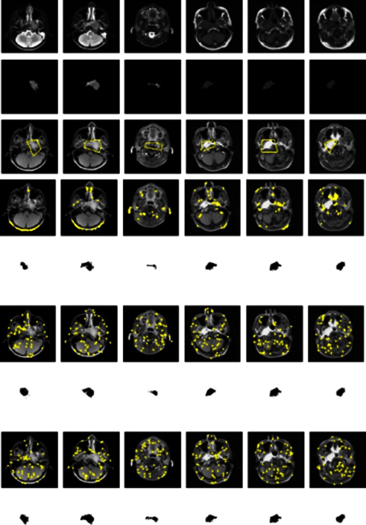
In clinical diagnosis of nasopharyngeal carcinoma (NPC) lesion, clinicians are often required to delineate boundaries of NPC on a number of tumor-bearing magnetic resonance images, which is a tedious and time-consuming procedure highly depending on expertise and experience of clinicians. Computer-aided tumor segmentation methods (either contour-based or region-based) are necessary to alleviate clinicians' workload. For contour-based methods, a minimal user interaction to draw an initial contour inside or outside the tumor lesion for further curve evolution to match the tumor boundary is preferred, but parameters within most of these methods require manual adjustment, which is technically burdensome for clinicians without specific knowledge. Therefore, segmentation methods with a minimal user interaction as well as automatic parameters adjustment are often favored in clinical practice. In this paper, two region-based methods with parameters learning are introduced for NPC segmentation. Two hundred fifty-three MRI slices containing NPC lesion are utilized for evaluating the performance of the two methods, as well as being compared with other similar region-based tumor segmentation methods. Experimental results demonstrate the superiority of adopting learning in the two introduced methods. Also, they achieve comparable segmentation performance from a statistical point of view.


Figures

Fig. 1
Fig. 1
An illustration of support vector machine classifier with a separating hyperplane (solid line) together with its two parallel hyperplanes (dashed lines), as well as samples from two classes in a simple 2-D feature space
Fig. 2
Fig. 2
An illustration of biased situations for PPV (left) and SEN (right)
Fig. 3
Fig. 3
NPC segmentation results from six pairs of MRI slices by all compared methods. (Rows: first—T1W (or T2W) MRI slices, second—CET1W MRI slices with fat suppression, third—NPC lesion ground truth manually traced by senior radiologists, fourth—NPC segmentation results by SVDD-based method, fifth—NPC segmentation results by SVM-based method, sixth—NPC segmentation results by Baseline; seventh—NPC segmentation results by the clustering-based method)
Fig. 4
Fig. 4
a Box-and-whisker plot of PPV among all four compared methods. b Box-and-whisker plot of SEN among all four compared methods
Fig. 5
Fig. 5
NPC segmentation results after applying different sampling strategies for negative training data in six pairs (columns) of cases. (Rows: first—T1W (or T2W) MRI slices, second—NPC lesion ground truth, third—ROI for each case, fourth, sixth, eighth—scatter plots of negative samples by applying a biased random sampling strategy, the random sampling strategy, and the stratified sampling strategy, respectively, fifth, seventh, ninth—corresponding NPC segmentation results after adopting a biased sampling strategy, the random sampling strategy, and the stratified sampling strategy in the proposed clustering-based method, respectively)

Similar articles

Cited by

References

    1. Chang E, Adami H. The enigmatic epidemiology of nasopharyngeal carcinoma. Canc Epidemiol Biomarkers Prev. 2006;15:1765–1777. doi: 10.1158/1055-9965.EPI-06-0353. - DOI - PubMed
    1. Parkin D, Bray F, Ferlay J, Pisani P. Global cancer statistics. CA Cancer J Clin. 2005;55:74–108. doi: 10.3322/canjclin.55.2.74. - DOI - PubMed
    1. Chong V, Fan V, Khoo J. Nasopharyngeal carcinoma with intracranial spread: CT and MR characteristics. J Comput Assist Tomogr. 1996;20(4):563–569. doi: 10.1097/00004728-199607000-00012. - DOI - PubMed
    1. Chua D, Sham J, Kwong D. Volumetric analysis of tumor extent in nasopharyngeal carcinoma and correlation with treatment outcome. Int J Radiat Oncol Biol Phys. 1997;39(3):711–719. doi: 10.1016/S0360-3016(97)00374-X. - DOI - PubMed
    1. Chen M, Chen T, Liu J, Chang C, Chie W. Better prediction of prognosis for patients with nasopharyngeal carcinoma using primary tumor volume. Cancer. 2004;100(10):2160–2166. doi: 10.1002/cncr.20210. - DOI - PubMed

MeSH terms