. 2012 Dec;80(12):2701-10.
doi: 10.1002/prot.24155. Epub 2012 Sep 15.

The mechanism of the converter domain rotation in the recovery stroke of myosin motor protein

Affiliations

The mechanism of the converter domain rotation in the recovery stroke of myosin motor protein

Andrij Baumketner. Proteins. 2012 Dec.

Abstract

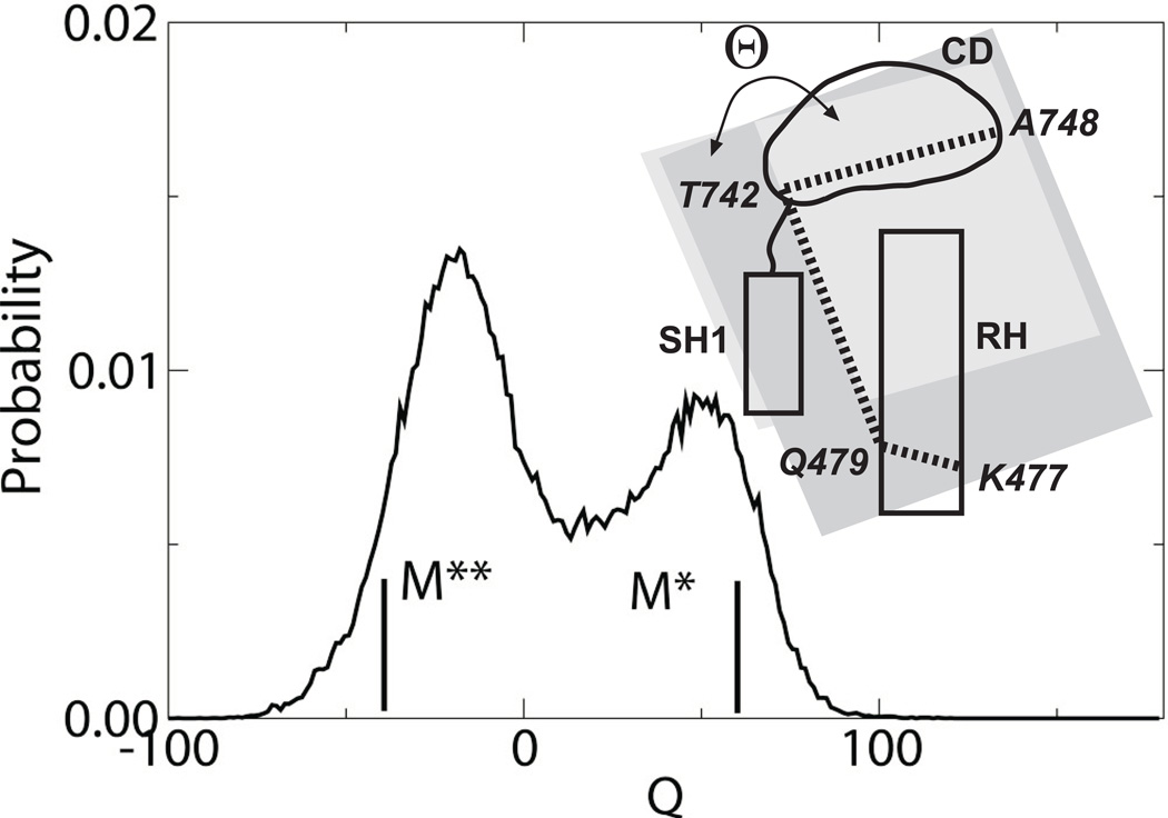
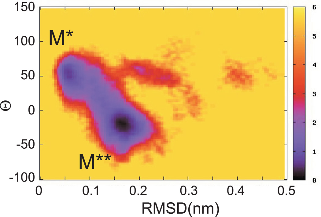
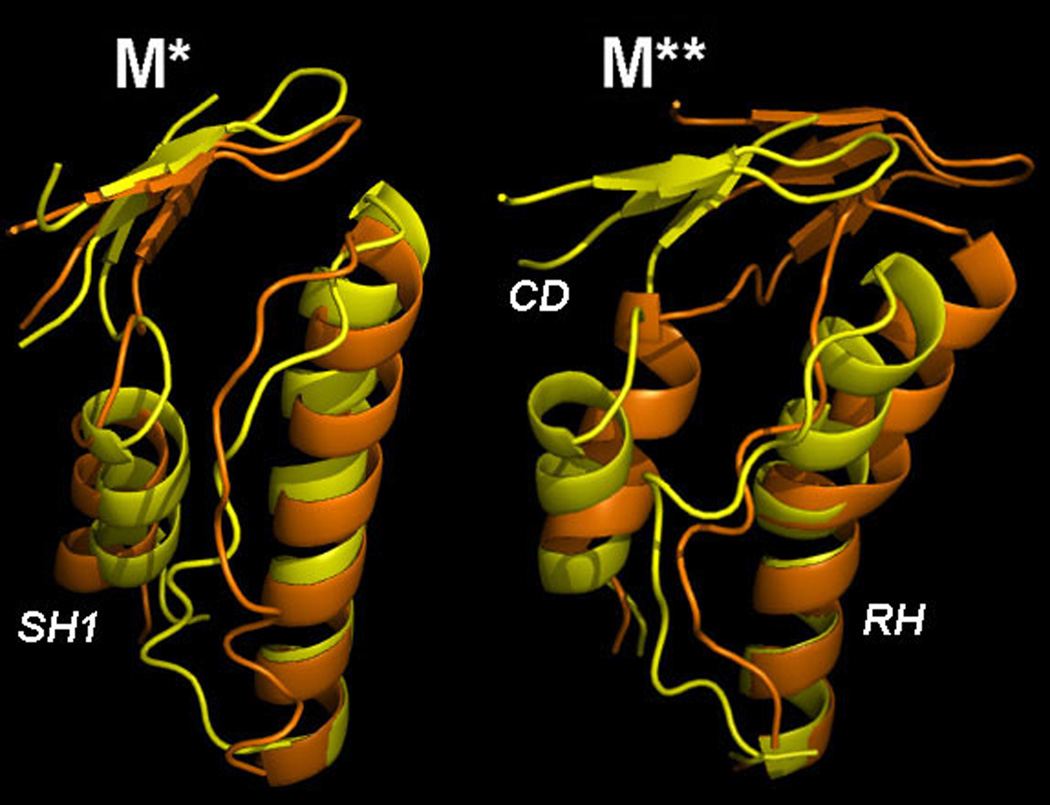
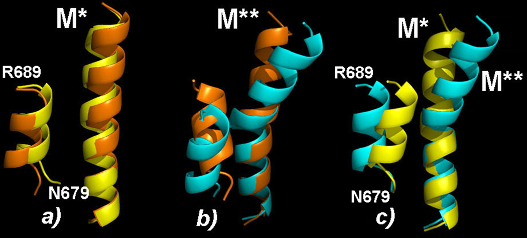
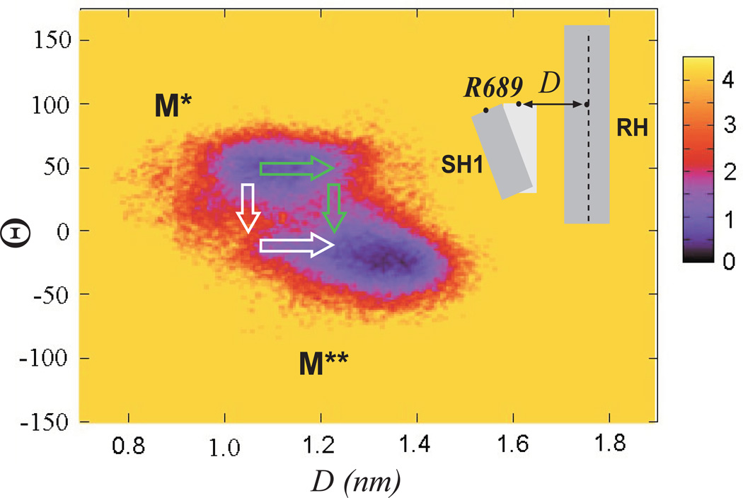
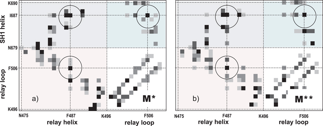
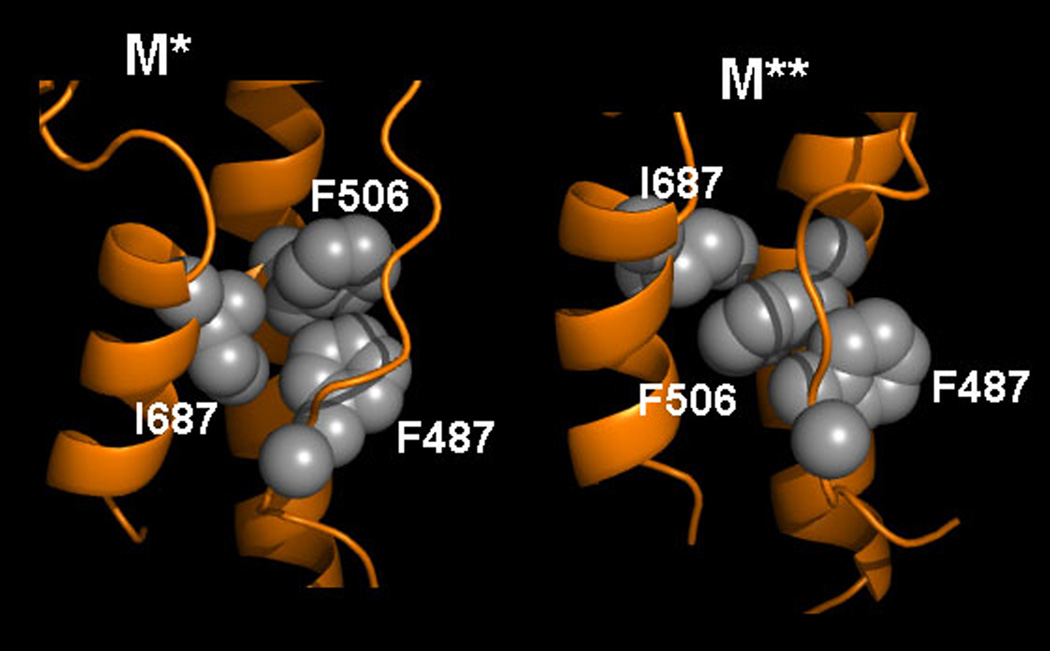
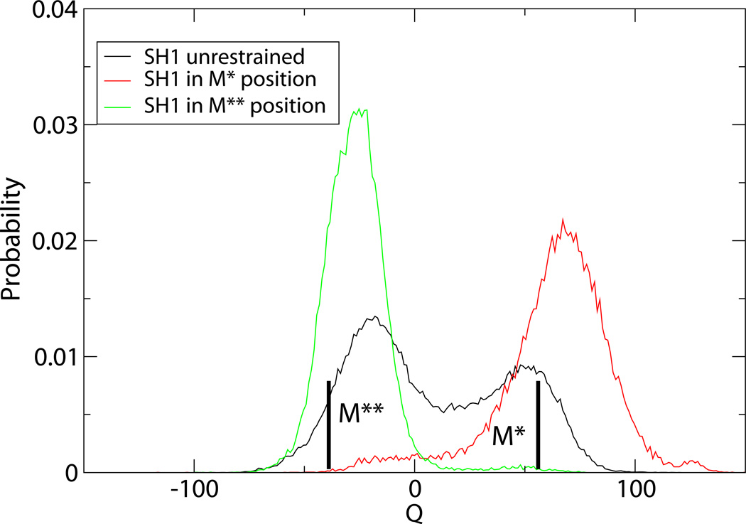
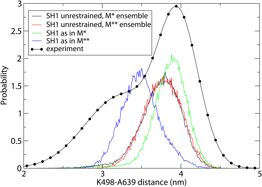
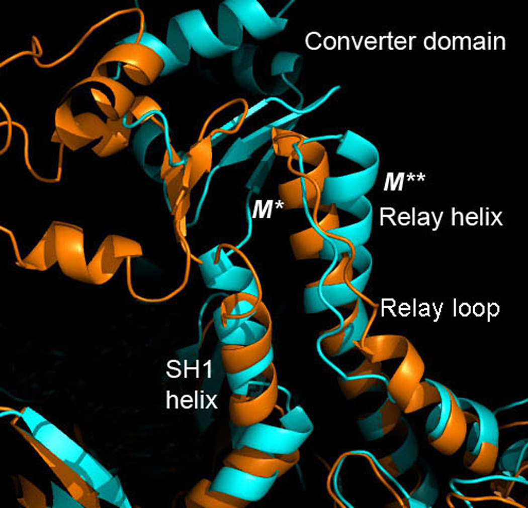
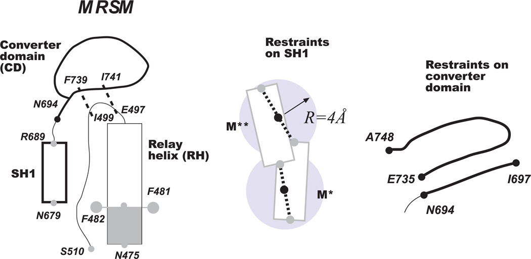
Upon ATP binding, myosin motor protein is found in two alternative conformations, prerecovery state M* and postrecovery state M**. The transition from one state to the other, known as the recovery stroke, plays a key role in the myosin functional cycle. Despite much recent research, the microscopic details of this transition remain elusive. A critical step in the recovery stroke is the rotation of the converter domain from "up" position in prerecovery state to "down" position in postrecovery state that leads to the swing of the lever arm attached to it. In this work, we demonstrate that the two rotational states of the converter domain are determined by the interactions within a small structural motif in the force-generating region of the protein that can be accurately modeled on computers using atomic representation and explicit solvent. Our simulations show that the transition between the two states is controlled by a small helix (SH1) located next to the relay helix and relay loop. A small translation in the position of SH1 away from the relay helix is seen to trigger the transition from "up" state to "down" state. The transition is driven by a cluster of hydrophobic residues I687, F487, and F506 that make significant contributions to the stability of both states. The proposed mechanism agrees well with the available structural and mutational studies.


Figures

Figure 1
Figure 1
Probability distribution of the torsional angle Θ as seen in the simulations of MRSM. The angle, formed by Cα of residues K477, Q479, T742 and A748, characterizes the orientation of the converter domain relative to the relay helix. Values of crystallographic M* and M** states are shown as thick lines.
Figure 2
Figure 2
Free energy map as a function of 1) RMSD over the relay helix from crystallographic M* conformation and 2) torsional angle Θ. Here and in other figures the energy is shown in units of kT, where k is the Boltzmann’s constant and T is the temperature. Two minima are seen with conformations associated with M* and M** states.
Figure 3
Figure 3
Most populated structures observed in our simulations for M* and M** ensembles, yellow, in comparison with the corresponding crystallographic structures, orange.
Figure 4
Figure 4
Orientation of SH1 helix in computational M* and M** clusters, shown in yellow and cyan respectively, in comparison with the corresponding crystallographic structures, shown in orange. In the computational M** state, SH1 is displaced by ˜2Å away from the relay helix.
Figure 5
Figure 5
Free energy map defined as a function of D, distance from Cα of R689 to RH, and the torsional angle Θ. Green arrows indicate the most likely scenario of M*-to M** transition, initiated by a displacement of SH1. White arrows illustrate an unlikely scenario where the converter domain rotates first and is followed by the displacement of SH1.
Figure 6
Figure 6
Contact probabilities of M* and M** ensembles observed in the reported simulations. A contact is considered formed between two residues if the shortest distance between any two atoms of their side chains is 6Å or less.
Figure 7
Figure 7
Configuration of critical hydrophobic residues I687, F487 and F506 in M* and M** conformations.
Figure 8
Figure 8
Distribution function of the torsional rotation angle obtained in the simulations of our model with SH1 a) unrestrained and b) restrained to its position in crystallographic M* and M** states.
Figure 9
Figure 9
Distribution function of the distance between K498 and A639 obtained in this work in various simulations as explained in the legend. Symbols indicate EPR data
Figure 10
Figure 10
Close up of M*, orange, and M**, cyan, conformations aligned along the N-terminal section of the relay helix. The kink visible in M** state occurs in the C-terminal part that interacts with relay loop, converter domain and SH1 helix. The latter is shifted by 4Å in M** relative to M*.
Figure 11
Figure 11
The minimal recovery stroke model (MSRM) introduced in this work. The model consists of relay helix (RH), relay loop (RL), converter domain (CD) and SH1 helix. Shaded areas correspond to the parts restrained to their initial crystallographic positions. Parts shown in bold lines are made to maintain their conformation by inter-residue restraints. Thin lines show completely flexible parts. SH1 helix is kept near RH by two restraints acting on N679 and R689 that permit both M** and M* conformations. The converter domain contains two fragments, kept in contact with RH by two restraints.

References

    1. Lymn RW, Taylor EW. Mechanism of Adenosine Triphosphate Hydrolysis by Actomyosin. Biochemistry. 1971;10(25):4617–4624. - PubMed
    1. Malnasi-Csizmadia A, Pearson DS, Kovacs M, Woolley RJ, Geeves MA, Bagshaw CR. Kinetic resolution of a conformational transition and the ATP hydrolysis step using relaxation methods with a Dictyostelium myosin II mutant containing a single tryptophan residue. Biochemistry. 2001;40(42):12727–12737. - PubMed
    1. Agafonov RV, Nesmelov YE, Titus MA, Thomas DD. Muscle and nonmuscle myosins probed by a spin label at equivalent sites in the force-generating domain. P Natl Acad Sci USA. 2008;105(36):13397–13402. - PMC - PubMed
    1. Sweeney HL, Houdusse A. Structural and Functional Insights into the Myosin Motor Mechanism. Annual Review of Biophysics, Vol 39. 2010;39:539–557. - PubMed
    1. Wray J, Urbanke C. A fluorescence temperature-jump study of conformational transitions in myosin subfragment 1. Biochemical Journal. 2001;358:165–173. - PMC - PubMed

Publication types

LinkOut - more resources