. 2012 Aug 1;4(145):145ra106.
doi: 10.1126/scitranslmed.3004140.

Inhibition of LTi cell development by CD25 blockade is associated with decreased intrathecal inflammation in multiple sclerosis

Affiliations

Inhibition of LTi cell development by CD25 blockade is associated with decreased intrathecal inflammation in multiple sclerosis

Justin S A Perry et al. Sci Transl Med. .

Abstract

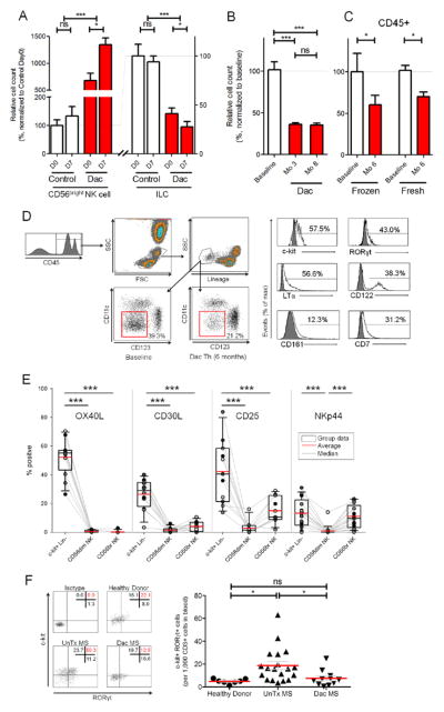
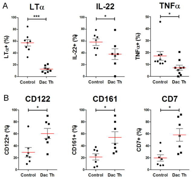
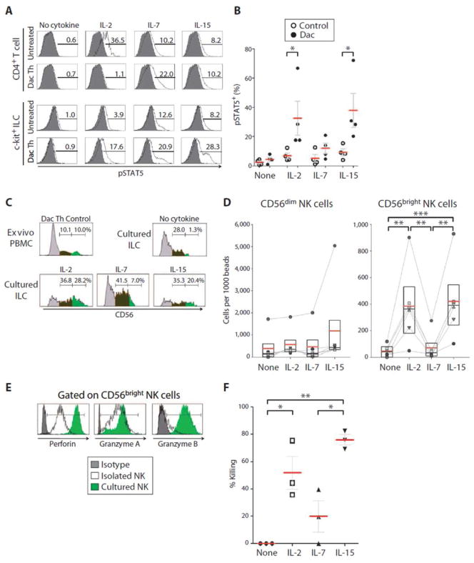
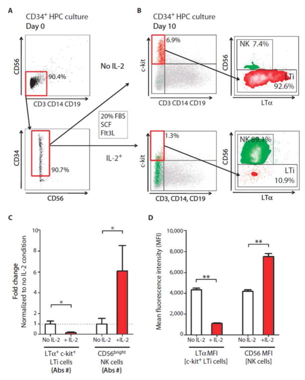
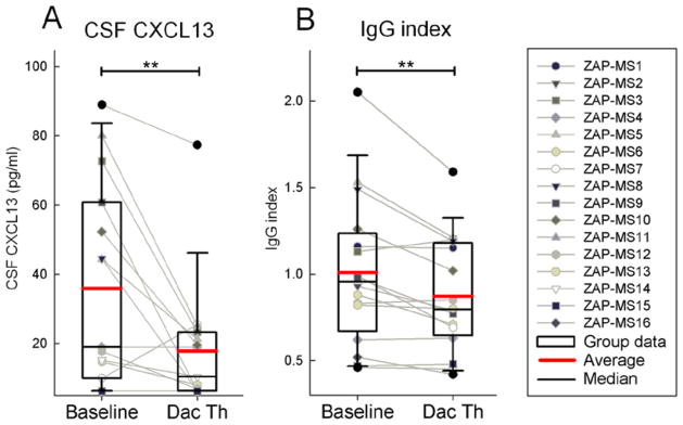
Genetic polymorphisms in the interleukin-2 receptor α (IL-2Rα) chain (CD25) locus are associated with several human autoimmune diseases, including multiple sclerosis (MS). Blockade of CD25 by the humanized monoclonal antibody daclizumab decreases MS-associated inflammation but has surprisingly limited direct inhibitory effects on activated T cells. The present study describes unexpected effects of daclizumab therapy on innate lymphoid cells (ILCs). The number of circulating retinoic acid receptor-related orphan receptor γt-positive ILCs, which include lymphoid tissue inducer (LTi) cells, was found to be elevated in untreated MS patients compared to healthy subjects. Daclizumab therapy not only decreased numbers of ILCs but also modified their phenotype away from LTi cells and toward a natural killer (NK) cell lineage. Mechanistic studies indicated that daclizumab inhibited differentiation of LTi cells from CD34⁺ hematopoietic progenitor cells or c-kit⁺ ILCs indirectly, steering their differentiation toward immunoregulatory CD56(bright) NK cells through enhanced intermediate-affinity IL-2 signaling. Because adult LTi cells may retain lymphoid tissue-inducing capacity or stimulate adaptive immune responses, we indirectly measured intrathecal inflammation in daclizumab-treated MS patients by quantifying the cerebrospinal fluid chemokine (C-X-C motif) ligand 13 and immunoglobulin G index. Both of these inflammatory biomarkers were inhibited by daclizumab treatment. Our study indicates that ILCs are involved in the regulation of adaptive immune responses, and their role in human autoimmunity should be investigated further, including their potential as therapeutic targets.


Conflict of interest statement

Competing interests: B.B. is a co-inventor on NIH patents related to daclizumab therapy and as such has received royalty payments based upon license to US patents 7,575,742 and 7,258,859 (and patent applications claiming priority to these patents) and other National Stage patents and patent applications claiming priority to PCT/US2002/038290 or PCT/US2003/020428. The other authors declare no competing interests.

Figures

Figure 1
Figure 1. Daclizumab therapy decreases numbers of pro-inflammatory ILCs
(A) Fresh PBMCs were analyzed before (D0) and 7 days after (D7) vaccination with Influenza haemagglutinin (Flu-HA) in daclizumab treated MS patients (Dac) and age/gender matched controls by flow cytometry. Data were normalized so that the baseline (D0) of the control group represented 100%, to which the baseline of daclizumab-treated MS patients and Flu-HA induced changes in both cohorts (D7) were compared. (n=5–8) (B) Cryopreserved PBMCs were analyzed similar to (A) and samples were from 17 MS patients at baseline (BL), after 3 months of treatment with daclizumab (Mo 3), and after 8 months of treatment with daclizumab (Mo 8). (C) Cryopreserved PBMCs (n=6) and fresh PBMCs (n=6) in the DAC HYP clinical trial were analyzed similar to (A and B) at baseline (BL) and after 6 months of treatment with daclizumab (Mo 6). Gating on CD45+ cells prevented inclusion of unlysed erythrocytes into the lineage-negative ILC gate. (D) Gating strategy for ILCs: ILCs were gated based on forward and side scatter, and subgated as CD45+ and lineage (CD3, CD14, CD19, CD20, CD56) negative, non-DCs (i.e. CD11c/CD123). The proportion of ILCs that expressed surface markers CD161, CD7, CD122 and c-kit and intracellular LTα and RORγt are depicted in representative sample. Dark gray histograms represent appropriate isotype controls. (E) Percentage of CD45+ c-kit+ ILCs, CD56dim and CD56bright NK cells which express OX40L, CD30L, CD25 and NKp44 were determined by flow cytometry. Fresh uncoagulated peripheral blood samples were used. (n=11–17) (F) Expression of RORγt and c-kit of ILCs in fresh peripheral blood from healthy donors, untreated MS patients (UnTx MS) and daclizumab-treated MS patients (Dac MS) was determined by flow cytometry using gating strategy depicted in (D). (G) Ratio of RORγt+/c-kit+ ILCs: CD3+ T cells was calculated from the peripheral blood samples of healthy donors, UnTx MS and Dac MS. The horizontal bars represent the mean of each group.
Figure 2
Figure 2. Daclizumab treatment modifies phenotype of ILCs away from LTi and toward NK cell lineage
(A) Percentage of ILCs which express inflammatory the cytokines LTα, IL-22, and TNF upon PMA (20ng/ml) and Ionomycin (1μM) stimulation for 3 hours was determined by flow cytometry. (B) Percentage of ILCs which express NK cell markers, CD161, CD7 and CD122 was determined. The horizontal bars represent the mean of each of the groups.
Figure 3
Figure 3. Enhanced intermediate IL-2/IL-15 signaling in daclizumab-treated patients promotes differentiation of ILCs toward functional NK cells
(A) Representative pSTAT5 levels of untreated or daclizumab (Dac Th) treated MS patients. Purified ILCs were stimulated with IL-2 (100IU/ml), IL-7 (10ng/ml), IL-15 (10ng/ml) or no cytokine for 10 minutes (optimal concentrations determined in pilot experiments). ILCs were then immediately fixed and stained for phosphorylated Stat5 production. (B) Group signaling data analogous to (A). (C) Purified c-kit+ ILCs cells were cultured for 7 days in media supplemented with SCF and Flt3L (both 10ng/ml) and in the presence of IL-2 (100IU/ml), IL-7 (10ng/ml), IL-15 (10ng/ml) or no cytokine control. At day 7, cultured cells were stained and analyzed by FACS for the presence of CD56+ NK cells (compared to ex vivo PBMCs of daclizumab-treated MS patients with significant expansion of CD56brightNK cells). (C) Raw data from a representative experiment and (D) represents group data of the number of CD56dim NK and CD56bright NK cells per 1,000 beads. (E) Purified c-kit+ ILCs were cultured as in panel C. At day 7, cultured cells were FACS stained for functional NK markers (Perforin, Granzyme A and B; compared to ex vivo PBMC of daclizumab-treated MS patients with significant expansion of CD56bright NK cells). FACS plots are representative of 4 replications. (F) Also on day 7, cultured cells were incubated for ~16hr with target GFP-tagged K562 cells at a 1:1 effector to target ratio (or K562 cell only control wells). K562 killing was then measured by flow cytometry. Killing was determined as the percentage of K562 cells that were lost relative to the target cells only condition. The horizontal bars represent the mean of each of the groups.
Figure 4
Figure 4. CD34+ HPCs differentiate in vitro to either LTi or CD56bright NK cells, depending on the presence of IL-2
(A and B) Purified CD34+ HPC cells were cultured with SCF and Flt3L (both 10ng/ml) with either no cytokine or IL-2 (100IU/ml). After 10–14 days of culture the proportion of LTi cells (lineage-/c-kit+/LTα+ and CD56+ NK cell was determined by flow cytometry. (B) Representative raw data (C) Fold change calculated from the absolute numbers of LTi and CD56bright NK cells and (D) MFI of LTα and CD56 in CD34 HPC cultures after 10–14 days of differentiation. The horizontal bars represent the mean of each of the groups.
Figure 5
Figure 5. Daclizumab treatment decreases CSF levels of CXCL13 and intrathecal production of IgG
(A) CSF CXCL13 concentration was measured by ELISA. (B) IgG index as measured by NIH clinical laboratory. Raw data for individual patients measured before (Baseline) and 6.5 months after initiation of daclizumab therapy (Dac Th) are depicted as gray dot and line blots and group data are depicted as box plots with mean highlighted as red and median as black horizontal line.

Comment in

References

    1. Waldmann TA, O’Shea J. The use of antibodies against the IL-2 receptor in transplantation. Curr Opin Immunol. 1998;10:507–512. - PubMed
    1. Nussenblatt RB, Thompson DJ, Li Z, Peterson JS, Robinson RR, Shames RS, Nagarajan S, Tang MT, Mailman M, Velez G, Roy C, Levy-Clarke GA, Suhler EB, Djalilian A, Sen HN, Al-Khatib S, Ursea R, Srivastava S, Bamji A, Mellow S, Sran P, Waldmann TA, Buggage RR. Humanized anti-interleukin-2 (IL-2) receptor alpha therapy: long-term results in uveitis patients and preliminary safety and activity data for establishing parameters for subcutaneous administration. J Autoimmun. 2003;21:283–293. - PubMed
    1. Bielekova B, Richert N, Howard T, Blevins G, Markovic-Plese S, McCartin J, Wurfel J, Ohayon J, Waldmann TA, McFarland HF, Martin R. Humanized anti-CD25 (daclizumab) inhibits disease activity in multiple sclerosis patients failing to respond to interferon-beta. Proc Natl Acad Sci U S A. 2004;101:8705–8708. - PMC - PubMed
    1. Wynn D, Kaufman M, Montalban X, Vollmer T, Simon J, Elkins J, O’Neill G, Neyer L, Sheridan J, Wang C, Fong A, Rose JW. Daclizumab in active relapsing multiple sclerosis (CHOICE study): a phase 2, randomised, double-blind, placebo-controlled, add-on trial with interferon beta. Lancet Neurol. 2010;9:381–390. - PubMed
    1. Rose JW, Watt HE, White AT, Carlson NG. Treatment of multiple sclerosis with an anti-interleukin-2 receptor monoclonal antibody. Ann Neurol. 2004;56:864–867. - PubMed

Publication types

MeSH terms